. 2020 Jun 29;17(2):e20200006.
doi: 10.1590/1984-3143-AR2020-0006.

Histomorphometric and vascular changes in equine endometrium after the infusion of conceptus fragments

Affiliations

Histomorphometric and vascular changes in equine endometrium after the infusion of conceptus fragments

Cesar Augusto Camacho et al. Anim Reprod. .

Abstract

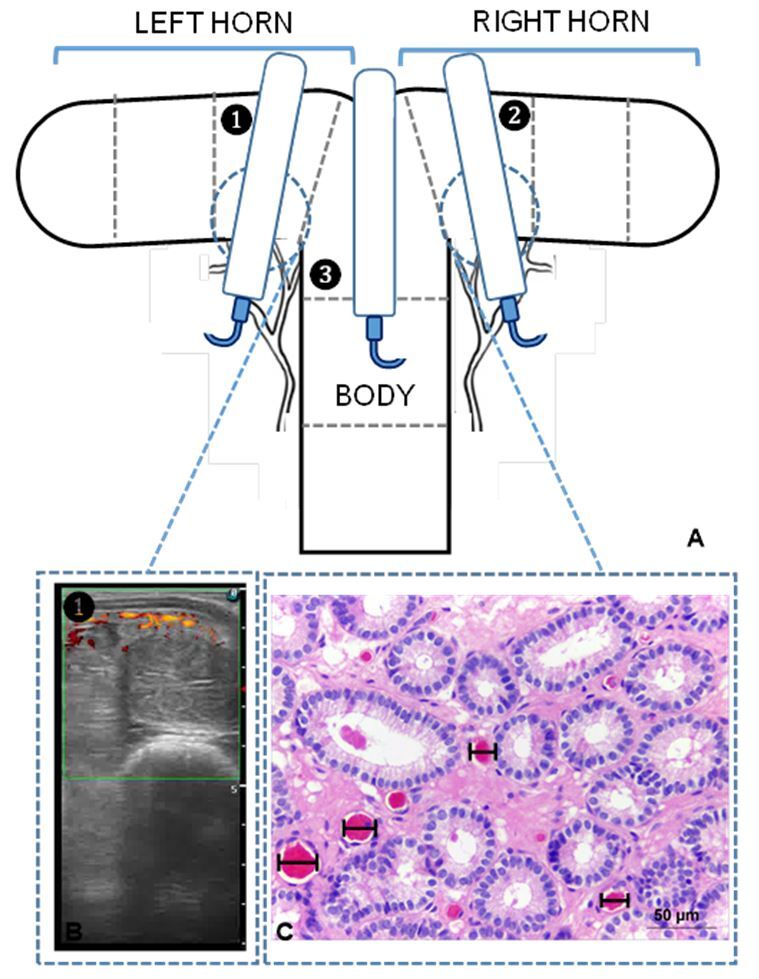
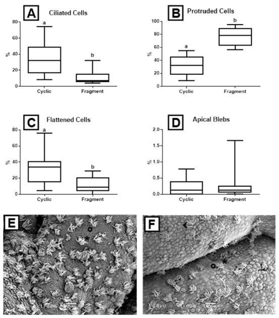
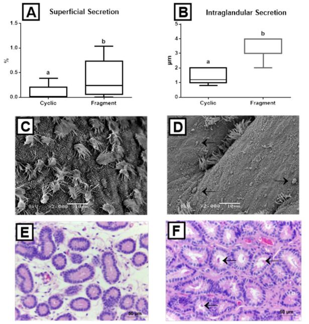
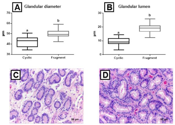
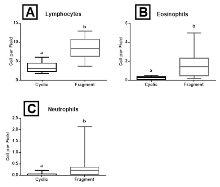
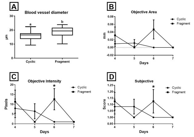
This experiment aimed to verify if the proteins present in a 13th day conceptus induce changes in the equine endometrial ultra-structure, histology, and vascularization, two days after its infusion. Ten healthy cyclic mares were used. Once estrus was confirmed, mares were examined daily to detect ovulation (day 0). After ovulation, mares were examined daily until day seven by transrectal palpation and B-mode and Doppler ultrasonography. In this first cycle, intrauterine biopsies were collected at day seven after ovulation, constituting the Cyclic group (n = 10). In the second cycle, the same mares daily were examined until ovulation was detected. After ovulation, mares were examined daily by transrectal palpation and B-mode and Doppler ultrasonography until day 7. On day 5, after ovulation, fragments from previously collected 13-day-old concepti were infused into the uterus of each mare. Intrauterine biopsies were collected at day 7 in all mares (n = 10), constituting the Fragment group. The percentage of ciliated and flattened cells decreased in the Fragment group. Protruded cells, superficial and intraglandular secretion, glandular lumen and diameter, blood vessel diameter, endometrial vascularization, and immune cells were higher in the Fragment group than in the Cyclic group. In summary, proteins of 13th day equine conceptus fragments infused at day five after ovulation signaled histological and vascular changes in the endometrium at the 7th day after ovulation.

Keywords: histological; ultrastructure; uterus; vascularization.


Conflict of interest statement

Conflicts of interest: The authors have no conflict of interest to declare.

Figures

Figure 1
Figure 1. (A) Diagram of the transrectal placement of a linear-array ultrasound transducer, showing the spatial relationships between the uterine horns and transducer. The numbers show the 3 uterine segments used in the experiment; (B) Ultrasonography image. The colored spots are Power-Doppler indicators of blood flow; (C) Histological section of an endometrial biopsy; black bars show blood vessel diameter.
Figure 2
Figure 2. Box-plots (whiskers represent min to max) of (A) ciliated cells; (B) protruded cells; (C) flattened cells; and (D) apical blebs of the Cyclic and Fragment groups. Different letters (a, b) represent significant difference (P < 0.002); (E) Scanning electron micrograph (1,000x, bar = 10 µm) of the endometrium of the Cyclic group; (F) Scanning electron micrograph (1,000x, bar = 10 µm) of the endometrium of the Fragment group. Asterisks (*) indicate ciliated cells, black arrows indicate protruded cells, and circles show flattened cells.
Figure 3
Figure 3. Box-plots (whiskers represent min to max) of (A) superficial secretion and (B) intraglandular secretion of the Cyclic and Fragment groups. Different letters (a, b) represent significant difference (P< 0.03); (C) (Cyclic group) Scanning electron micrograph (2,000x, bar = 10 µm) of the endometrium showing ciliated cells, protruded secretory cells, flattened secretory cells, and secretion; (D) (Fragment group) Scanning electron micrograph (2,000x, bar = 10 µm) of the endometrium showing ciliated cells, protruded secretory cells, flattened secretory cells, and secretion; (E) (Cyclic group) Histological section of endometrial glands (400x, bar = 20 µm); (F) (Fragment group) Histological section of endometrial glands (400x, bar = 20 µm). Black arrow shows superficial and intraglandular secretion.
Figure 4
Figure 4. Box-plots (whiskers represent min to max) of (A) glandular diameter and (B) glandular lumen of the Cyclic and Fragment groups. Different letters (a, b) represent significant difference (P < 0.001); (C) (Cyclic group) histological section of endometrial glands (400x, bar = 20 µm); (D) (Fragment group) histological section of endometrial glands (400x, bar = 20 µm), equine endometrial tissues.
Figure 5
Figure 5. Box-plots (whiskers represent min to max) of (A) lymphocytes; (B) eosinophils; and (C) neutrophils of the Cyclic and Fragment groups in the equine endometrial tissues. Different letters (a, b) represent significant difference (P = 0.001).
Figure 6
Figure 6. Box-plots (whiskers represent min to max) of (A) blood vessel diameter of the Cyclic and Fragment groups (7th day). Different letters (a, b) represent significant difference (P = 0,022); (B) (4th to 7th day) Power Doppler objective endometrial vascularization area; (C) (4th to 7th day) Power Doppler objective intensity of endometrial vascularization; (D) (4th to 7th day) Subjective endometrial vascularization. Asterisks (*) represent significant difference (P < 0.05).

Similar articles

References

    1. Aurich C, Budik S. Early pregnancy in the horse revisited: does exception prove the rule? J Anim Sci Biotechnol. 2015;6(1):50. doi: 10.1186/s40104-015-0048-6. - DOI - PMC - PubMed
    1. Ball B, Altschul M, Hillman R. Luteal maintenance in mares after transfer of equine trophoblastic vesicles. Equine Vet J. 1989;21(S8):21–24. doi: 10.1111/j.2042-3306.1989.tb04667.x. - DOI
    1. Bastos HBA, Martinez MN, Camozzato GC, Estradé MJ, Barros E, Vital CE, Vidigal PMP, Meikle A, Jobim MIM, Gregory RM, Mattos RC. Proteomic profile of histotroph during early embryo development in mares. Theriogenology. 2019;125:224–235. doi: 10.1016/j.theriogenology.2018.11.002. - DOI - PubMed
    1. Bazer FW, Roberts RM, Thatcher WW. Actions of hormones on the uterus and effect on conceptus development. J Anim Sci. 1979;49(Suppl 2):35–45. doi: 10.1093/ansci/49.Supplement_II.35. - DOI - PubMed
    1. Bazer FW, Vallet JL, Roberts RM, Sharp DC, Thatcher WW. Role of conceptus secretory products in establishment of pregnancy. J Reprod Fertil. 1986;76(2):841–850. doi: 10.1530/jrf.0.0760841. - DOI - PubMed

LinkOut - more resources