. 2020 Jun 8;7(14):2000532.
doi: 10.1002/advs.202000532. eCollection 2020 Jul.

Structural Basis of Human Helicase DDX21 in RNA Binding, Unwinding, and Antiviral Signal Activation

Affiliations

Structural Basis of Human Helicase DDX21 in RNA Binding, Unwinding, and Antiviral Signal Activation

Zijun Chen et al. Adv Sci (Weinh). .

Abstract

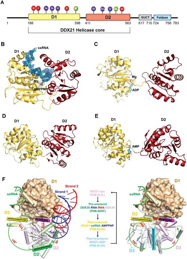
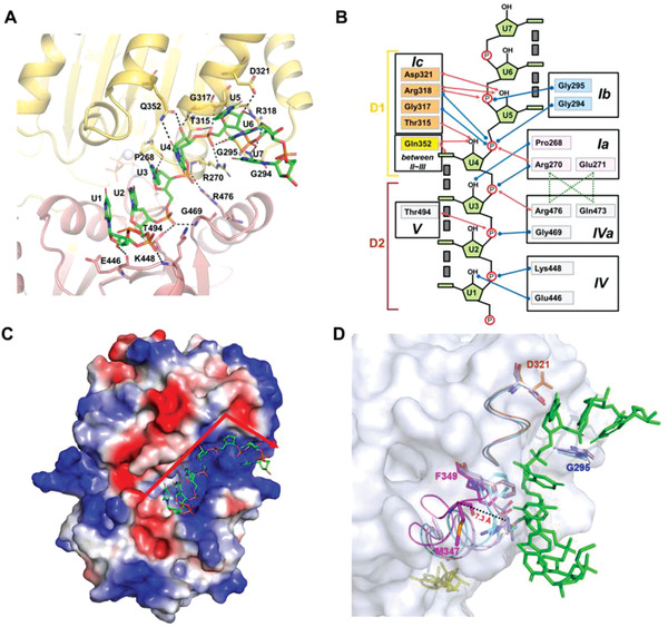
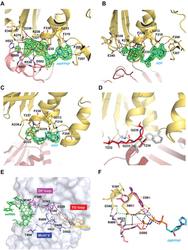
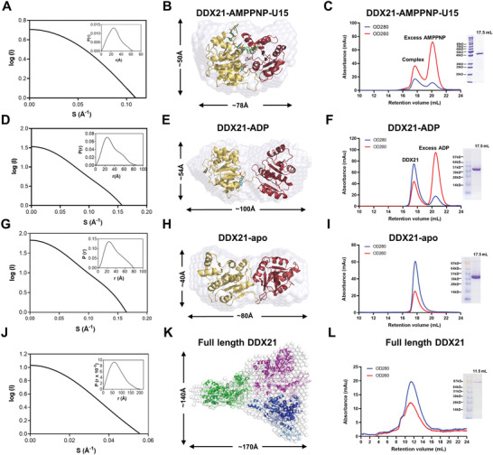
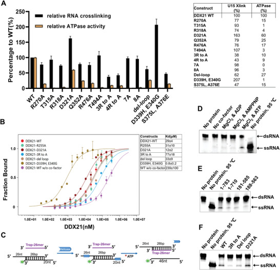
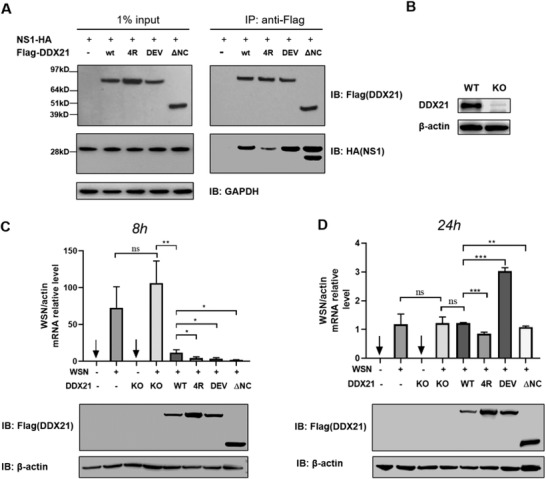
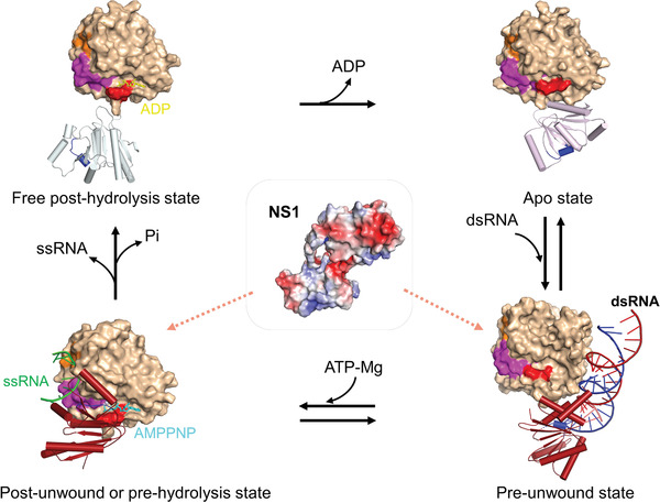
RNA helicase DDX21 plays vital roles in ribosomal RNA biogenesis, transcription, and the regulation of host innate immunity during virus infection. How DDX21 recognizes and unwinds RNA and how DDX21 interacts with virus remain poorly understood. Here, crystal structures of human DDX21 determined in three distinct states are reported, including the apo-state, the AMPPNP plus single-stranded RNA (ssRNA) bound pre-hydrolysis state, and the ADP-bound post-hydrolysis state, revealing an open to closed conformational change upon RNA binding and unwinding. The core of the RNA unwinding machinery of DDX21 includes one wedge helix, one sensor motif V and the DEVD box, which links the binding pockets of ATP and ssRNA. The mutant D339H/E340G dramatically increases RNA binding activity. Moreover, Hill coefficient analysis reveals that DDX21 unwinds double-stranded RNA (dsRNA) in a cooperative manner. Besides, the nonstructural (NS1) protein of influenza A inhibits the ATPase and unwinding activity of DDX21 via small RNAs, which cooperatively assemble with DDX21 and NS1. The structures illustrate the dynamic process of ATP hydrolysis and RNA unwinding for RNA helicases, and the RNA modulated interaction between NS1 and DDX21 generates a fresh perspective toward the virus-host interface. It would benefit in developing therapeutics to combat the influenza virus infection.

Keywords: ATPases; DDX21; RNA helicases; crystal structures; viral protein NS1.


Conflict of interest statement

The authors declare no conflict of interest.

Figures

Figure 1
Figure 1
The overall structure of DDX21 helicase core in different unwinding states. A) Domain structure of human DDX21 and conserved sequence motifs of the helicase core. The DDX21 helicase core (D1D2 core, residue 188‐563) was used for structural studies. The 12 highly conserved DDX sequence motifs were colored for their primary functions: red, ATP binding; blue, RNA binding; and green, inter‐domain interaction. See also Figure S1 (Supporting Information). B) Crystal structure of the D1D2 core (yellow/red) bound with ssRNA and AMPPNP‐Mg2+. DDX21 is shown in cartoon model, whereas ssRNA is in a stick model with an electron density map contoured to 3.0 σ at the F oF c map. C) Crystal structure of the D1D2 core (yellow/red) bound with ADP‐Mg2+. D) Crystal structure of the D1D2 core (yellow/red) in its apo‐state. E) Crystal structure of the D1D2 core (yellow/red) bound with AMP. The disordered part (residues Ile400 – Ile409) connecting the two domains is indicated by a broken line. F) Conformational comparison between the DDX21‐apo (in pink, PDB: 6L5L), DDX3X‐dsRNA (in green, PDB: 6O5F; and only one DDX3X molecule is shown for clarity), DDX21‐ssRNA‐AMPPNP (in yellow, PDB: 6L5N), and DDX21‐ADP (in cyan, PDB: 6L5O) structures. The left panel shows the conformational difference between the apo state, pre‐unwound state and post‐unwound state. The right panel shows the conformational difference between the post‐wound state, post‐hydrolysis and apo state, combining into a four‐step unwinding cycle elucidated in the middle panel. The structures were aligned based on their D1 domains. The D1 of the DDX21‐ssRNA‐AMPPNP structure is illustrated as a molecular surface, and those of other structures are not shown for clarity. The D2 of all four structures are shown as cartoon models (helices as cylinders, strands as arrows, and loops as tubes). The orientation of D2, relative to the fixed D1 at each state, is indicated with a black arrow on the same α10 helix in D2 and a color gradient for emphasis. See also Figure 7 and Figure S3 (Supporting Information).
Figure 2
Figure 2
RNA binding and bending in DDX21‐ssRNA‐AMPPNP complex. A) Details of the RNA binding site in DDX21‐RNA‐AMPPNP complex structure. B) Schematic representation of the RNA binding site. Blue and red arrows indicated interactions through the main and the side chains of different residues, respectively. Gray boxes indicate base stacking. C) The electrostatic potential surface of the DDX21‐RNA‐AMPPNP complex. ssRNA is shown in the stick model. The red arrow shows the bent orientation of RNA around the motif Ic. Blue: positive charge; Red: negative charge. D) Comparison of the RNA binding pocket in three DDX21 structures with the surface representation of DDX21‐apo (gray). The four resides from the three motifs that had steric clashes with ssRNA (green) in apo‐state (light blue), AMP‐bound state (light pink) and ADP‐bound state (cyan) were shown as stick models, with the “DF loop” (D321‐F349; magenta), motif Ic (orange), motif Ib (GG; blue), and AMPPNP (yellow). The “DF loop” shifted 7.3 Å between DDX21‐apo and DDX21‐ssRNA‐AMPPNP, measured with the M347 residue.
Figure 3
Figure 3
The ATPase sites of DDX21. A) Representation of the ATPase sites in DDX21‐RNA‐AMPPNP complex, with the electron density map (F oF c) for AMPPNP‐Mg2+ and the presumptive catalytic water rendered at 3.0 σ. The broken red line connects the probable attacking water and the γ‐phosphorus atom. Arg204 is omitted for clarity. B) The ADP binding sites in DDX21‐ADP complex, with the electron density map (F oF c) for ADP‐Mg2+ rendered at 3.0 σ. Arg204 is omitted for clarity. C) The AMP binding sites in DDX21‐AMP complex, with the electron density map (F oF c) for AMP rendered at 3.0 σ. D) The potential ATP binding sites in the DDX21‐apo structure. The “TG loop” is colored in red. The modeled AMPPNP is colored in gray. E) Stereo view of the link between ATP binding pocket and RNA binding pocket, with the surface representation of DDX21‐ssRNA‐AMPPNP (gray). In the DDX21‐ssRNA‐AMPPNP structure, the “DF loop” is indicated in magenta, motif V (and Va) in blue, the “TG loop” in red, AMPPNP in yellow and RNA in green. In the DDX21‐apo structure, the “DF loop” is colored in light blue, and the “TG loop” is colored in red. Both of the “DF loop” and “TG loop” were colored in cyan in the DDX21‐ADP structure and light pink in the DDX21‐AMP structure. Green broken lines show the inter‐domain interactions. F) Superimposition of D339H and E340G mutants. D339H forms a new salt bridge (purple) with ATP phosphate and new cooperative bond (purple) with Mg2+ ion by the His substitution, while E340G mutation lacks the inter‐domain interactions with Arg499 and His523. Yellow broken lines show the interaction of Asp339 with Mg2+, mediated by water. Green broken lines show the inter‐domain interactions of Glu340 with Arg499 and His523 and inter‐domain interaction of Asp342 with Arg499. The broken red line shows the interaction between the presumptive attacking water and the γ‐phosphorus atom.
Figure 4
Figure 4
SAXS models and gel‐filtration profiles of full‐length DDX21 and the helicase core. A) The experimental scattering curve and the distance distribution function curve (inset) for DDX21 helicase core (188‐563) with AMPPNP and U15 ssRNA. B) Crystal structure of DDX21‐AMPPNP‐ssRNA (PDB: 6L5N) was fitted into the ab initio envelope obtained from SAXS. C) Gel filtration profile and the SDS‐PAGE of DDX21 helicase core (188‐563) with AMPPNP and U15 ssRNA. The helicase core and U15 ssRNA plus AMPPNP were coeluted out at the retention volume 17.6 mL from the superdex200 10/300 column (GE healthcare, Boston, USA), and excess AMPPNP was eluted out lately. D) The experimental scattering curve and the distance distribution function curve (inset) for DDX21 helicase core (188‐563) with ADP. E) Crystal structure of DDX21 (del‐loop)‐ADP (PDB: 6L5O) was fitted into the ab initio envelope obtained from SAXS. F) Gel filtration profile and the SDS‐PAGE of DDX21 helicase core (188‐563) with ADP. The helicase core with ADP was eluted out at the retention volume 17.5 mL from the superdex200 10/300 column (GE healthcare, Boston, USA), and excess ADP was eluted out lately. G) The experimental scattering curve and the distance distribution function curve (inset) for DDX21 helicase core (188‐563). H) Crystal structure of DDX21 (del‐loop)‐apo (PDB: 6L5L) was fitted into the ab initio envelope obtained from SAXS. I) Gel filtration profile and the SDS‐PAGE of DDX21 helicase core (188‐563). The helicase core was eluted out at the retention volume 17.6 mL from the superdex200 10/300 column (GE healthcare, Boston, USA), corresponding with a molecular weight around 30 kD, indicating the possible monomer status in solution. J) The experimental scattering curve and the distance distribution function curve (inset) for full‐length DDX21. (K) The modeled structure of full‐length DDX21 was fitted into the SAXS ab initio envelope with three DDX21 molecules. The modeled DDX21 structure was obtained using the Phyre2 web portal (http://www.sbg.bio.ic.ac.uk/~phyre/). L) Gel filtration profile and the SDS‐PAGE of full‐length DDX21 (1‐783). DDX21 was eluted out at the retention volume 11.6 mL from the superdex200 10/300 column (GE healthcare, Boston, USA), corresponding with a molecular weight around 340 kD, indicating the possible trimer or tetramer status in solution.
Figure 5
Figure 5
Mutational analysis of key residues involved in ATP hydrolysis and RNA binding. A) Histogram (left panel) and detailed values (right panel) of the RNA‐crosslinking and RNA‐stimulated ATPase activities, which are shown as percentages of the wide‐type DDX21 activity. All presented data represent means ±S.D. of three independent determinations. B) The U15 binding affinities of WT and mutant DDX21. Error bars represent the standard error between three replicate experiments. C) Schematic diagram of the unwinding process with 5’‐tailed RNA duplex and trap RNA. D) Full‐length DDX21 unwinds RNA duplex with or without different cofactors. The final products of unwinding reaction were separated by a 4–20% gradient Novex TBE Gel (Thermo Fisher Scientific, Waltham, USA). DDX21 unwound 5′‐tailed RNA duplex effectively with ATP and MgCl2, while very weak bands of ssRNA were still observed from other reaction pools without ATP. E) RNA unwinding with full length and different truncated forms of DDX21 by using a 4–20% gradient Novex TBE Gel (Thermo Fisher Scientific, Waltham, USA). The unwinding reactions were proceeded with 2 × 10−3 m ATP and 2 × 10−3 m MgCl2 by using 2 × 10−6 m DDX21 full‐length or truncations, 0.2 × 10−6 m 5′‐tailed RNA duplex, and 0.8 × 10−6 m unlabeled trap RNA. F) RNA unwinding with the full‐length WT and mutant DDX21, by using a 4–20% gradient Novex TBE Gel (Thermo Fisher Scientific, Waltham, USA), and the same reaction conditions described above in (E).
Figure 6
Figure 6
NS1 counteracts DDX21 activity in the cells. A) Coimmunoprecipitation of NS1 with WT and mutated DDX21 from cotransfected HEK293T cells. B) Immunoblotting analysis for endogenous DDX21 in HEK293T cells. C,D) Viral RNA synthesis level determination with quantitative real‐time RT‐PCR. qRT‐PCR analysis of viral RNA in wild‐type and DDX21 knock‐out HEK293T cells that transfected with indicated plasmids at C) 8 h post‐infection and D) 24 h post‐infection with WSN. *P < 0.05, **P < 0.01, ***P < 0.001, and ns, not significant (Student's t‐test, error bars, s.e.m., n = 3).
Figure 7
Figure 7
A proposed model for DDX21 unwinding cycle associated with NS1. A four‐step unwinding cycle represented by the apo‐ (PDB: 6L5L, this work), pre‐unwound (PDB: 6O5F), post‐unwound (PDB: 6L5N, this work), and post‐hydrolysis (PDB: 6L5O, this work) states. Full‐length NS1 might bind to DDX21 through RNA in its pre‐unwound state or post‐unwound state. The D1 of all the structures are illustrated as a molecular surface (light orange), and the D2 domains are shown as cartoon models (helices as cylinders, strands as arrows, and loops as tubes). On the D1 surface, the orange color represents motif Ic (the “wedge” helix); the magenta color represents the “DF loop” spanning from DEVD box within motif II to the residue Phe349; the red color represents the “TG loop” within the motif I; the blue color in D2 domain represents the motif V (the “sensor”). DDX3X‐dsRNA structure (PDB: 6O5F) is used here to represent DDX21 pre‐unwound state and only one DDX3X molecule is shown for clarity. The corresponding motifs in the DDX3X structure are shown in the same colors as for DDX21. All these motifs within both D1 and D2 have significant conformational changes during the unwinding cycle.

References

    1. a) Caruthers J. M., Mckay D. B., Curr. Opin. Struct. Biol. 2002, 12, 123; - PubMed
    2. b) Gilman B., Tijerina P., Russell R., Biochem. Soc. Trans. 2017, 45, 1313. - PMC - PubMed
    1. a) Linder P., Nucleic Acids Res. 2006, 34, 4168; - PMC - PubMed
    2. b) Martin R., Straub A. U., Doebele C., Bohnsack M. T., RNA Biol. 2013, 10, 4; - PMC - PubMed
    3. c) Sloan K. E., Leisegang M. S., Doebele C., Ram A. S., Simm S., Safferthal C., Kretschmer J., Schorge T., Markoutsa S., Haag S., Karas M., Ebersberger I., Schleiff E., Watkins N. J., Bohnsack M. T., Nucleic Acids Res. 2015, 43, 553. - PMC - PubMed
    1. Fuller‐pace F. V, Nicol S. M., Methods Enzymol. 2012, 511, 347. - PubMed
    1. Steimer L., Klostermeier D., RNA Biol. 2012, 9, 751. - PubMed
    1. Chou C. F., Lin W. J., Lin C. C., Luber C. A., Godbout R., Mann M., Chen C. Y., PLoS One 2013, 8, 73752. - PMC - PubMed

LinkOut - more resources