. 2020 Jun 26:8:630.
doi: 10.3389/fbioe.2020.00630. eCollection 2020.

Adaptation and Survival of Burkholderia cepacia and B. contaminans During Long-Term Incubation in Saline Solutions Containing Benzalkonium Chloride

Affiliations

Adaptation and Survival of Burkholderia cepacia and B. contaminans During Long-Term Incubation in Saline Solutions Containing Benzalkonium Chloride

Mariana Tavares et al. Front Bioeng Biotechnol. .

Abstract

The Burkholderia cepacia complex (Bcc) is a group of opportunistic pathogenic bacteria with a remarkable metabolic capacity and broad genotypic/phenotypic plasticity, allowing their adaptation to hostile conditions, including nutrient depleted solutions containing antimicrobial agents. Bcc bacteria are feared contaminants in pharmaceutical industries and cause nosocomial outbreaks, posing health threats to immunocompromised individuals and cystic fibrosis (CF) patients. In this study, the adaptation and survival of B. cepacia and B. contaminans isolates was investigated after long-term incubation in nutrient depleted saline solutions supplemented with increasing concentrations of the biocidal preservative benzalkonium chloride (BZK), recreating the storage conditions of pharmaceutical products. These epidemiologically related isolates were recovered from intrinsically contaminated saline solutions for nasal application and from two CF patients. Long-term incubation in saline solutions containing BZK led to the development of bacterial sub-populations that survived for at least 16 months, despite an initial 2-3 log decrease in viability, displaying a progressive dose-dependent decrease of colony and cell size, including the appearance of small colony variants (SCVs). Bacterial colonies lost pigmentation, changed the morphotype from rough to smooth and produced more spherical cells during extended incubation with BZK. The development of macroscopically visible cellular aggregates, rich in polysaccharide and harboring viable cells in their interior was triggered by BZK. The existence of a metabolic pathway for BZK degradation was confirmed through genome analysis. This study reveals mechanisms underlying the prevalence of Bcc bacteria as contaminants of pharmaceutical products containing BZK, which often lead to false-negative results during quality control and routine testing.

Keywords: Burkholderia cepacia; Burkholderia cepacia complex; Burkholderia contaminans; benzalkonium chloride adaptation; nutrient starvation adaptation; pharmaceutical products' contamination; small colony variants.


Figures

Figure 1
Figure 1
Effect of long-term incubation in saline solutions (A1,A2) and saline solutions supplemented with 0.0053% (B1,B2) or 0.05% BZK (C1,C2) on the viability of five B. cepacia isolates, obtained from contaminated batches of saline solutions (IST612 and IST701) and from the sputum of a CF patient (IST4152, IST4168, and IST4222). Cellular viability was assessed as CFUs/mL, obtained through colony counts from three separate plates, corresponding to a range of three different serial dilutions. The inserted panels show results from two independent incubation experiments for B. cepacia IST612, during the first month of incubation in each experimental condition (a, b, c).
Figure 2
Figure 2
Effect of long-term incubation in saline solutions (A1,A2) and saline solutions supplemented with 0.0053% (B1,B2) or 0.05% BZK (C1,C2) on the viability of four B. contaminans isolates, obtained from contaminated batches of saline solutions (IST601) and from the sputum of a CF patient (IST4148, IST4241, and IST4224). Cellular viability was determined as CFUs/mL, obtained through colony counts from three separate plates, corresponding to a range of three different serial dilutions. The inserted panels show results from two independent incubation experiments for B. contaminans IST601, during the first month of incubation in each experimental condition (a,b,c).
Figure 3
Figure 3
Different colony morphotypes exhibited by B. cepacia IST612 and B. contaminans IST601 cellular populations incubated in saline solutions without BZK, and in saline solutions supplemented with 0.0053% or 0.05% BZK, after 16 months of incubation. Colony morphologies were compared with those obtained for the same bacterial isolates at initial inoculation (“day-zero”) in the same conditions. It is possible to observe differences in the colony size, morphology, and pigmentation.
Figure 4
Figure 4
Evolution of cell viability (formula image0.05% BZK) and percentage of smaller-sized colonies (formula image% Smaller-sized colonies 0.05% BZK) obtained for the B. cepacia bacterial populations during 16 months of incubation in saline solutions containing 0.05% BZK. The percentage of smaller-sized colonies only started to be registered at the 82nd day. Cell viability values hereby plotted correspond to those represented in Figure 1.
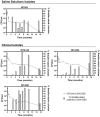
Figure 5
Figure 5
Evolution of cell viability (formula image 0.05% BZK) and percentage of smaller-sized colonies (formula image % Smaller-sized colonies 0.05% BZK) obtained for the B. contaminans bacterial populations during 16 months of incubation in saline solutions containing 0.05% BZK. The percentage of smaller-sized colonies only started to be registered at the 82nd day. Cell viability values hereby plotted correspond to those represented in Figure 2.
Figure 6
Figure 6
Colony diameter distribution of the cell populations corresponding to B. cepacia IST612 (A) and B. contaminans IST601 (B) incubated for 16 months under three different stress conditions. The colony diameters of the original isolates, grown on LB medium, were also assessed and used as a control. From 30 to 260 individual colonies were selected for measurement. Mean colony diameters ± SD are also plotted. The results of the Mann–Whitney U-test (****P < 0.0001, n.s. not significant) are indicated.
Figure 7
Figure 7
Scatter plot of cell length and width of B. cepacia (IST612, IST4152, and IST4168) and B. contaminans (IST601, IST4241, and IST4224) populations recovered from saline solutions containing 0.05% BZK after 3 months of incubation, corresponding to the normal sized clonal variant (dark gray) and to the small clonal variant (light gray). The results of the Mann-Whitney u-test (***P ≤ 0.001, n.s. not significant) are indicated.
Figure 8
Figure 8
Microscopic observation of the structures formed by B. cepacia IST612 and B. contaminans IST601, after 16 months of incubation in saline solutions supplemented with increasing concentrations of BZK. (A1,B1) Control cells, incubated in saline solution (NaCl 0.9% (w/v)); (A2,B2) Cells incubated in saline solution supplemented with 0.0053% (w/v) of BZK; (A3,B3) Cells incubated in saline solution supplemented with 0.05% (w/v) of BZK. Cellular viability was assessed through co-staining of cells with SYTO 9 (green) and propidium iodide (red). Images were acquired using a fluorescence microscope (the same settings were applied for each image) and are equally scaled to allow direct comparison.
Figure 9
Figure 9
Levels of polysaccharide (A), protein (B) and the ratio of polysaccharide vs. protein (C) produced by B. cepacia IST612 and the B. contaminans IST601, assessed by the phenol-sulphuric and biuret methods, respectively, and expressed in mg per mL of bacterial sample, at initial incubation (“Time = 0”) and after, 1 and 18 months of incubation in saline solutions and saline solutions supplemented with 0.0053% (w/v) or 0.05% (w/v) BZK. The results are means of two independent experiments with three replicates each. At initial inoculation (“Time = 0”) no polysaccharide content was detected. The results of the 2-way ANOVA test (*P < 0.05, **P ≤ 0.01, ***P ≤ 0.001, ****P < 0.0001, ns not significant) are indicated.
Figure 10
Figure 10
Genomic organization and transcriptional orientation of the 15 homologs of the B. cenocepacia AU1054 BZK catabolism genes, identified within the genomes of B. cepacia IST612 and IST701. Genes are represented by labeled arrows (A) and their respective locus-tag, products, percentage DNA identity, and percentage protein identity and positives (from BLASTp tool) between reference and IST isolates are listed below (B). The IST701 BZK catabolism genes are colored in green and red, depending on whether the genes are encoded in the same or reversed transcriptional orientation of that of IST612, respectively. Gene size, distance and positioning in the chromosomes and scaffolds are not to scale.

Similar articles

Cited by

References

    1. Ahn Y., Kim J. M., Ahn H., Lee Y. J., LiPuma J. J., Hussong D., et al. . (2014). Evaluation of liquid and solid culture media for the recovery and enrichment of Burkholderia cenocepacia from distilled water. J. Ind. Microbiol. Biotechnol. 41, 1109–1118. 10.1007/s10295-014-1442-3 - DOI - PubMed
    1. Ahn Y., Kim J. M., Kweon O., Kim S. J., Jones R. C., Woodling K., et al. . (2016). Intrinsic resistance of Burkholderia cepacia complex to benzalkonium chloride. mBio 7:e01716–16. 10.1128/mBio.01716-16 - DOI - PMC - PubMed
    1. Ahn Y., Kim J. M., Lee Y. J., LiPuma J., Hussong D., Marasa B., et al. . (2017). Effects of extended storage of chlorhexidine gluconate and benzalkonium chloride solutions on the viability of Burkholderia cenocepacia. J. Microbiol. Biotechnol. 27, 2211–2220. 10.4014/jmb.1706.06034 - DOI - PubMed
    1. Ali M. (2016). Burkholderia cepacia in pharmaceutical industries. Int. J. Vaccines Vaccin. 3:00064 10.15406/ijvv.2016.03.00064 - DOI
    1. Avgeri S. G., Matthaiou D. K., Dimopoulos G., Grammatikos A. P., Falagas M. E. (2009). Therapeutic options for Burkholderia cepacia infections beyond co-trimoxazole: a systematic review of the clinical evidence. Int. J. Antimicrob. Agents 33, 394–404. 10.1016/j.ijantimicag.2008.09.010 - DOI - PubMed

LinkOut - more resources