Review
. 2021 Feb;46(2):544-561.
doi: 10.1007/s00261-020-02642-z. Epub 2020 Jul 26.

Typical imaging finding of hepatic infections: a pictorial essay

Affiliations
Review

Typical imaging finding of hepatic infections: a pictorial essay

Sonaz Malekzadeh et al. Abdom Radiol (NY). 2021 Feb.

Abstract

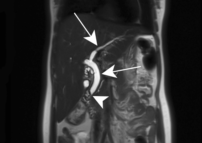
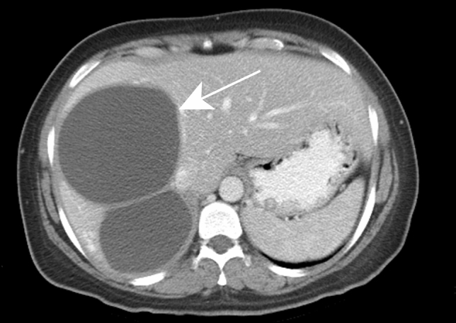
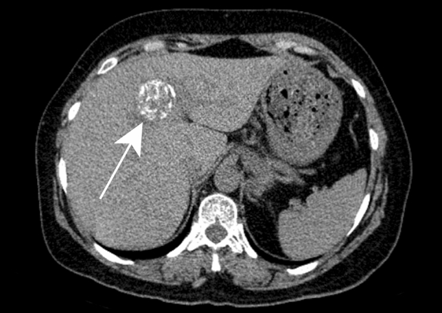
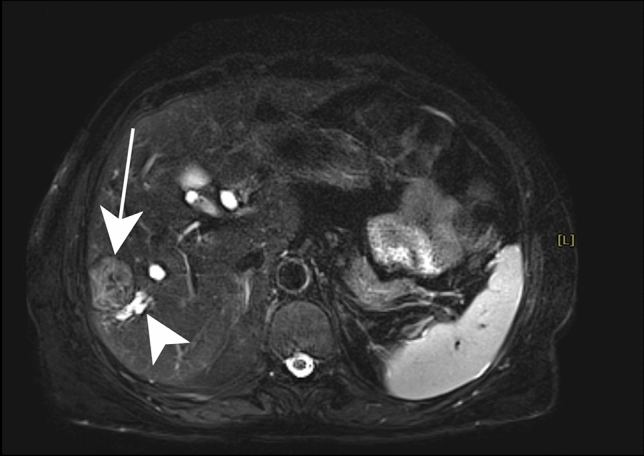
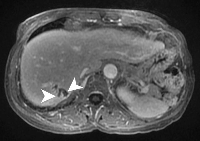
Hepatic infections are frequent in clinical practice. Although epidemiological, clinical and laboratory data may suggest hepatic infection in certain cases, imaging is nearly always necessary to confirm the diagnosis, assess disease extension and its complications, evaluate the response to treatment, and sometimes to make differential diagnoses such as malignancies. Ultrasound (US) is usually the first-line investigation, while computed tomography (CT) and magnetic resonance imaging (MRI) provide better characterization and a more precise assessment of local extension, especially biliary and vascular. The purpose of this article is to describe the typical features and main complications of common hepatic infections. Familiarity with the radiological features of this entity can help suggest the correct diagnosis and the need for further studies as well as determine appropriate and timely treatment.

Keywords: Hepatic infection; Liver imaging; Liver infection.


Figures

Fig. 1
Fig. 1
Pyogenic abscess in a 60-year-old female patient with a history of chronic pancreatitis who presented with asthenia and fever. Axial arterial phase (a) and portal venous phase (b) contrast-enhanced CT show small clustering lesions with a dominant hypoattenuating lesion in segment V of the liver (white arrows) corresponding to pyogenic hepatic abscesses. Altered perfusion disorder is observed as geographic areas of hyperattenuation peripheral to the hepatic abscesses (white arrowheads), clearly visualized on arterial phase (a)
Fig. 2
Fig. 2
Liver tuberculosis in a 49-year-old female patient with asthenia and loss of weight without fever. Ultrasound a demonstrates a focal well-defined subcapsular hypoechoic lesion in segment III (white arrow). Enlarged lymph node (white arrowhead) is also observed in the porta hepatis (b) (Courtesy of Dr. Suzan Elhakiem, Ibn Sina Hospital, Khartoum, Sudan)
Fig. 3
Fig. 3
Brucellosis in a 42-year-old male patient with evening fever and sweating. Contrast-enhanced axial CT image a shows a heterogeneous lesion with enhanced contours (black arrow), showing a central hypoechoic fluid component and a calcium deposit (white arrow). Axial contrast-enhanced T1-weighted image b shows enhancement of the peripheral tissular areola (black arrow) and central saccular formation with fluid, surrounded by an intermediary heterogeneous component. Reprinted from Sisteron et al. [27], with permission from Elsevier
Fig. 4
Fig. 4
Bartonellosis in a 65-year-old female patient treated for autoimmune hepatitis. Axial contrast-enhanced CT demonstrates hypoattenuating ill-defined lesions are scattered throughout the liver parenchyma (white arrowheads)
Fig. 5
Fig. 5
Acute viral hepatitis in a 59-year-old male patient with jaundice and elevated liver enzymes due to hepatitis A infection. Axial contrast-enhanced CT shows a contracted gallbladder with a thick hypoattenuating edematous wall and an enhancing mucosal layer (white arrow)
Fig. 6
Fig. 6
HIV-related cholangiopathy in a 16-year-old female patient. Coronal T2-weighted image demonstrates intrahepatic and extrahepatic bile duct dilatation (white arrows) due to papillary stenosis (white arrowhead), a common finding in this disease
Fig. 7
Fig. 7
Candidiasis in a 65-year-old male patient with acute myeloblastic leukemia. Axial contrast-enhanced T1-weighted image on hepatobiliary phase illustrates multiple tiny hypointense lesions throughout the liver parenchyma (white arrowheads) (Courtesy of Dr. Luisa Paulatto, Beaujon Hospital, Clichy, France)
Fig. 8
Fig. 8
Cystic echinococcosis type CL in a 32-year-old female patient with upper abdominal discomfort. Axial contrast-enhanced CT image shows two large cystic lesions with thin walls in segment VII and VIII of the liver (white arrow). Ultrasound (not shown) revealed an anechoic cyst with a double layered wall, no internal daughter cyst or detached membrane
Fig. 9
Fig. 9
Cystic echinococcosis type CE3a in a 27-year-old female patient with cough. Axial contrast-enhanced CT of the lower pulmonary parenchyma a reveals a pulmonary cyst in the middle lobe (white arrow) associated with a partial consolidation of the right lower lobe. Axial contrast-enhanced CT of the upper abdomen b shows two hepatic cystic lesions (white arrow) with internal detached membranes (“water-lily” sign) (white arrowhead)
Fig. 10
Fig. 10
Cystic echinococcosis type CE3b in a 54-year-old male patient with right upper quadrant pain. Axial contrast-enhanced CT reveals a large hepatic cyst in right hepatic lobe (black arrow) with solid matrix in the center (asterisk) and peripherally located daughter cysts (white arrow)
Fig. 11
Fig. 11
Cystic echinococcosis type CE5 in a 50-year-old female patient with an incidental solid mass reported on ultrasound. Axial precontrast CT shows a round highly calcified lesion in segment IV (white arrow)
Fig. 12
Fig. 12
Cystic echinococcosis type CE4 in a 62-year-old female patient with upper abdominal pain. Axial fat-suppressed T2-weighted image shows a well-defined subcapsular moderately hyperintense lesion (white arrow) with a characteristic “ball of wool” sign. An adjacent dilated intrahepatic bile duct is also noted (white arrowhead)
Fig. 13
Fig. 13
Peritonitis due to a ruptured hydatid cyst in a 45-year-old female patient with acute upper abdominal pain. Axial contrast-enhanced CT of the upper abdomen (a) demonstrates two hepatic cysts with irregular borders in the right and left liver lobe (white arrows); the latter reaches the anterior liver surface. Axial contrast-enhanced CT of lower abdomen (b) shows a significant amount of intraperitoneal fluid with a thickened enhancing peritoneum (white arrowheads)
Fig. 14
Fig. 14
Budd-Chiari syndrome secondary to compression of liver out-flow by the hydatid cyst in a 20-year-old female patient. Hypertrophy of the caudate lobe with inhomogeneous mottled liver appearance is indicative of Budd-Chiari syndrome. Axial delayed phase contrast-enhanced CT shows a deformed cystic lesion in the deep portion of segments VII and VIII of the liver (white arrow) associated with pneumobilia secondary to previous sphincterotomy (white arrowhead)
Fig. 15
Fig. 15
Alveolar echinococcosis incidentally detected in a 75-year-old male patient. Axial contrast-enhanced CT a shows an ill-defined subcapsular hypoattenuating lesion in segment VII of the liver (black arrow). Axial fat-suppressed T2-weighted image b further characterizes this lesion as multiple tiny cystic lesions (white arrowhead) surrounding a solid component corresponding to type 3 of alveolar echinococcosis
Fig. 16
Fig. 16
Amebic abscess in a 57-year-old male patient who presented with fever of unknown origin and right upper abdominal pain with a recent history of travel to Africa. Ultrasound a demonstrates a large relatively well-delineated lesion with a heterogeneous solid-appearing content (white arrow). Axial contrast-enhanced CT shows the “double target sign” (black arrow)
Fig. 17
Fig. 17
Hepatic schistosomiasis in a 20-year-old male patient with a history of gastrointestinal bleeding from 1 year ago. Ultrasound demonstrates a marked diffuse periportal thickening as a hyperechoic mantle encompassing the anechoic portal vein (white arrowheads) (Courtesy of Dr. Suzan Elhakiem, Ibn Sina Hospital, Khartoum, Sudan)
Fig. 18
Fig. 18
Fascioliasis in a 42-year-old female patient with right upper abdominal pain and low-grade fever. Axial contrast-enhanced CT (a, b) show patchy ill-defined hypoattenuating lesions with subcapsular (white arrowheads) and periportal distribution (black arrow)
Fig. 19
Fig. 19
Ascariasis in a 36-year-old male patient. Axial and coronal reformatted contrast-enhanced CT (a, b) show intrahepatic and extrahepatic bile duct dilatation (black arrows). Note intrahepatic bile ducts filled with structures more attenuating than bile (black arrowhead), indicating adult worms. Oblique coronal single-shot fast spin-echo MR cholangiogram (c) shows adult worms as serpiginous and nodular filling defects in the left intrahepatic and extrahepatic bile ducts (white arrows)
Fig. 20
Fig. 20
Clonorchiasis in a 74-year-old female patient with recurrent cholangitis. Axial contrast-enhanced fat-suppressed T1-weighted image shows intrahepatic bile ducts dilatation in segment VII reaching the subcapsular zone (white arrowheads)

References

    1. Rahimian J, Wilson T, Oram V, Holzman RS (2004) Pyogenic liver abscess: recent trends in etiology and mortality. Clin Infect Dis 39 (11):1654-1659. 10.1086/425616 - PubMed
    1. Liu Y, Wang JY, Jiang W (2013) An Increasing Prominent Disease of Klebsiella pneumoniae Liver Abscess: Etiology, Diagnosis, and Treatment. Gastroenterol Res Pract 2013:258514. 10.1155/2013/258514 - PMC - PubMed
    1. Seeto RK, Rockey DC (1996) Pyogenic liver abscess. Changes in etiology, management, and outcome. Medicine (Baltimore) 75 (2):99-113. 10.1097/00005792-199603000-00006 - PubMed
    1. Okano H, Shiraki K, Inoue H, Kawakita T, Yamamoto N, Deguchi M, Sugimoto K, Sakai T, Ohmori S, Murata K, Nakano T (2002) Clinicopathological analysis of liver abscess in Japan. Int J Mol Med 10 (5):627-630 - PubMed
    1. Siu LK, Yeh KM, Lin JC, Fung CP, Chang FY (2012) Klebsiella pneumoniae liver abscess: a new invasive syndrome. Lancet Infect Dis 12 (11):881-887. 10.1016/s1473-3099(12)70205-0 - PubMed

Publication types