Review
. 2020 Jun 19;127(1):51-72.
doi: 10.1161/CIRCRESAHA.120.316363. Epub 2020 Jun 18.

Molecular Basis of Atrial Fibrillation Pathophysiology and Therapy: A Translational Perspective

Affiliations
Review

Molecular Basis of Atrial Fibrillation Pathophysiology and Therapy: A Translational Perspective

Stanley Nattel et al. Circ Res. .

Abstract

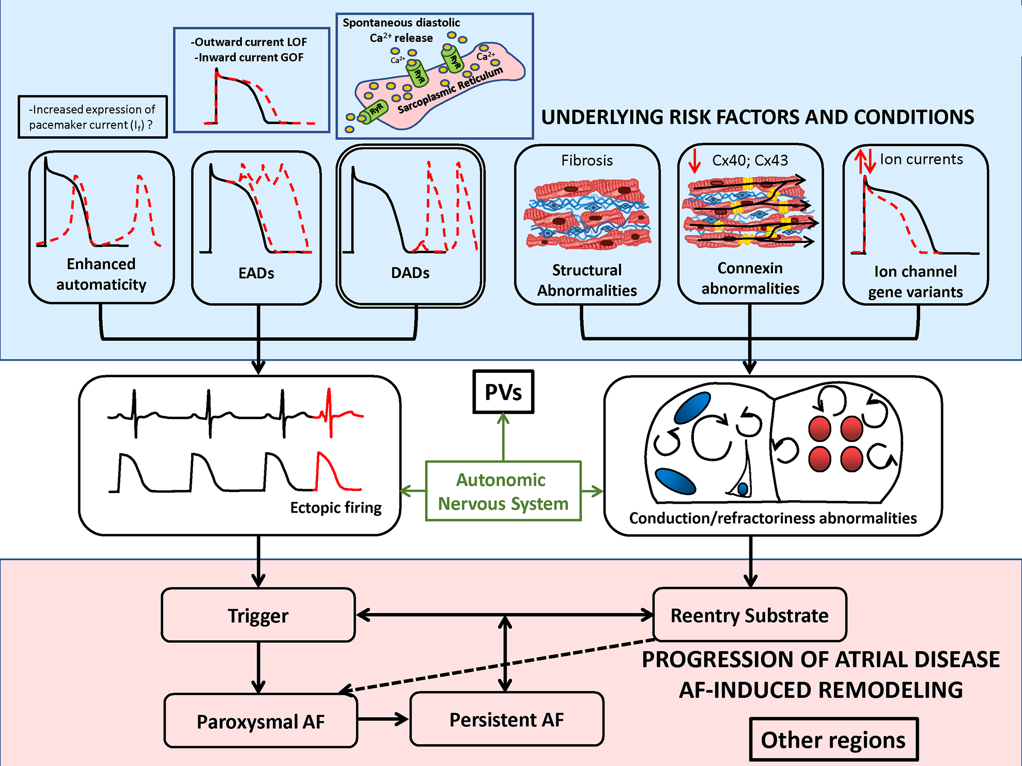
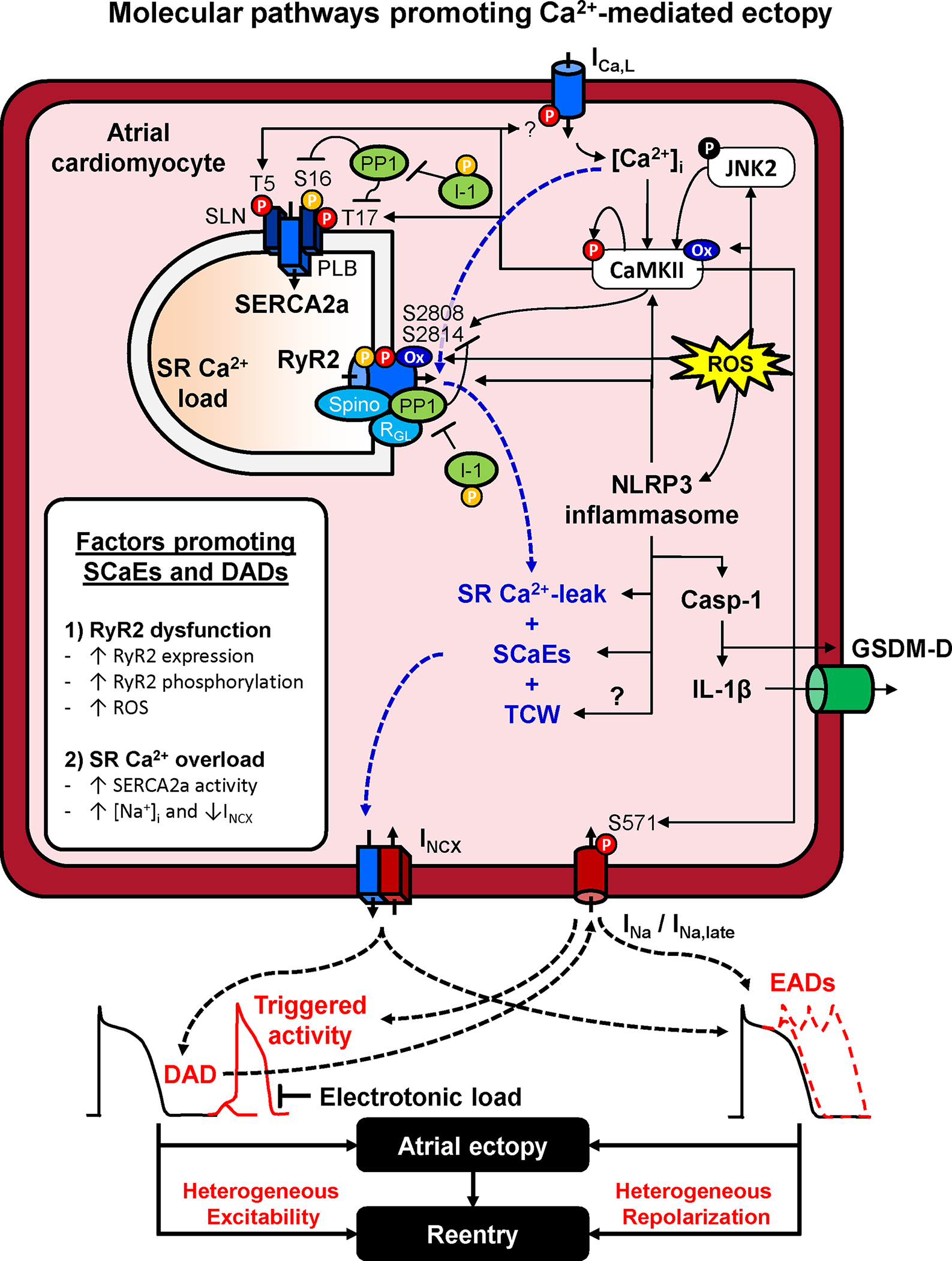
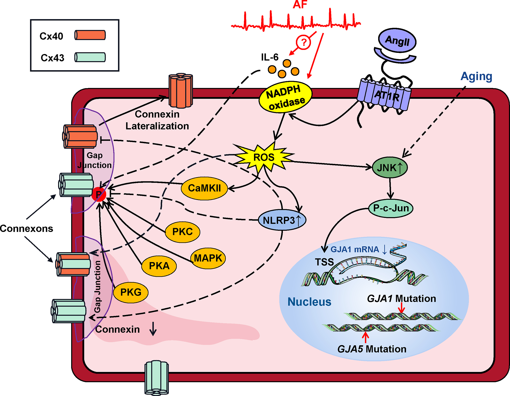
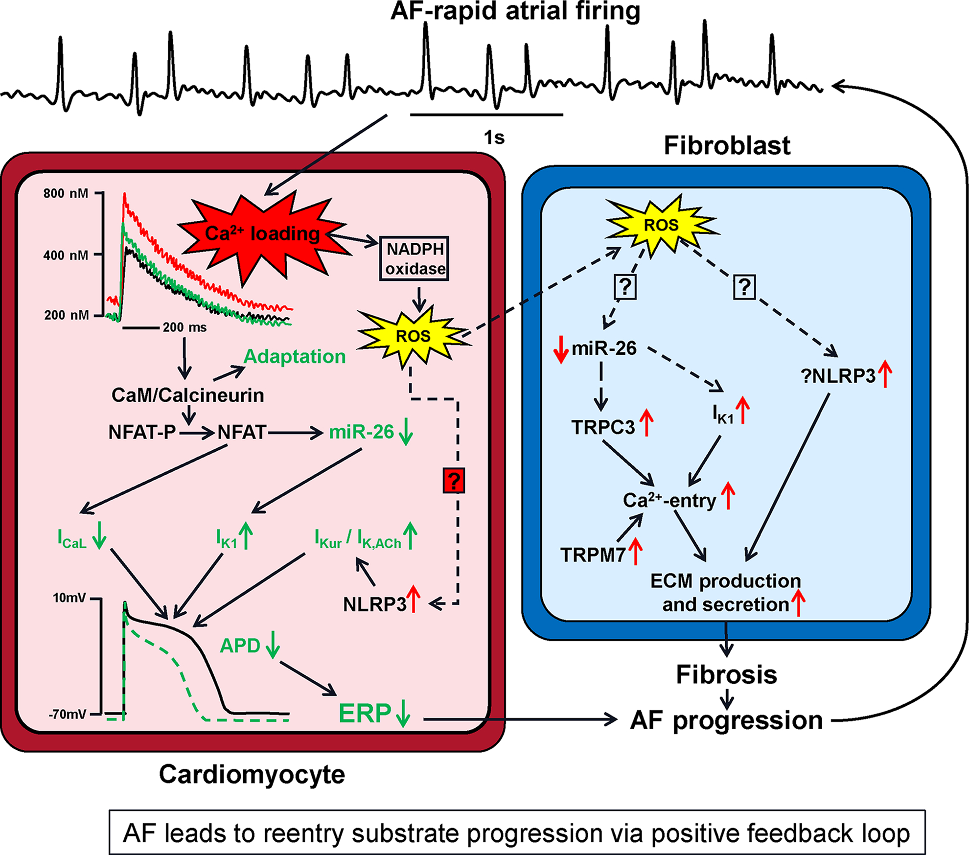
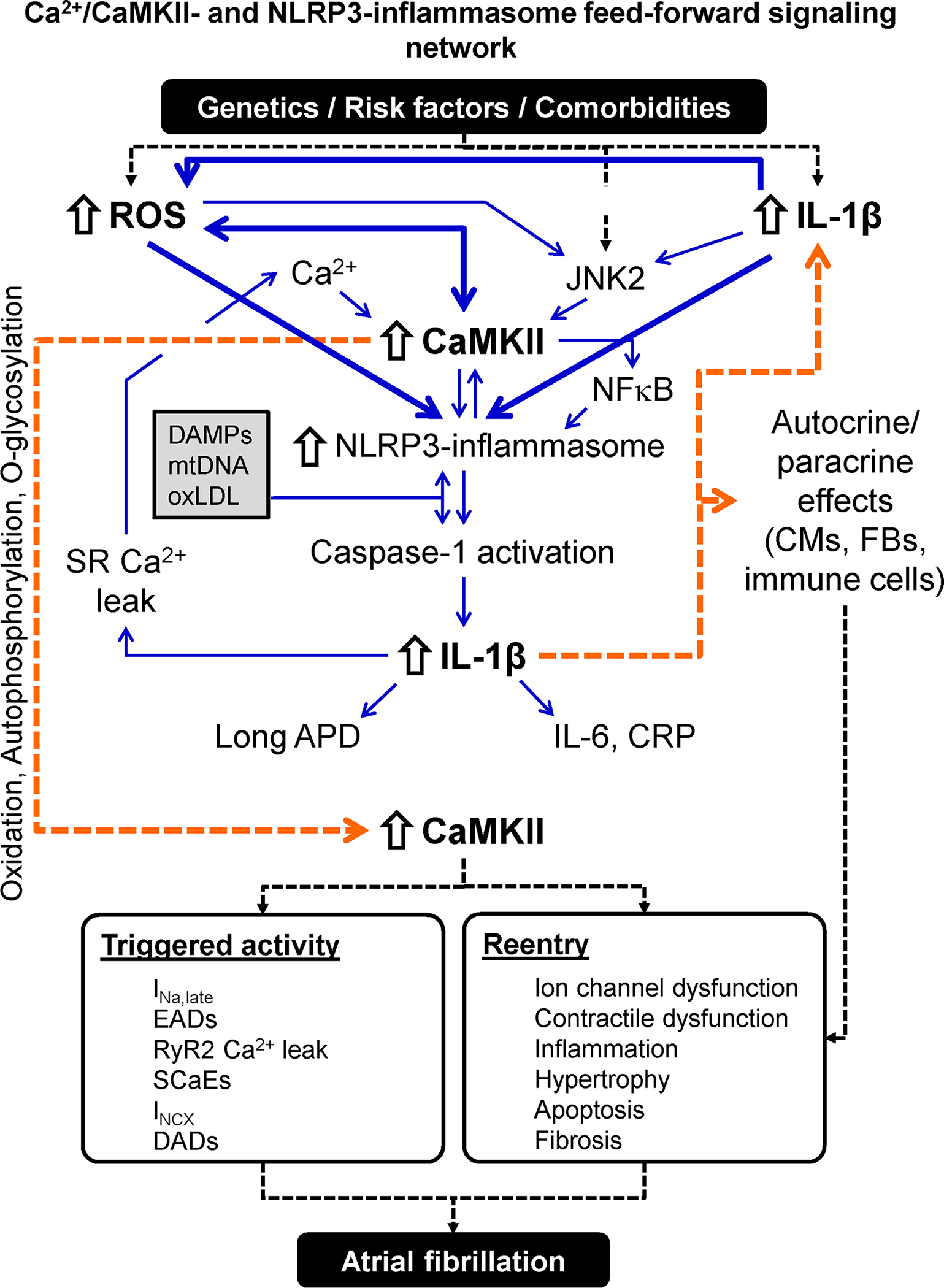
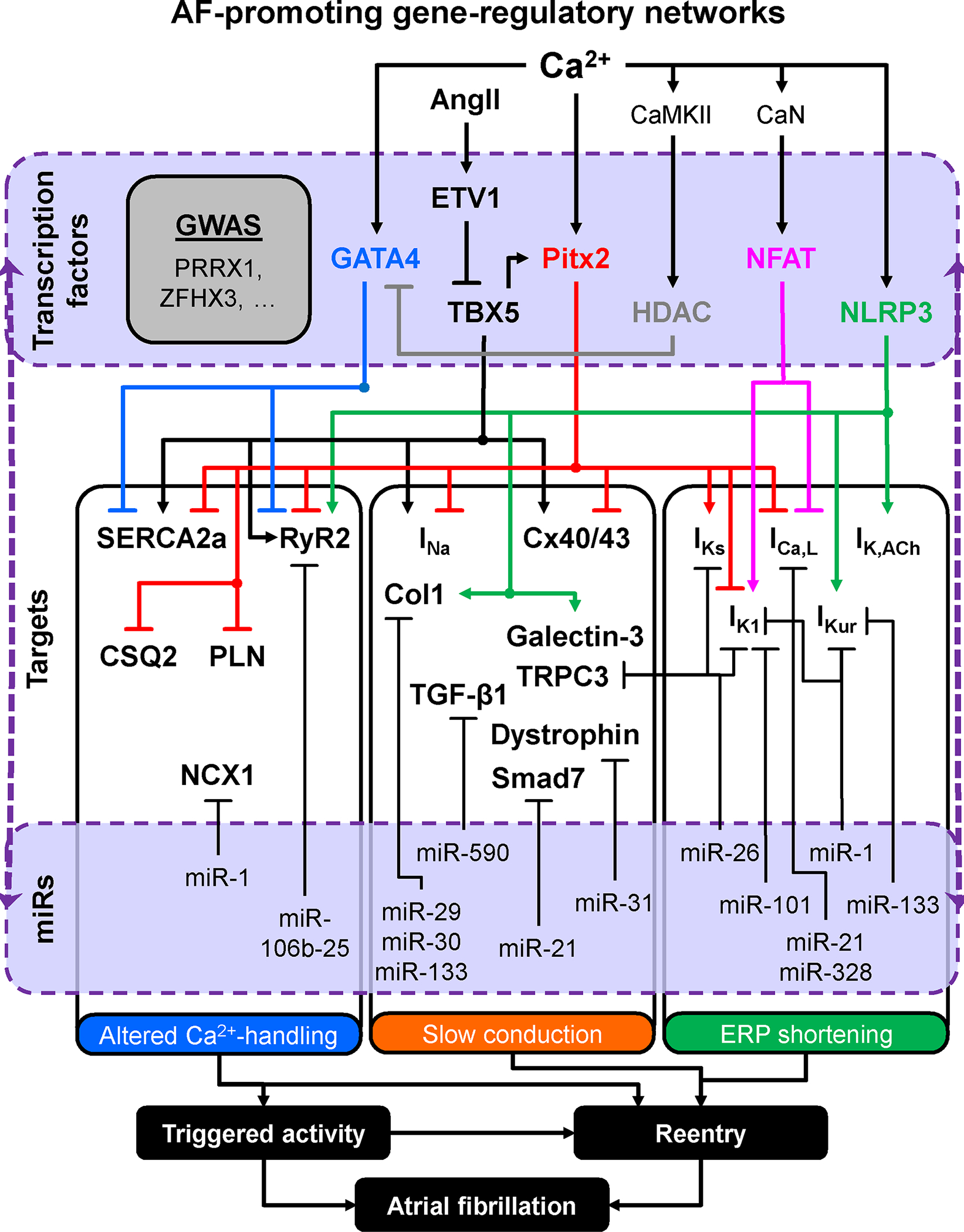
Atrial fibrillation (AF) is a highly prevalent arrhythmia, with substantial associated morbidity and mortality. There have been significant management advances over the past 2 decades, but the burden of the disease continues to increase and there is certainly plenty of room for improvement in treatment options. A potential key to therapeutic innovation is a better understanding of underlying fundamental mechanisms. This article reviews recent advances in understanding the molecular basis for AF, with a particular emphasis on relating these new insights to opportunities for clinical translation. We first review the evidence relating basic electrophysiological mechanisms to the characteristics of clinical AF. We then discuss the molecular control of factors leading to some of the principal determinants, including abnormalities in impulse conduction (such as tissue fibrosis and other extra-cardiomyocyte alterations, connexin dysregulation and Na+-channel dysfunction), electrical refractoriness, and impulse generation. We then consider the molecular drivers of AF progression, including a range of Ca2+-dependent intracellular processes, microRNA changes, and inflammatory signaling. The concept of key interactome-related nodal points is then evaluated, dealing with systems like those associated with CaMKII (Ca2+/calmodulin-dependent protein kinase-II), NLRP3 (NACHT, LRR, and PYD domains-containing protein-3), and transcription-factors like TBX5 and PitX2c. We conclude with a critical discussion of therapeutic implications, knowledge gaps and future directions, dealing with such aspects as drug repurposing, biologicals, multispecific drugs, the targeting of cardiomyocyte inflammatory signaling and potential considerations in intervening at the level of interactomes and gene-regulation. The area of molecular intervention for AF management presents exciting new opportunities, along with substantial challenges.

Keywords: atrial fibrillation; calcium; inflammasome; myocytes, cardiac; transcription factors.


Figures

Figure 1.
Figure 1.. Overview of mechanisms linked to AF-occurrence.
AF AF-triggers result from focal ectopic firing. Ectopic activity is most clearly linked to spontaneous diastolic Ca2+-release from the sarcoplasmic reticulum Ca2+-stores via leaky ryanodine-receptor (RyR2) Ca2+-release channels. Early afterdepolarizations (EADs) due to loss-of-function (LOF) outward-current mutations or gain-of-function (GOF) inward-current mutations have also been linked to spontaneous ectopy. Enhanced automaticity, for example due to pacemaker current expression, is another possible cause of ectopic activitty, but has not been definitively demonstrated. AF-persistence is linked to AF-maintaining reeentry that requires both trigger and a vulnerable reentrant substrate. The latter can be caused by abbreviated refractoriness (e.g. due to a GOF K+-channel mutation or to enhanced vagal tone) or by conduction abnormalities due to tissue fibrosis, connexin (Cx) dysfunction or LOF Na+-channel mutations. Ectopic firing typically originates from the pulmonary veins (PVs), but the PVs are also a priviledged site for reentry susceptibility.
Figure 2.
Figure 2.. Atrial ectopy. Molecular pathways promoting Ca2+-mediated ectopy.
Increased sarcoplasmic reticulum (SR) Ca2+ leak and spontaneous SR Ca2+-release events (SCaEs) primarily result from dysfunction of the cardiac ryanodine receptor type-2 (RyR2) channel or SR Ca2+-overload. RyR2 dysfunction is promoted by increased RyR2 expression, hyperphosphorylation (e.g., due to increased Ca2+/calmodulin-dependent protein kinase-II, CaMKII, activity or improper targeting of protein phosphatase-1, PP1), or RyR2 oxidation due to increased reactive oxygen species (ROS). ROS mediated NLRP3 inflammasome activation amplifies the Ca2+-handling abnormalities and activates caspase-1 (Casp-1) which increases interleukin (IL)-1β generation and the formation of gasdermin-D-derived plasmamembrane-pores, allowing the release of IL-1β out of the cell, spreading inflammatory signaling. SR Ca2+-overload is promoted by increased activity of the SR Ca2+-ATPase-2a (SERCA2a) or elevated intracellular Na+, reducing Ca2+-extrusion via the Na+/Ca2+ exchanger type-1 (NCX1). SR Ca2+ overload also promotes L-type Ca2+-current (ICa,L)-dependent triggered Ca2+ waves (TCW). SCaEs and TCW activate a transient-inward current mediated by NCX (INCX) resulting in DADs or EADs, depending on their timing relative to the atrial action potential. DADs and EADs can promoted atrial ectopy, as well as reentry through increased heterogeneity of excitability and repolarization. Abbreviations: GSDM-D, N-terminal Gasdermin-D fragment; I-1, inhibitor-1 of PP1; JNK2, c-Jun N-terminal kinases-2; NLRP3, NACHT, LRR and PYD domains-containing protein 3; PLB, phospholamban; SLN, sarcolipin.
Figure 3.
Figure 3.. Molecular determinants of tissue fibrosis.
The main pathways governing profibrotic signaling. Extracellular profibrotic signaling molecules like angiotensin-II (Ang-II), transforming growth factor-β1 (TGFβ), platelet-derived growth-factor (PDGF) and connective- tissue growth-factor (CTGF) activate membrane receptors coupled to downstream signaling which leads to enhanced gene-transcription to increase extracellular matrix (ECM) production. Fibroblast ion-channels control Ca2+-entry to regulate fibroblast activation. For additional discussion, see text. Abbreviations: bb, integrin receptor oblast activation; Ang-II, angiotensin- II; AP, activator-protein; AT1R, angiotensin-II type-1 receptor; CTGF, connective-tissue growth- factor; ER, endoplasmic reticulum; ERK1/2, extracellular signal-related kinase-1/2; ERK-P, phosphorylated extracellular signal-related kinase; Grb2, growth-factor receptor binding-protein 2; IK1, inward-rectifier K+-channel; IP3, inositol 1,4,5-trisphosphate; JAK, Janus kinase; JNK, c-jun N-terminal kinase; LOX, lysyl oxidase; MAPK, mitogen-activated protein kinase; MMP, matrix metalloproteinase; NADPH, nicotine adenine dinucleotide-phosphate; NF-κB, nuclear factor-kappa B; NLRP3, NACHT-, LRR- and PYD domains-containing protein 3; PKC, protein-kinase C; PDGF, platelet-derived growth factor; PDGFR, PDGF-receptor; PIP2, phosphatidylinositol bisphosphate; PLC, phospholipase-C; ROC, receptor-operated channel; ROS, reactive-oxygen species; Shc, src homologous and collagen protein; SMAD, sma- and mad-related proteins; SOC, store-operated channel; SOS, son of sevenless protein; Src, sarcoma proto-oncogene tyrosine kinase; STAT, signal transducers and activators of transcription; TAK1, TGFTGF TAK1, ed kinase-1; TF, transcription factor; TGFβR, transforming growth factor β receptor; TIMP, tissue inhibitor of matrix metalloproteinase; TRP, transient receptor potential; TSS, transcriptional start site.
Figure 4.
Figure 4.. Connexin dysregulation in AF.
Dysfunction of the connexins that ensure cell-to-cell coupling in gap junctions results from connexin (Cx) downregulation or lateralization to transverse cell-borders. The moecular mechanisms governing these changes are illustrated. Abbreviations: Ang-II, angiotensin-II, AT1R, angiotensin-receptor type-1; CaMKII, Ca2+-calmodulin dependent kinase type-II; JNK, c-Jun N-terminal kinase; MAPK, mitogen-activated protein-kinase; P, phosphate; PKA, PKC, PKG, protein-kinases A, C, G respectively; ROS, reactive-oxygen species.
Figure 5.
Figure 5.. Molecular pathways involved in AF-progression.
The rapid atrial firing in AF leads to cellular Ca2+-loading, which engages compensatory mechanisms (shown in green) that attenuate Ca2+-loading at the price of action-potential duration (APD) abbreviation that favors reentry. Ca2+-loading is a proximal signal to this and other processes resulting ultimately in APD and refractoriness-abbreviation in cardiomyocytes, along with enhanced collagen-production in fibroblasts, to cause progression of the reentry substrate and greater resistance of AF to therapy. A positive feedback loop results, wherein AF causes changes that increase AF-vulnerability and perpetuate the events that cause progression. Abbreviations: ECM, extracellular matrix; ERP, effective refractory period; ICa,L, L-type Ca2+-current; IK1, inward-rectifier K+-current; miR, microRNA; NLRP3, NACHT-, LRR- and PYD domains-containing protein 3; TRPC3, transient-receptor channel potential current canonical type-3; TRPM7, transient-receptor channel potential current melastatin type-7; ROS, reactive-oxygen species.
Figure 6.
Figure 6.. Atrial fibrillation-promoting Ca2+/calmodulin-dependent protein kinase-II (CaMKII)- and NACHT, LRR and PYD domains-containing protein 3 (NLRP3)-inflammasome feed-forward signaling network.
Risk factors and comorbidities create an environment in which danger-associated molecular patterns (DAMPs), mitochondrial DNA (mtDNA) and oxLDL activates the atrial NLRP3 inflammasome. Cardiac-restricted increases in reactive oxygen species (ROS) production and c-Jun N-terminal kinase-2 (JNK2) activity further stimulate the NLRP3 inflammasome via CaMKII-dependent and -independent pathways. The resulting stimulation of caspase-1 maturates interleukin (IL)-1β, which leaves the cell, thereby spreading the inflammatory signaling and increasing the synthesis of IL-6 and C-reactive protein (CRP). IL-1β amplifies the NLRP3 inflammatory signaling and promotes sarcoplasmic reticulum (SR) Ca2+ leak and action potential duration (APD) changes in cardiomyocytes (CMs), creating a feedforward signaling network. IL-1β also exerts paracrine effects on cardiac fibroblasts (CFs) and immune cells causing hypertrophy, apoptosis and fibrosis. Activation and perpetuation of this feedforward Ca2+/CaMKII/NLRP3-inflammasome signaling network promotes triggered activity and reentry and increases AF susceptibility. Abbreviations: DADs, delayed afterdepolarizations; EADs, early afterdepolarizations; INa,late, Persistent/late Na+ current; INCX, Na+-Ca2+-exchanger current; NFκB, nuclear factor kappa-light-chain-enhancer of activated B cells; RyR2, ryanodine receptor type-2; SCaEs, spontaneous SR Ca2+-release events.
Figure 7.
Figure 7.. Atrial fibrillation-promoting gene-regulatory networks.
Multiple gene-regulatory networks interact to fine-tune the expression levels of key proteins shaping the effective refractory period (ERP), enabling proper impulse conduction and governing Ca2+-handling processes. Transcriptional alterations in the level of key regulator genes disrupt the network balance, increasing the likelihood of ERP, conduction and Ca2+-handling abnormalities, along with superimposed post-transcriptional changes due to abnormal microRNA (miR) function, leading to the formation of AF triggers and substrates. Abbreviations: Ang-II, angiotensin-II; CaMKII, Ca2+/calmodulin-dependent protein kinase-II; CaN, calcineurin; Col1, collagen-1; CSQ2, calsequestrin-2; Cx40/43, connexin 40/43; ETV1, ETS translocation variant 1 transcription factor; HDAC, histone deacetylase; ICa,L, L-type Ca2+-current; IK1, inward-rectifier K+-current; IK,ACh, acetylcholine-activated inward-rectifier K+-current; IKs, slow delayed-rectifier K+-current; IKur, ultra-rapid delayed-rectifier K+-current; INa, Na+-current; NCX1, Na+-Ca2+-exchanger type-1; NFAT, nuclear factor of activated T-cells; NLRP3, NACHT, LRR and PYD domains-containing protein-3; PITX2, paired Like Homeodomain 2; PLN, phopsholamban; RyR2, ryanodine receptor channel type-2; SERCA2a, SR Ca2+-ATPase type-2a; Smad7, mothers against decapentaplegic homolog 7; TBX5, T-box transcription factor 5; TGF-β1, transforming growth factor β1; TRPC3 transient-receptor potential channel canonical type-3.

References

    1. Andrade J, Khairy P, Dobrev D, Nattel S. The clinical profile and pathophysiology of atrial fibrillation: relationships among clinical features, epidemiology, and mechanisms. Circ Res. 2014;114:1453–1468 - PubMed
    1. Heijman J, Algalarrondo V, Voigt N, Melka J, Wehrens XH, Dobrev D, Nattel S. The value of basic research insights into atrial fibrillation mechanisms as a guide to therapeutic innovation: a critical analysis. Cardiovasc Res. 2016;109:467–479 - PMC - PubMed
    1. Nattel S, Guasch E, Savelieva I, et al. Early management of atrial fibrillation to prevent cardiovascular complications. Eur Heart J. 2014;35:1448–1456 - PubMed
    1. Lau DH, Nattel S, Kalman JM, Sanders P. Modifiable Risk Factors and Atrial Fibrillation. Circulation. 2017;136:583–596 - PubMed
    1. Wijffels MC, Kirchhof CJ, Dorland R, Allessie MA. Atrial fibrillation begets atrial fibrillation. A study in awake chronically instrumented goats. Circulation. 1995;92:1954–1968 - PubMed

Publication types

MeSH terms