Review
. 2021 Jan:80:100877.
doi: 10.1016/j.preteyeres.2020.100877. Epub 2020 Jul 24.

Plasmacytoid dendritic cells in the eye

Affiliations
Review

Plasmacytoid dendritic cells in the eye

Arsia Jamali et al. Prog Retin Eye Res. 2021 Jan.

Abstract

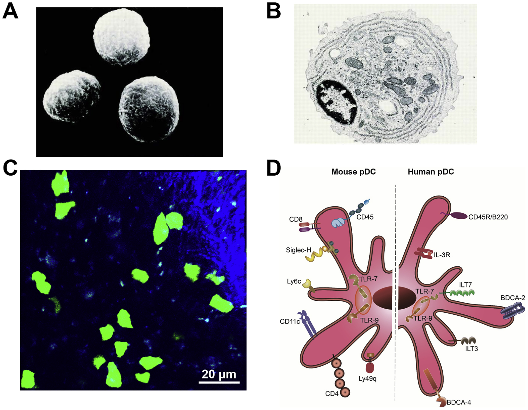
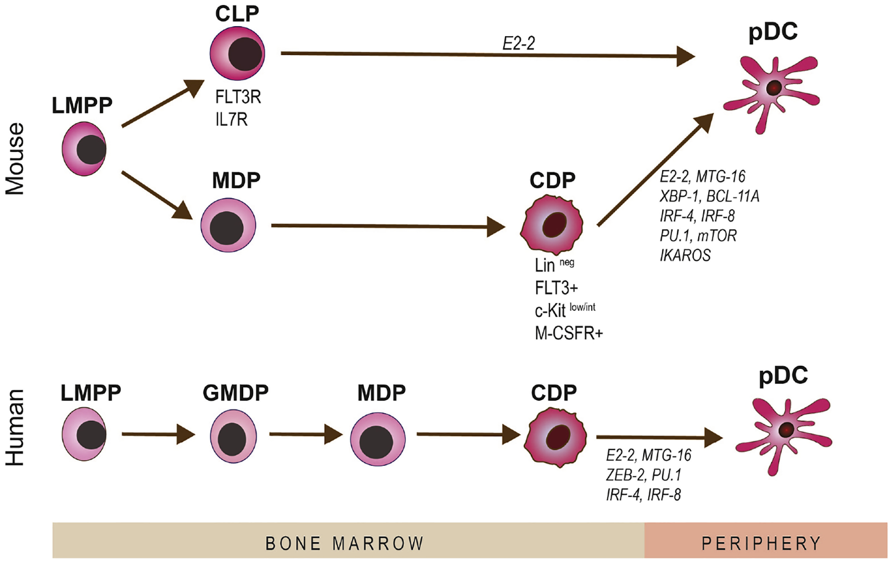
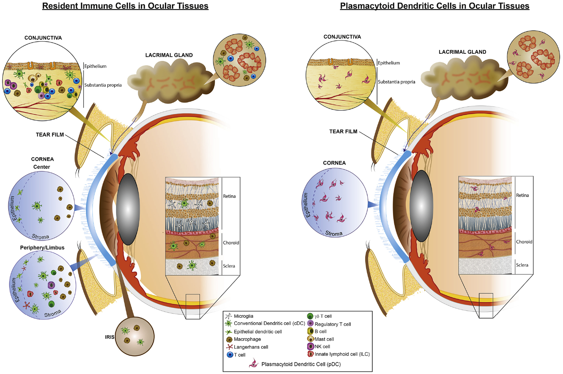
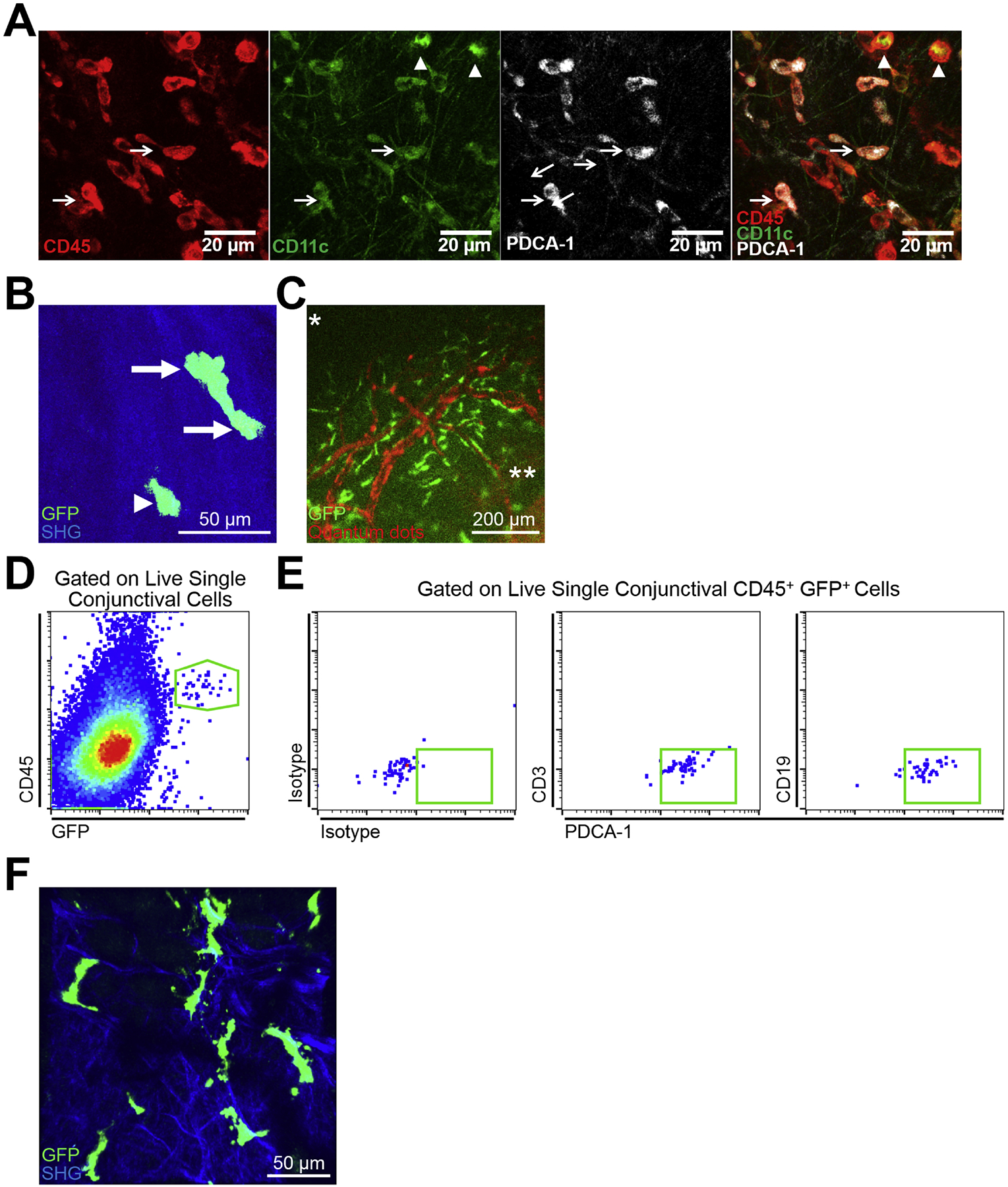
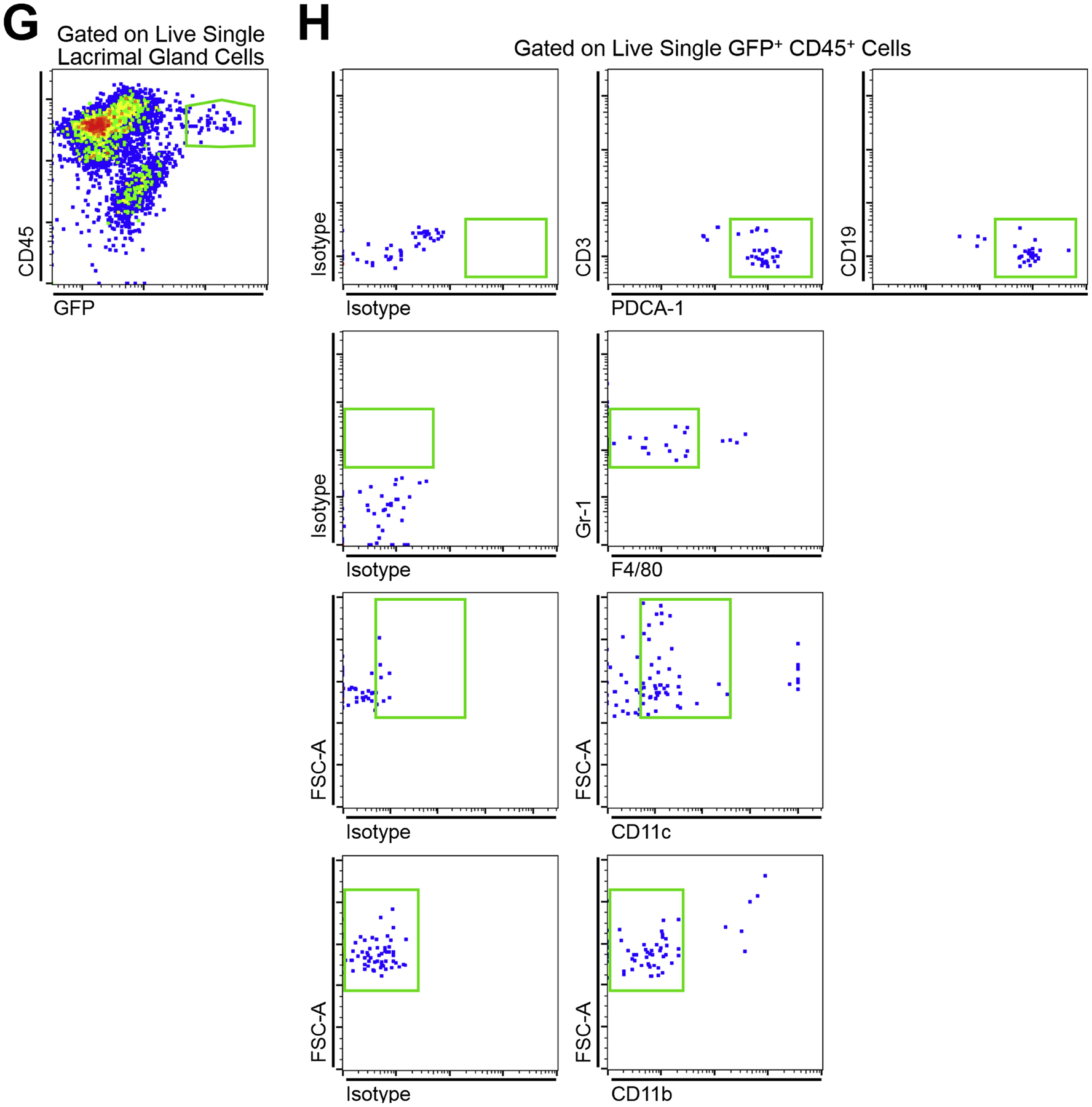
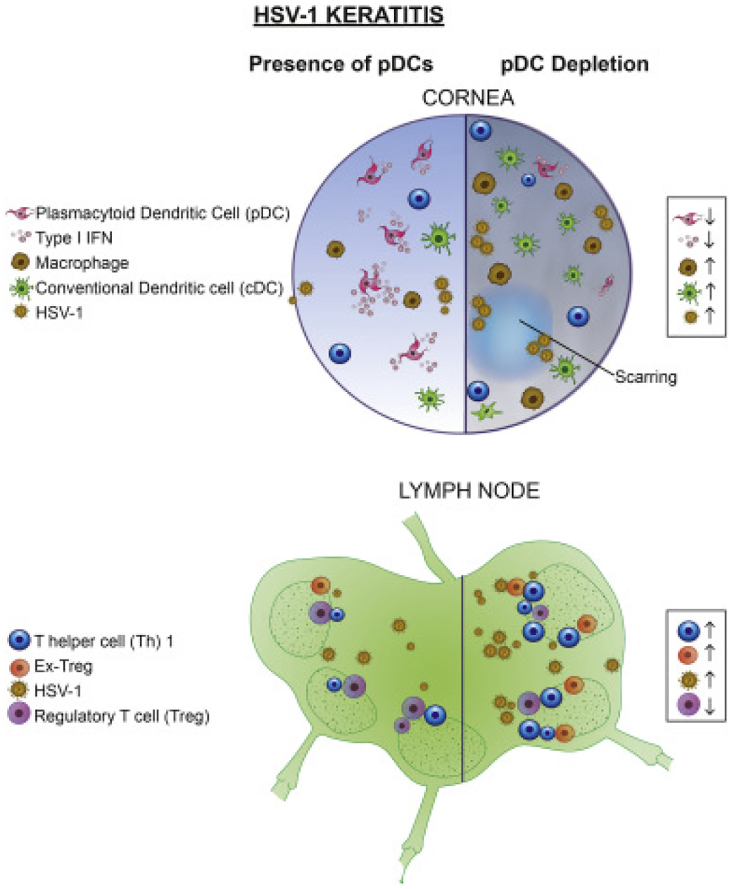
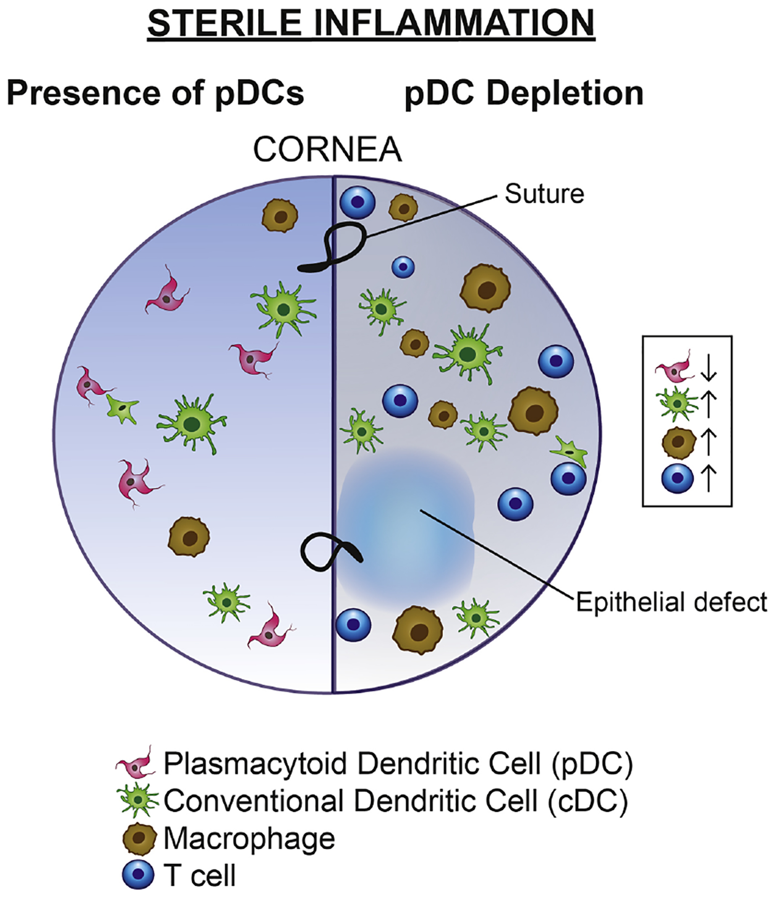
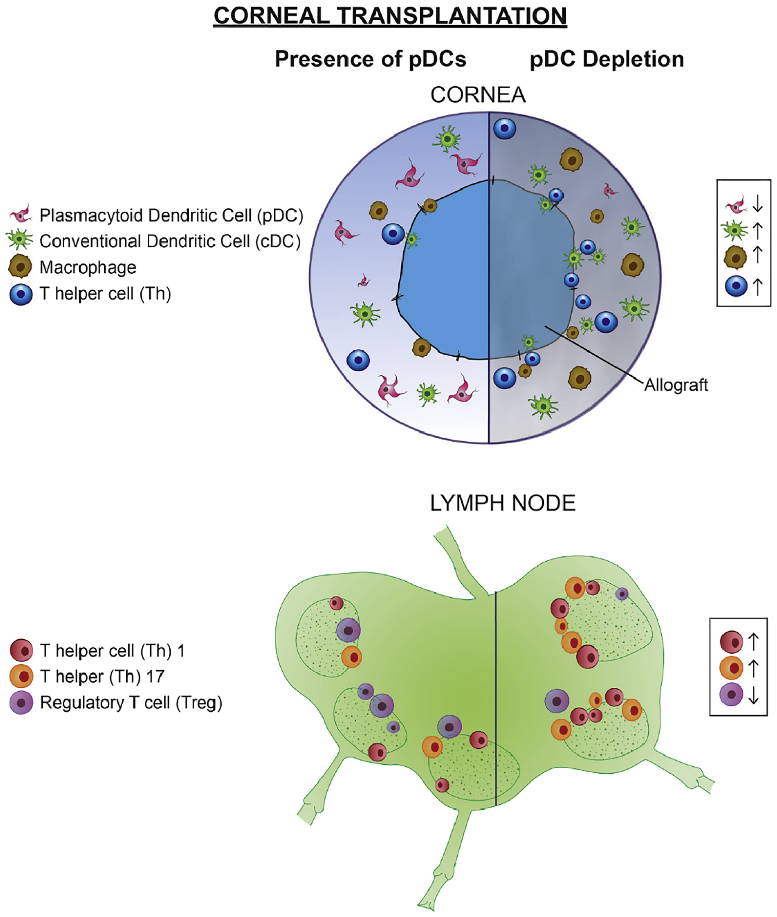
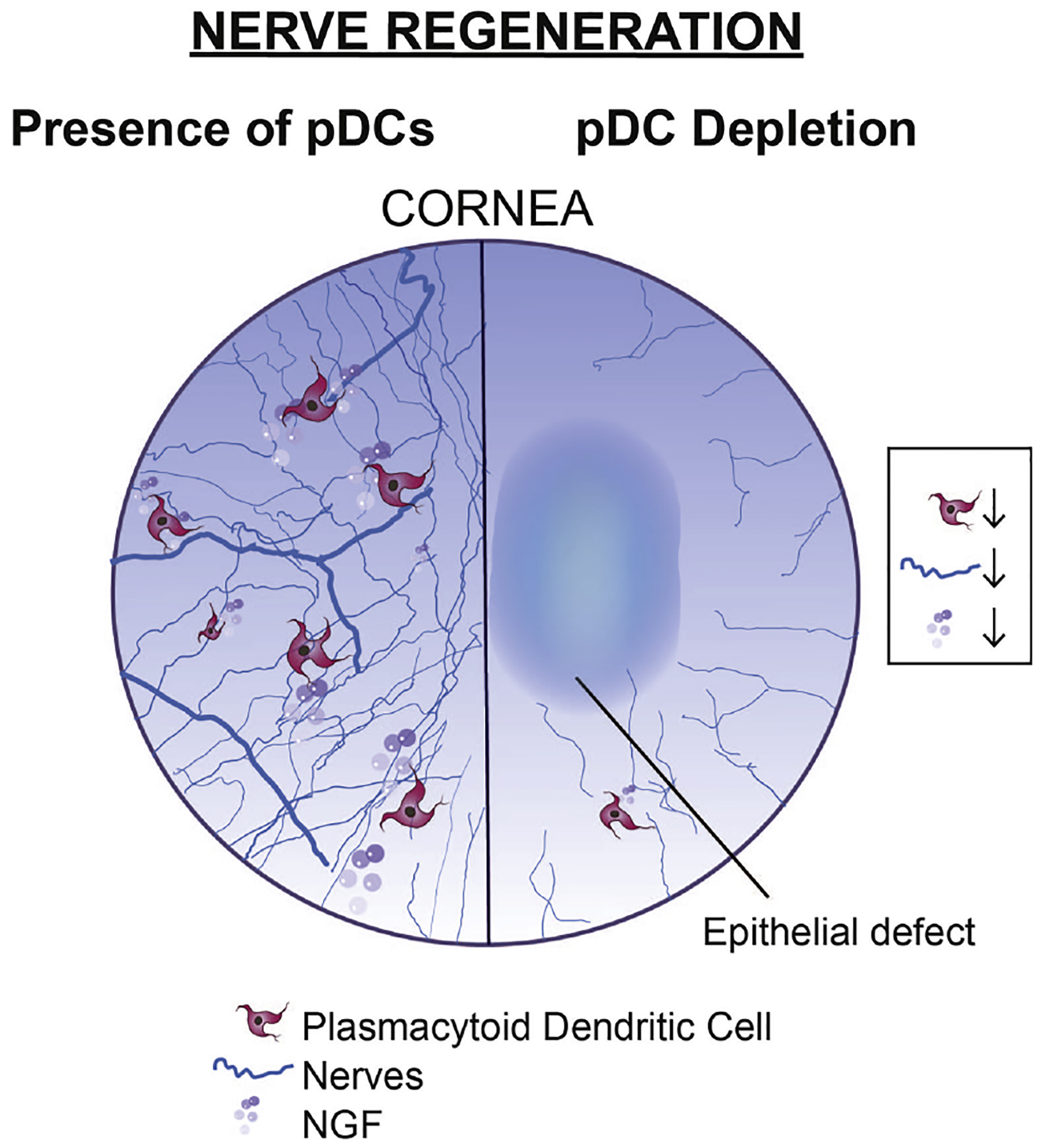
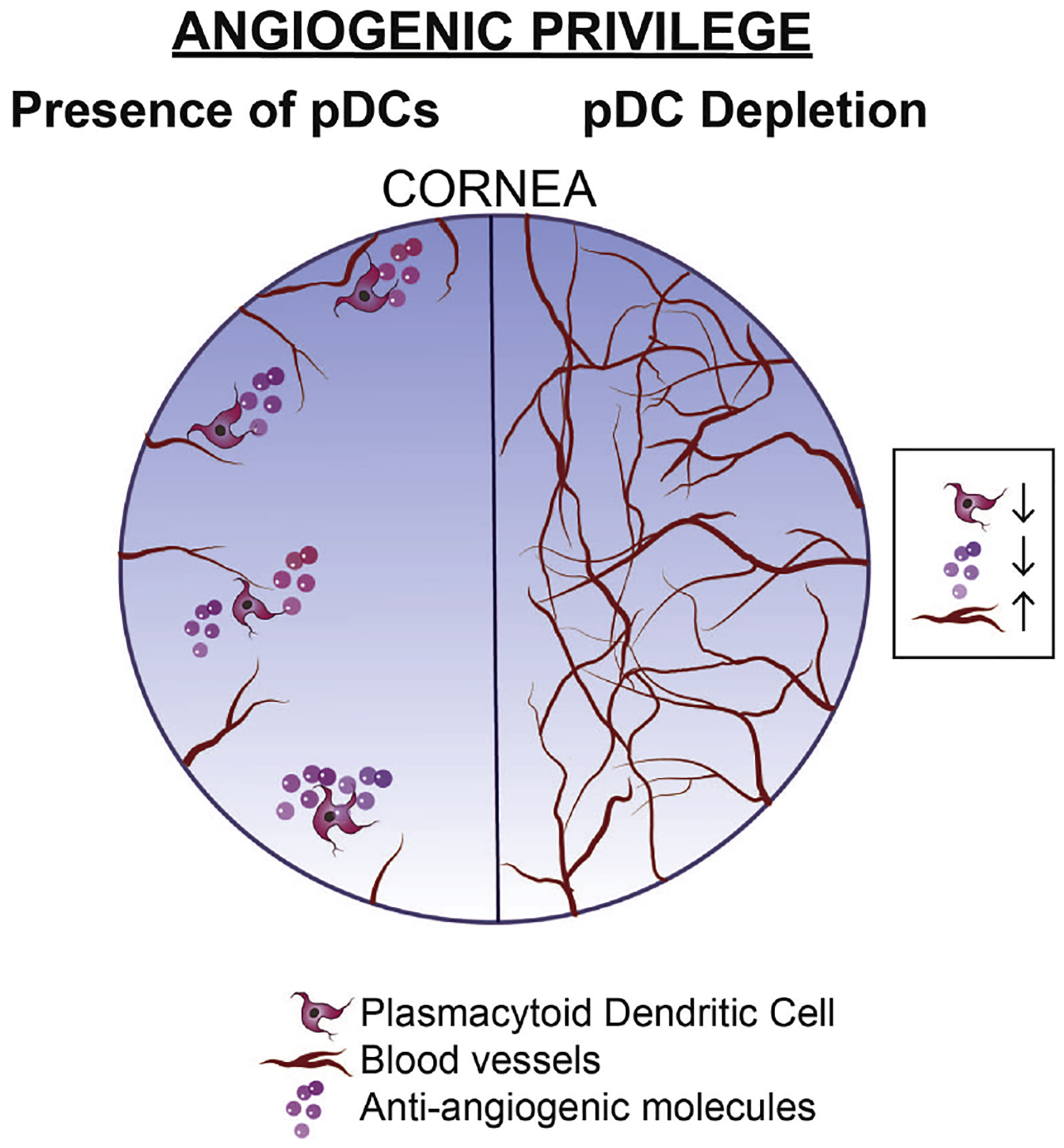
Plasmacytoid dendritic cells (pDCs) are a unique subpopulation of immune cells, distinct from classical dendritic cells. pDCs are generated in the bone marrow and following development, they typically home to secondary lymphoid tissues. While peripheral tissues are generally devoid of pDCs during steady state, few tissues, including the lung, kidney, vagina, and in particular ocular tissues harbor resident pDCs. pDCs were originally appreciated for their potential to produce large quantities of type I interferons in viral immunity. Subsequent studies have now unraveled their pivotal role in mediating immune responses, in particular in the induction of tolerance. In this review, we summarize our current knowledge on pDCs in ocular tissues in both mice and humans, in particular in the cornea, limbus, conjunctiva, choroid, retina, and lacrimal gland. Further, we will review our current understanding on the significance of pDCs in ameliorating inflammatory responses during herpes simplex virus keratitis, sterile inflammation, and corneal transplantation. Moreover, we describe their novel and pivotal neuroprotective role, their key function in preserving corneal angiogenic privilege, as well as their potential application as a cell-based therapy for ocular diseases.

Keywords: Angiogenesis; Neuroprotection; Plasmacytoid dendritic cells; Tolerance; Transplantation; Viral keratitis.


Figures

Figure 1.
Figure 1.. Illustration of plasmacytoid dendritic cells.
(A, B) Scanning electron micrograph (A) and transmission electron micrograph (B) of human pDCs isolated from peripheral blood. Magnification: ×3,500 in (A) and ×8,000 in (B); ©1997 Grouard et al. Originally published in J Exp Med. https://doi.org/10.1084/jem.185.6.1101. (C) Representative image of splenic pDCs in a DPE-GFP×RAG1−/− mouse with GFP-tagged pDCs. Scale bar: 20 μm. (D) Schematic representation of pDC markers in mice and humans. In humans, pDCs express the specific markers BDCA-2 and ILT-7, and share expression of BDCA-4, IL-3Rα, and ILT-3 with other immune cells. In mice, pDCs express PDCA-1, Siglec-H, CD11c, CD45R/B220, Ly6C, and Ly49Q. Both human and murine pDCs express intracellular receptors TLR-7 and TLR-9.
Figure 2.
Figure 2.. Schematic diagram on development of plasmacytoid dendritic cells.
As bone marrow-derived cells, pDCs can origin from both myeloid and lymphoid precursors, in mice. The cellular precursors of pDCs and transaction factors involved in development of pDCs are shown.
Figure 3.
Figure 3.. Schematic illustration of distribution of various resident immune cells in the ocular tissues.
(A) Resident immune cells are located in different parts of the ocular system. In the conjunctiva, cDCs, macrophages, B cell, and T cells are detected; in cornea, cDCs and macrophages comprise the main resident immune cells; in choroid, cDCs and macrophages and in retina microglia, perivascular macrophages, and cDCs are considered the main resident immune cells; in the lacrimal gland, cDCs, macrophages, and B cells are predominant resident immune cells. (B) To date, resident pDCs are reported in the central and peripheral cornea, limbus, bulbar conjunctiva, choroid, retina, and lacrimal gland
Figure 4.
Figure 4.. Presence of resident plasmacytoid dendritic cells in ocular tissues.
(A) Representative confocal micrographs of the limbus of a wild-type C57BL/6 mouse indicting CD45+ PDCA-1neg CD11chigh cDCs (arrow heads) as well as CD45+ PDCA-1+ CD11clow pDCs (arrows) during steady state. Scale bar: 20 μm. (B) Representative reconstructed multiphoton micrograph of cornea of a transgenic DPE-GFP×RAG1−/− mouse with specifically GFP-tagged plasmacytoid dendritic cells (green), highlighting typical morphology of resident corneal plasmacytoid dendritic cells with knob-like extensions (arrows) as well as less common morphology of corneal plasmacytoid dendritic cells with a round cell body without long stellates (arrow head). Second harmonic generation (SHG; blue) delineates corneal stroma. Scale bar: 50 μm. (C) Representative fluorescent microscopy image of the limbus in a transgenic DPE-GFP×RAG1−/− mouse receiving intravenous injection of quantum dots (red), reveals strategic localization of plasmacytoid dendritic cells (green) in close proximity to vessels in the limbus. Scale bar: 200 μm. (D) Representative flow cytometric dot plot on pooled conjunctiva of DPE-GFP×RAG1−/− mice during steady state indicating presence of CD45+ GFP+ cells among conjunctival single cells following gating out debris, dead cells, and debris (not shown). (E) Representative flow cytometric dot plots gated on live single CD45+ GFP+ cells of pooled conjunctiva of DPE-GFP×RAG1−/− mice during steady state depicting the identity of the CD45+ GFP+ cells as mainly plasmacytoid dendritic cells based on expression of PDCA-1 and lack of expression of CD3 and CD19. (F) Representative reconstructed multiphoton micrograph of the lacrimal gland of a transgenic DPE-GFP×RAG1−/− mouse, illustrating presence of resident lacrimal gland plasmacytoid dendritic cells. Second harmonic generation (SHG; blue) delineates lacrimal gland stroma. Scale bar: 50 μm. (G) Representative flow cytometric dot plot on lacrimal gland of a DPE-GFP×RAG1−/− mouse during steady state validating the presence of CD45+ GFP+ cells among lacrimal gland single cells following gating out debris, dead cells, and debris (not shown). (H) Representative flow cytometric dot plots gated on live single CD45+ GFP+ cells of lacrimal gland of a DPE-GFP×RAG1−/− mouse during steady state. Flow plots demonstrate that the majority of the CD45+ GFP+ cells are plasmacytoid dendritic cells based on expression of PDCA-1, moderate to low levels of CD11c, Gr-1 as well as lack of expression of CD3, CD19, CD11b, and F4/80; nevertheless, a minor population lack expression of PDCA-1 or express F4/80, CD11b, and/or high levels of CD11c.
Figure 5.
Figure 5.. Schematic illustration on the role of plasmacytoid dendritic cells during herpes simplex virus-1 keratitis.
During HSV-1 keratitis, local depletion of pDCs in the cornea is accompanied by increased infiltration of cellular members of innate and adaptive immunity, including cDCs, macrophages, and ex-Tregs, enhanced viral load, and reduced IFN-α level. In the draining lymph nodes, corneal pDC depletion leads to re-programming of Tregs to effector ex-Tregs, enhanced density of Th1 cells and decreased Tregs.
Figure 6.
Figure 6.. Schematic illustration on the role of plasmacytoid dendritic cells during sterile corneal inflammation.
During sterile corneal inflammation induced by suture placement, local depletion of pDCs leads to increased accumulation of immune cells in particular cDCs and macrophages.
Figure 7.
Figure 7.. Schematic illustration on the role of plasmacytoid dendritic cells in corneal allograft.
During allogeneic corneal transplantation, local depletion of pDCs prior to the procedure, enhances recruitment of innate immune cells including cDCs, macrophages, and CD4+ Th cells to the cornea and leads to enhanced generation of effector Th1 and Th17 and reduced density of Tregs in the draining lymph nodes.
Figure 8.
Figure 8.. Schematic illustration on the role of plasmacytoid dendritic cells in homeostasis of corneal nerves.
Local depletion of pDCs in the cornea during steady state is accompanied by decreased levels of neurotrophic molecule NGF in the cornea, leading to corneal nerve degeneration, and compromise of epithelial integrity.
Figure 9.
Figure 9.. Schematic illustration on the role of plasmacytoid dendritic cells in corneal angiogenic privilege.
While during steady state cornea enjoys angiogenic privilege, local depletion of pDCs in the cornea is accompanied by decreased levels of anti-angiogenic molecules, leading to break down of corneal angiogenic privilege.
Figure 10.
Figure 10.. Representative confocal micrograph of whole-mounted cornea showing successful local adoptive transfer of plasmacytoid dendritic cells.
The figure illustrates a representative image of the paracentral cornea, 48 h following adoptive transfer of 10,000 GFP+ pDCs isolated from the spleen of DPE-GFP×RAG1−/− mouse. Scale bar: 50 μm.

References

    1. Abb J, et al. (1983). “Phenotype of human alpha-interferon producing leucocytes identified by monoclonal antibodies.” Clin Exp Immunol 52(1): 179–184. - PMC - PubMed
    1. Abe M, et al. (2005). “Plasmacytoid dendritic cell precursors induce allogeneic T-cell hyporesponsiveness and prolong heart graft survival.” Am J Transplant 5(8): 1808–1819. - PubMed
    1. Abou-Slaybi A, et al. (2019). “Analysis of leukocyte populations and nerves in developing murine corneas.” The Journal of Immunology 202(1 Supplement): 117.115–117.115.
    1. Abtin A, et al. (2014). “Perivascular macrophages mediate neutrophil recruitment during bacterial skin infection.” Nat Immunol 15(1): 45–53. - PMC - PubMed
    1. Agrawal T, et al. (2009). “Recruitment of myeloid and plasmacytoid dendritic cells in cervical mucosa during Chlamydia trachomatis infection.” Clin Microbiol Infect 15(1): 50–59. - PubMed

Publication types