

Evolution of viral quasispecies during SARS-CoV-2 infection
- PMID: 32717416
- PMCID: PMC7378485
- DOI: 10.1016/j.cmi.2020.07.032
Evolution of viral quasispecies during SARS-CoV-2 infection
Abstract
Objectives: Studies are needed to better understand the genomic evolution of the recently emerged severe acute respiratory syndrome coronavirus 2 (SARS-CoV-2). This study aimed to describe genomic diversity of SARS-CoV-2 by next-generation sequencing (NGS) in a patient with longitudinal follow-up for SARS-CoV-2 infection.
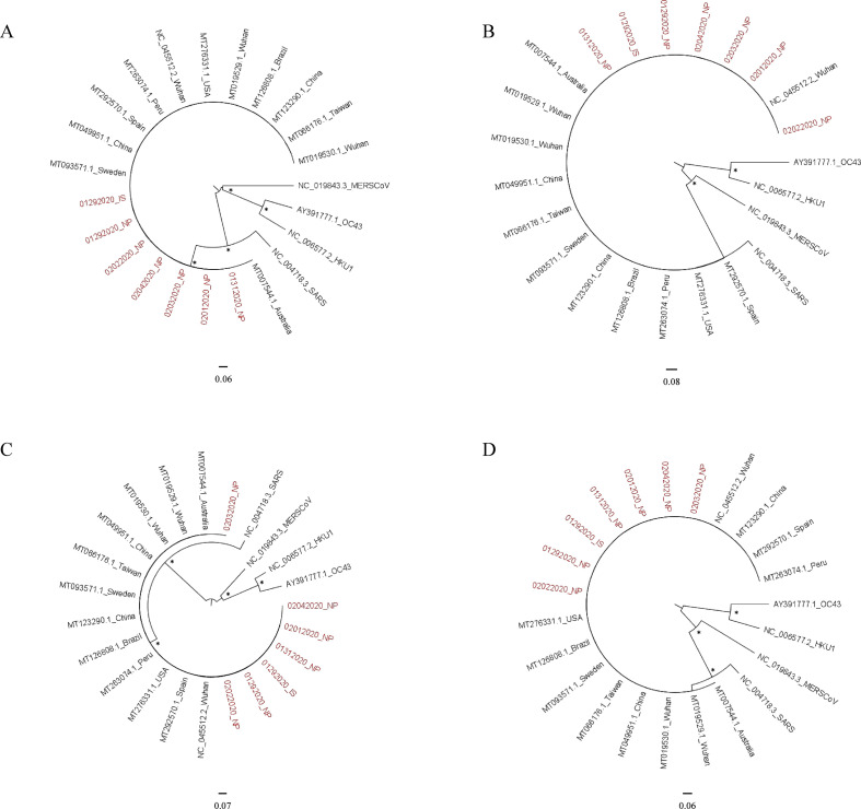
Methods: Sequential samples collected between January 29th and February 4th, 2020, from a patient infected by SARS-CoV-2 were used to perform amplification of two genome fragments-including genes encoding spike, envelope, membrane and nucleocapsid proteins-and NGS was carried out with Illumina® technology. Phylogenetic analysis was performed with PhyML and viral variant identification with VarScan.
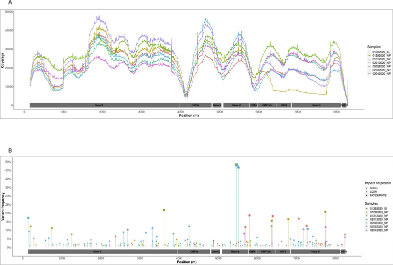
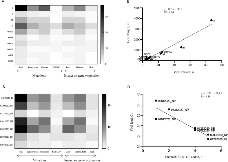
Results: Majority consensus sequences were identical in most of the samples (5/7) and differed in one synonymous mutation from the Wuhan reference sequence. We identified 233 variants; each sample harboured in median 38 different minority variants, and only four were shared by different samples. The frequency of mutation was similar between genes and correlated with the length of the gene (r = 0.93, p = 0.0002). Most of mutations were substitution variations (n = 217, 93.1%) and about 50% had moderate or high impact on gene expression. Viral variants also differed between lower and upper respiratory tract samples collected on the same day, suggesting independent sites of replication of SARS-CoV-2.
Conclusions: We report for the first time minority viral populations representing up to 1% during the course of SARS-CoV-2 infection. Quasispecies were different from one day to the next, as well as between anatomical sites, suggesting that in vivo this new coronavirus appears as a complex and dynamic distributions of variants.
Keywords: Infection follow-up; Minority variants; NGS; Quasispecies; SARS-CoV-2.
Copyright © 2020 European Society of Clinical Microbiology and Infectious Diseases. Published by Elsevier Ltd. All rights reserved.
Figures
References
-
- Chan J.F.-W., Kok K.-H., Zhu Z., Chu H., To K.K.-W., Yuan S., et al. Genomic characterization of the 2019 novel human-pathogenic coronavirus isolated from a patient with atypical pneumonia after visiting Wuhan. Emerg Microbe. Infect. 2020;9:221–236. doi: 10.1080/22221751.2020.1719902. - DOI - PMC - PubMed
-
- To K.K.-W., Tsang O.T.-Y., Leung W.-S., Tam A.R., Wu T.-C., Lung D.C., et al. Temporal profiles of viral load in posterior oropharyngeal saliva samples and serum antibody responses during infection by SARS-CoV-2: an observational cohort study. Lancet Infect Dis. 2020;20:565–574. doi: 10.1016/S1473-3099(20)30196-1. - DOI - PMC - PubMed
MeSH terms
Substances
LinkOut - more resources
Full Text Sources
Other Literature Sources
Miscellaneous
