Review
. 2020 Jul 23;12(8):1633.
doi: 10.3390/polym12081633.

Enzymatic Protein Biopolymers as a Tool to Synthetize Eukaryotic Messenger Ribonucleic Acid (mRNA) with Uses in Vaccination, Immunotherapy and Nanotechnology

Affiliations
Review

Enzymatic Protein Biopolymers as a Tool to Synthetize Eukaryotic Messenger Ribonucleic Acid (mRNA) with Uses in Vaccination, Immunotherapy and Nanotechnology

Fabiola Urbina et al. Polymers (Basel). .

Abstract

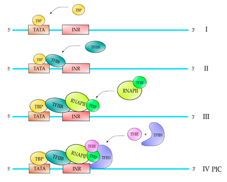
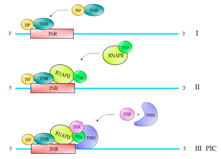
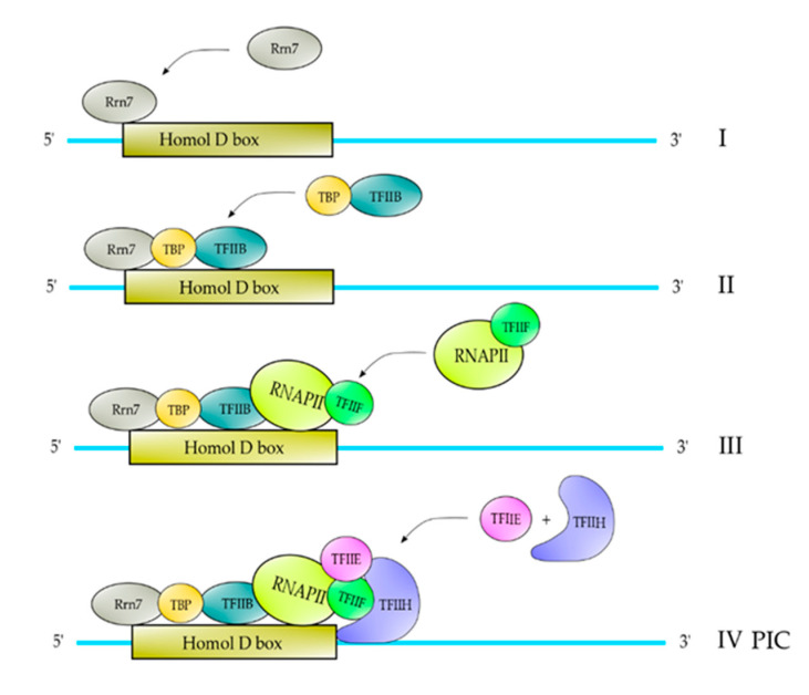
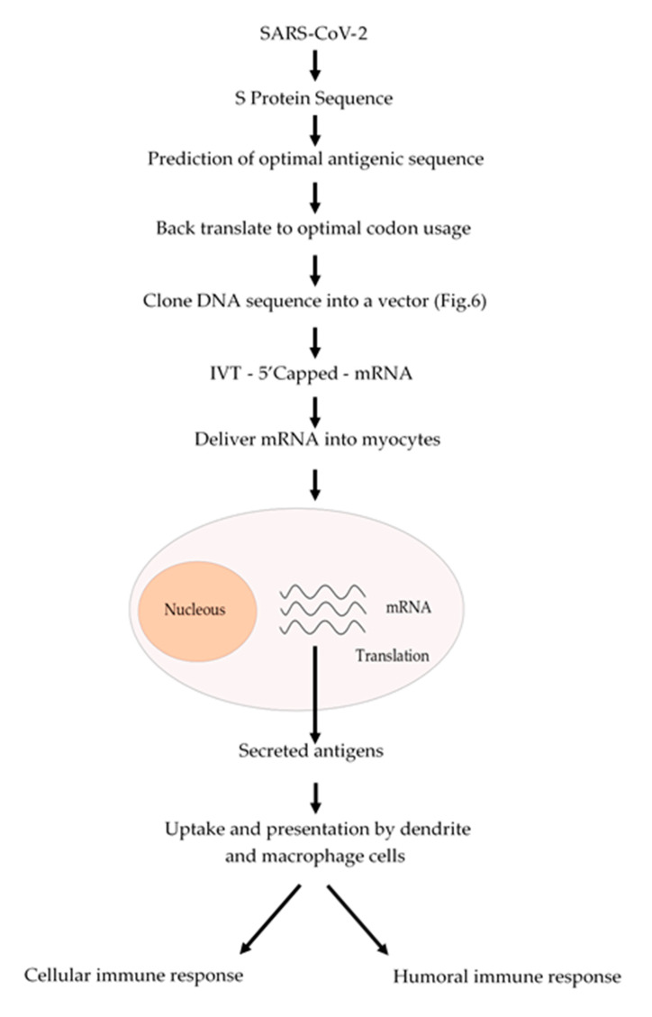
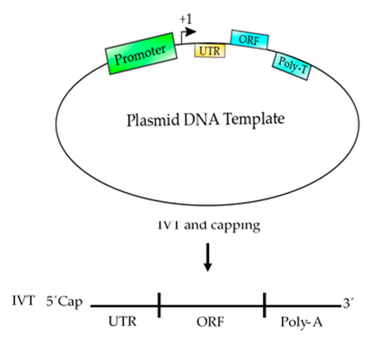
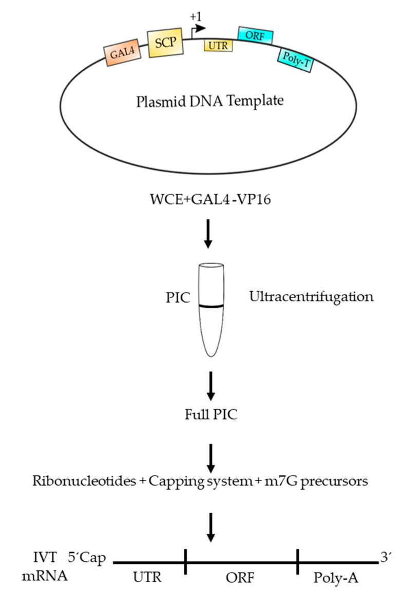
Multi-subunit enzymes are protein biopolymers that are involved in many cellular processes. The enzyme that carries out the process of transcription of mRNAs is RNA polymerase II (RNAPII), which is a multi-subunit enzyme in eukaryotes. This protein biopolymer starts the transcription from specific sites and is positioned by transcription factors, which form a preinitiation complex (PIC) on gene promoters. To recognize and position the RNAPII and the transcription factors on the gene promoters are needed specific DNA sequences in the gene promoters, which are named promoter elements. Those gene promoter elements can vary and therefore several kinds of promoters exist, however, it appears that all promoters can use a similar pathway for PIC formation. Those pathways are discussed in this review. The in vitro transcribed mRNA can be used as vaccines to fight infectious diseases, e.g., in immunotherapy against cancer and in nanotechnology to deliver mRNA for a missing protein into the cell. We have outlined a procedure to produce an mRNA vaccine against the SARS-CoV-2 virus, which is the causing agent of the big pandemic, COVID-19, affecting human beings all over the world. The potential advantages of using eukaryotic RNAPII to synthetize large transcripts are outlined and discussed. In addition, we suggest a method to cap the mRNA at the 5' terminus by using enzymes, which might be more effective than cap analogs. Finally, we suggest the construction of a future multi-talented RNAPII, which would be able to synthetize large mRNA and cap them in the test tube.

Keywords: immunotherapy; mRNA; nanotechnology; protein biopolymer; transcription; vaccine.


Conflict of interest statement

The authors declare no conflict of interest. The funders had no role in the design of the study; in the collection, analyses, or interpretation of data; in the writing of the manuscript, or in the decision to publish the results.

Figures

Figure 1
Figure 1
Description of the DNA template. The top strand is the coding strand or RNA-like strand, while the bottom strand is the template strand. +1 is the transcription start site.
Figure 2
Figure 2
PIC formation on TATA-containing promoters. The first step of PIC formation on this kind of promoters is the binding of TBP to the TATA element (I), which follows the binding of TFIIB through interactions with TBP and the promoter near to the transcription start site (II). After the binding of TFIIB, the complex RNAPII-TFIIF is assembled into the complex (III), which follows the binding of TFIIE and TFIIH to form a PIC on the promoter (IV). Only the top strand of the promoter is shown in the figure for didactic purposes, but the PIC is formed on the double stranded promoter. This is also valid for Figure 3 and Figure 4.
Figure 3
Figure 3
PIC formation on an INR-containing promoter. In those promoters that contain an INR, the complex TBP-TFIIB binds to this element (I) and after the complex RNAPII-TFIIF enters the complex (II), which follows the binding of TFIIE and TFIIH to complete the PIC formation (III). This model of PIC formation has not been published yet, but we have determined this pathway using pure RNAPII and recombinant factors from fission yeast and the nmt1 promoter in which the TATA box was deleted.
Figure 4
Figure 4
PIC formation on HomolD-box-containing promoters. In the first step of PIC formation, the transcription factor Rrn7 binds to the HomolD-box (I), which in turn is able to recruit the TBP-TFIIB complex (II). After TBP-TFIIB are recruited to the complex, it follows the binding of RNAPII-TFIIF (III), and then the TFIIE and TFIIH are recruited to the complex to form the complete PIC (IV).
Figure 5
Figure 5
Schematic procedure to produce an mRNA vaccine against SARS-CoV-2. From the analysis of the amino acid sequence of the S protein an antigenic region can be predicted. Whenever possible, a region of the S protein which is less prone to mutations should be selected. The amino acid sequence of the selected region can be back translated to DNA nucleotide sequence and it can be chemically synthetized according to the codon usage. Afterwards, this DNA sequence can be cloned into a DNA plasmid vector, as described in Figure 6, and used for in vitro transcription (IVT) by using a RNA polymerase to produce an mRNA that can be 5′ capped to be fully functional. This mRNA can be coated with lipid nanoparticles and delivered to the myocytes to be translated by the cell ribosomes. The resulting antigens will be secreted and take up by macrophages and dendritic cells, which will process and present the antigen to the immune system cells to activate both humoral (antibodies) and cellular (cytotoxic) immune responses.
Figure 6
Figure 6
Schematic representation of a plasmid DNA template used to produce an mRNA-based vaccine. This template contains the basic elements to produce a functional mRNA. A promoter is needed for the RNA polymerase to start transcription at the +1 site. The 5′ UTR is added to enhance the translation and the stability of the mRNA and the ORF contains the coding sequence of the antigen, which could be synthetized according to the codon usage to enhance the translation process. Finally, a poly T of at least 200 nucleotides is needed to produce a poly A tail on the mRNA. After, the template is transcribed (in vitro transcription, IVT) using RNA polymerase and ribonucleotides to produce an mRNA, which should be 5′ capped to produce a functional mRNA to be used as vaccine.
Figure 7
Figure 7
Outline of a procedure to produce a functional mRNA using eukaryotic RNAPII. The plasmid DNA template contains a super core promoter (SCP) and an upstream GAL4 binding site. Transcription starts at the +1 site, which is followed by an UTR (untranslated region) and an ORF containing the coding region to produce the polypeptide chain. A poly T tail is included in the template to produce a poly adenylated mRNA. The template is mixed with a highly active whole cell extract (WCE) from fission yeast, plus GAL4-VP16 activator and subjected to ultracentrifugation through a glycerol gradient (5–15%). Afterwards, those templates containing a fully assembled PIC are selected and used for transcription by adding ribonucleotides, together with methyltransferase and capping enzymes (capping system) plus the necessary precursors to form a m7G cap. The produced mRNA can be used as an mRNA vaccine.

References

    1. Hahn S. Structure and mechanism of the RNA polymerase II transcription machinery. Nat. Struct. Mol. Boil. 2004;11:394–403. doi: 10.1038/nsmb763. - DOI - PMC - PubMed
    1. Cramer P. Multisubunit RNA polymerases. Curr. Opin. Struct. Boil. 2002;12:89–97. doi: 10.1016/S0959-440X(02)00294-4. - DOI - PubMed
    1. Bushnell D.A., Cramer P., Kornberg R.D. Structural basis of transcription: Alpha-amanitin-RNA polymerase II cocrystal at 2.8 A resolution. Proc. Natl. Acad. Sci. USA. 2002;99:1218–1222. doi: 10.1073/pnas.251664698. - DOI - PMC - PubMed
    1. Phatnani H.P., Greenleaf A.L. Phosphorylation and functions of the RNA polymerase II CTD. Genes Dev. 2006;20:2922–2936. doi: 10.1101/gad.1477006. - DOI - PubMed
    1. Meinhart A., Kamenski T., Hoeppner S., Baumli S., Cramer P. A structural perspective of CTD function. Genes Dev. 2005;19:1401–1415. doi: 10.1101/gad.1318105. - DOI - PubMed

LinkOut - more resources