Review
. 2020 Sep;40(3):257-270.
doi: 10.1016/j.cll.2020.04.002. Epub 2020 Jun 16.

Proteopathic Seed Amplification Assays for Neurodegenerative Disorders

Affiliations
Review

Proteopathic Seed Amplification Assays for Neurodegenerative Disorders

Natália do Carmo Ferreira et al. Clin Lab Med. 2020 Sep.

Abstract

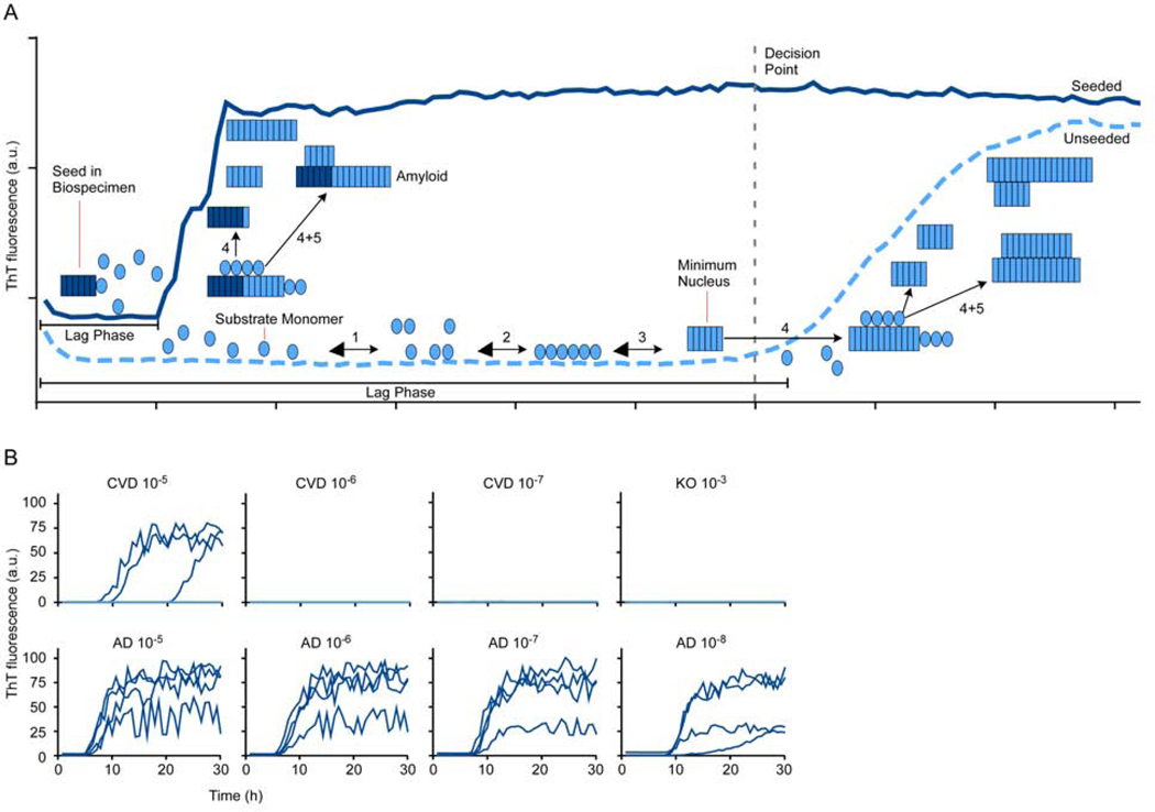
The need for etiological biomarkers for neurodegenerative diseases involving protein aggregation has prompted development of ultrasensitive cellular and cell-free assays based on the prion-like seeding capacity of such aggregates. Among them, prion RT-QuIC assays allow accurate antemortem Creutzfeldt-Jakob disease diagnosis using cerebrospinal fluid and nasal brushings. Analogous assays for synucleinopathies (e.g., Parkinson disease and dementia with Lewy bodies) provide unprecedented diagnostic sensitivity using cerebrospinal fluid. Biosensor cell and tau RT-QuIC assays can detect and discriminate tau aggregates associated with multiple tauopathies (e.g., Alzheimer disease and frontotemporal degeneration). An expanding panel of seed amplification assays should improve diagnostics and therapeutics development.

Keywords: Biomarkers; PMCA; Prion; RT-QuIC; Seed; Synuclein; Tau; β-Amyloid.


Conflict of interest statement

Disclosure B. Caughey is an inventor on patents or patent applications relating to prion, αSyn, and tau RT-QuIC assays. N.C. Ferreira has nothing to disclose.

Figures

Figure 1.
Figure 1.. Diagram of seeded polymerization in RT-QuIC reactions.
A. Reactions are set up in multiwell plates by diluting a biospecimen into reaction mixture containing a vast stoichiometric excess of an appropriate protein monomer (substrate) for the type of disease-associated seed being detected. The reaction also contains the amyloid-sensitive fluorescent dye, thioflavin T (ThT). When seeds are present in the biospecimen (left), they immediately start to grow (4) by recruiting new monomers. Elongated fibrils can promote secondary nucleation (5), i.e. the production of new seeding surfaces either by fragmentation or by providing lateral surfaces that can facilitate the ordering and conformational conversion of monomers into additional fibrils. Secondary nucleation contributes to the exponential growth of the new amyloid that enhances ThT fluorescence (dark blue trace). The lag phase in a seeded reaction represents the time it takes for the seeded amyloids to accumulate to levels that are detectable with ThT. Eventually, the reaction plateaus when all of the available monomer is converted to amyloid. In the absence of seeds in a negative control biospecimen (light blue trace), spontaneous nucleation (1–3) may occur, but only after a prolonged lag phase during which the kinetically unfavorable process of forming minimal stable nuclei (in this case a 6-mer) occurs . A key to developing an effective assay is finding substrates and assay conditions giving the greatest fold-separation between the lag phases of seeded versus unseeded reactions (e.g. see ). B. Representative primary AD tau RT-QuIC data comparing seeding by serial 10-fold dilutions of human familial AD (age 44) and cerebrovascular disease (CVD; age 53) brain tissue with reference to a 100 dilution being solid brain tissue. Tau knock-out (KO) mouse brain served as a completely tau-free negative control. Traces from quadruplicate wells for each brain dilution are shown. AD brain could be diluted at least 105-fold and 103-fold further than the KO and CVD brains, respectively, and still seed positive reactions. Although the CVD brain was not recorded as having tau pathology by immunohistochemistry, the typically higher sensitivity of RT-QuIC assays likely allowed detection of tau aggregates that were below the detection limit of immunohistochemistry.

References

    1. Caughey B, Lansbury PT. Protofibrils, pores, fibrils, and neurodegeneration: separating the responsible protein aggregates from the innocent bystanders. AnnuRevNeurosci. 2003;26:267–298. - PubMed
    1. Chiti F, Dobson CM. Protein Misfolding, Amyloid Formation, and Human Disease: A Summary of Progress Over the Last Decade. Annu Rev Biochem. 2017;109:27–68. - PubMed
    1. Silveira JR, Raymond GJ, Hughson AG, et al. The most infectious prion protein particles. Nature. 2005;437(7056):257–261. - PMC - PubMed
    1. Diaz-Villanueva JF, Diaz-Molina R, Garcia-Gonzalez V. Protein Folding and Mechanisms of Proteostasis. Int J Mol Sci. 2015;16(8):17193–17230. - PMC - PubMed
    1. Powers ET, Morimoto RI, Dillin A, Kelly JW, Balch WE. Biological and chemical approaches to diseases of proteostasis deficiency. AnnuRevBiochem. 2009;78:959–991. - PubMed

Publication types