

Case Reports
doi: 10.1016/j.carpath.2020.107227.
Epub 2020 May 12.
Fatal Pulmonary Thromboembolism in SARS-CoV-2-Infection
Affiliations
- PMID: 32718733
- PMCID: PMC7214296
- DOI: 10.1016/j.carpath.2020.107227
Item in Clipboard
Case Reports
Fatal Pulmonary Thromboembolism in SARS-CoV-2-Infection
Cardiovasc Pathol.
2020 Sep-Oct.
No abstract available
Figures
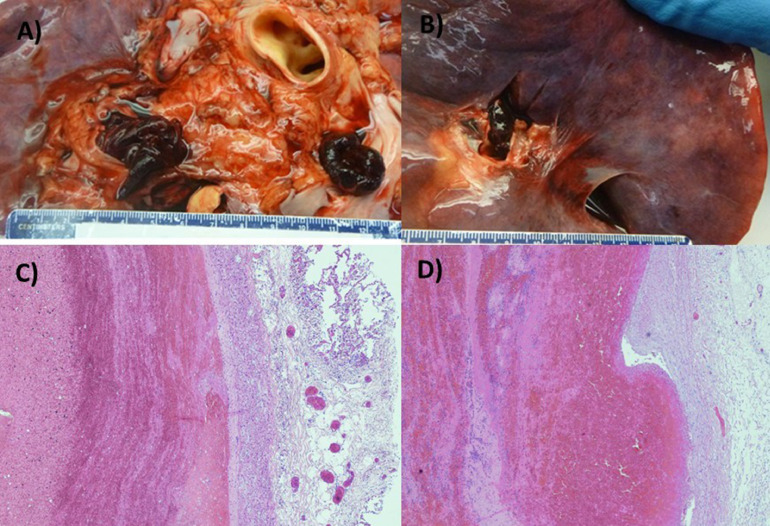
Pulmonary Emboli Occluding the Main Pulmonary Arteries (A) Gross autopsy image showing pulmonary thromboembolism occluding right and left main pulmonary arteries (Patient 1) and (B) right main pulmonary artery (Patient 2). Pulmonary embolism: alternating areas of pale pink (fibrin) and red (erythrocytes) layers forming "lines of Zahn" in patient 1 (C) and 2 (D).
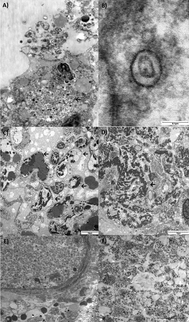
Electron microscopy and histopathological lung findings of SARS-CoV-2. Lung sections were initially placed in neutral buffered formalin and later transferred to 3% buffered glutaraldehyde. Following post-fixation in 1% osmium tetroxide, tissues were serially dehydrated and embedded in epoxy resin in standard fashion. One micron toluidine-stained scout sections were prepared for light microscopic orientation and 80nm ultrathin sections for electron microcopy were stained with uranyl acetate and lead citrate and examined in an Hitachi 7650 transmission electron microscope at 80kV. (A) Transmission electron microscopy image showing an infected pneumocyte. Note the cytoplasmic vacuole loaded with viral particles in various stages of bud formation (←), and shedding into the luminal compartment (*) (3000x). (B) Typical morphology of Coronavirus (SARS-CoV-2) showing a symmetrical arrangement, outer envelope and surrounding electron dense shell with central zone. Note the distinctive projections conferring a solar corona appearance. (C) Multiple capillaries showing deposits of fibrin deposition (←). (D) Coarse electron dense areas of cytoplasmic fibrin deposition. (E) Pure platelet thrombus within the arterial lumen and (F) Detail on platelet thrombi. Scale bars are shown at the bottom right of each figure.
References
-
- Porcheduu R., Serra C., Kelvin D., Kelvin N., Rubino S. Similarity in case fatality rates (CFR) of COVID-19/SARS-COV-2 in Italy and China. J Infect Dev Ctries. 2020;14:125–128. - PubMed
Publication types
MeSH terms
LinkOut - more resources
Full Text Sources
Medical
Miscellaneous
