. 2020 Sep 11;295(37):13031-13046.
doi: 10.1074/jbc.RA120.013396. Epub 2020 Jul 21.

Quantitating denaturation by formic acid: imperfect repeats are essential to the stability of the functional amyloid protein FapC

Affiliations

Quantitating denaturation by formic acid: imperfect repeats are essential to the stability of the functional amyloid protein FapC

Line Friis Bakmann Christensen et al. J Biol Chem. .

Abstract

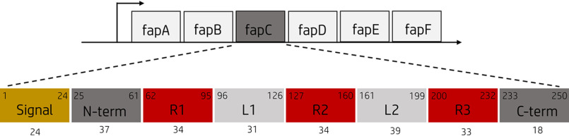
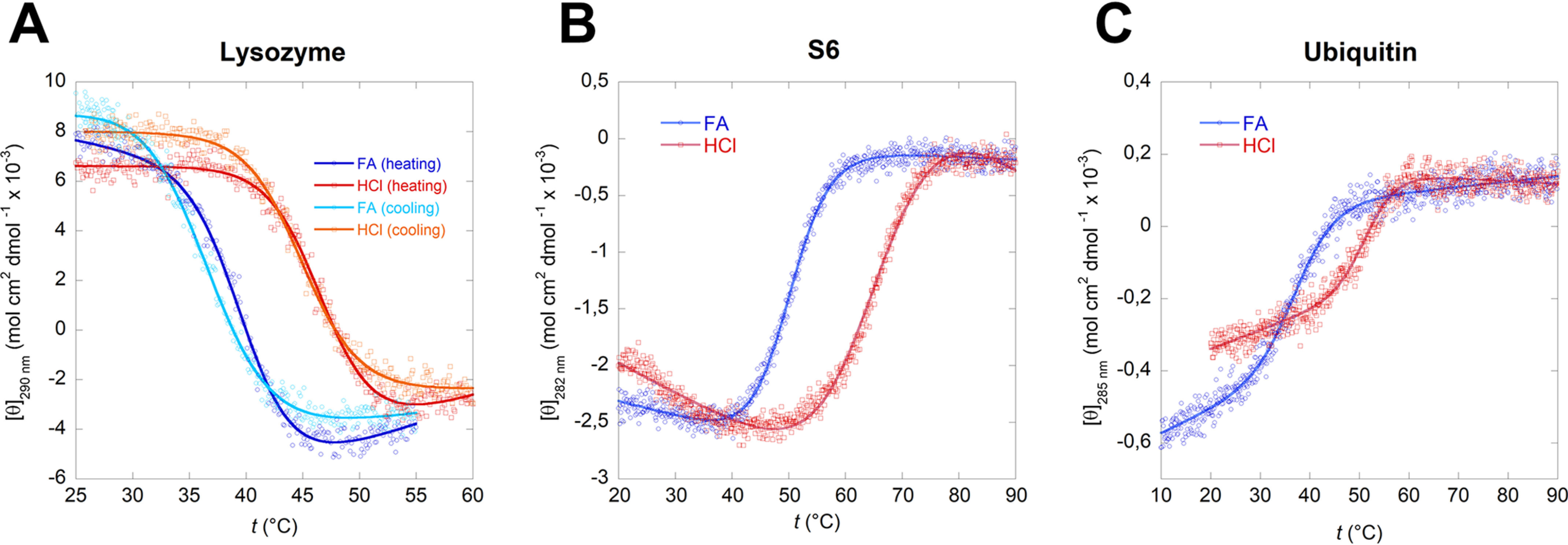
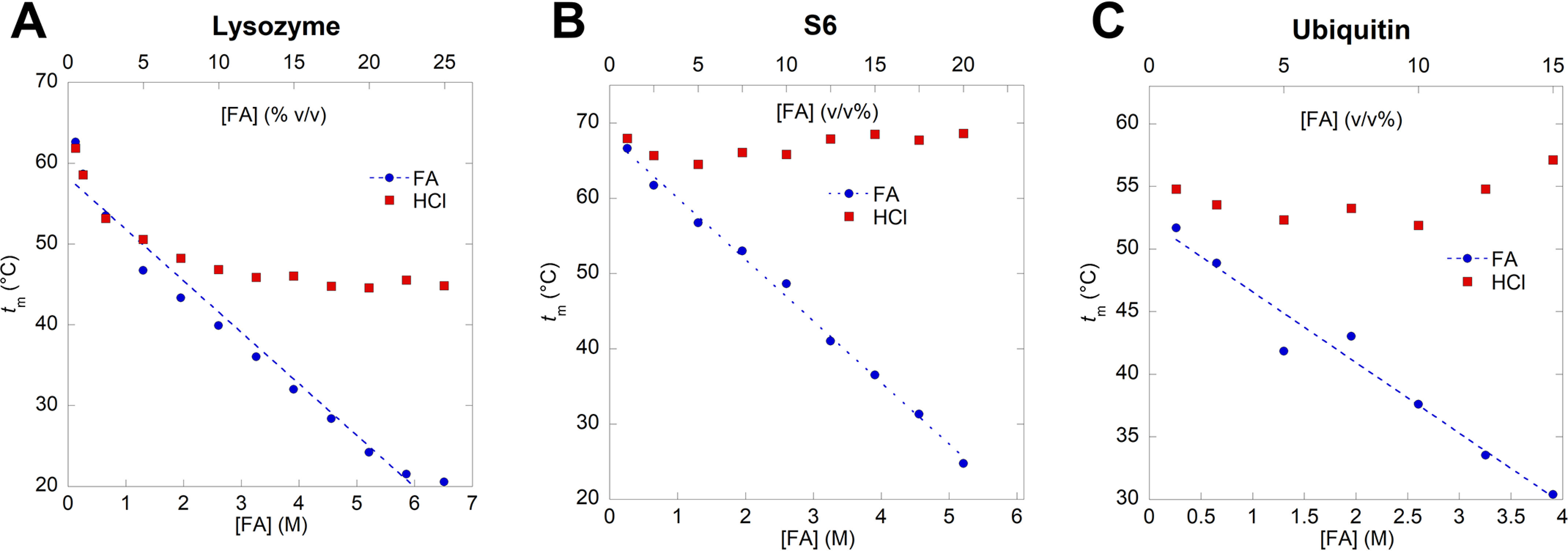
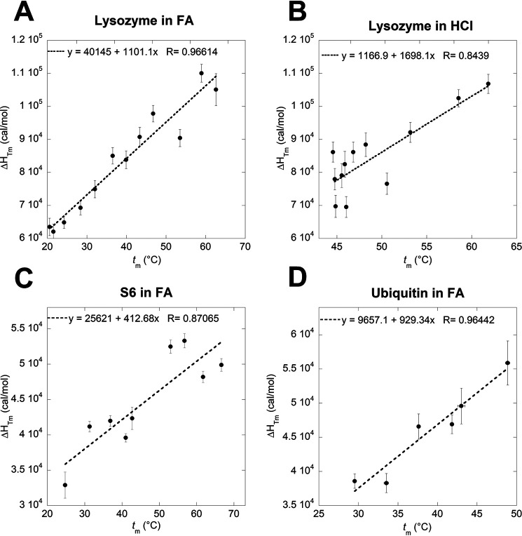
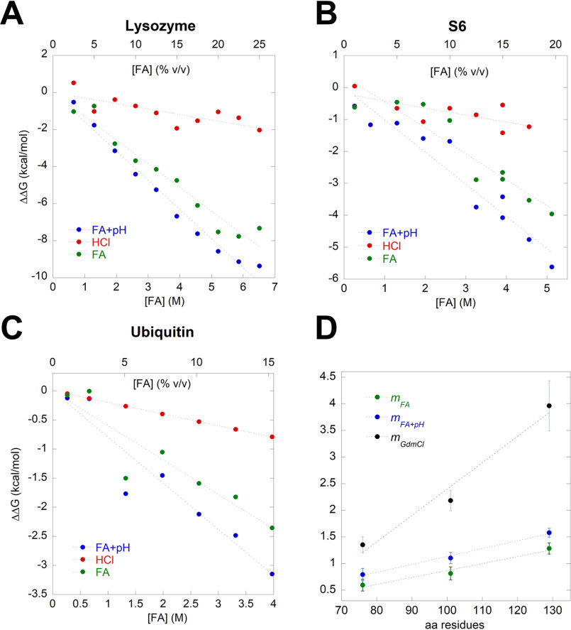
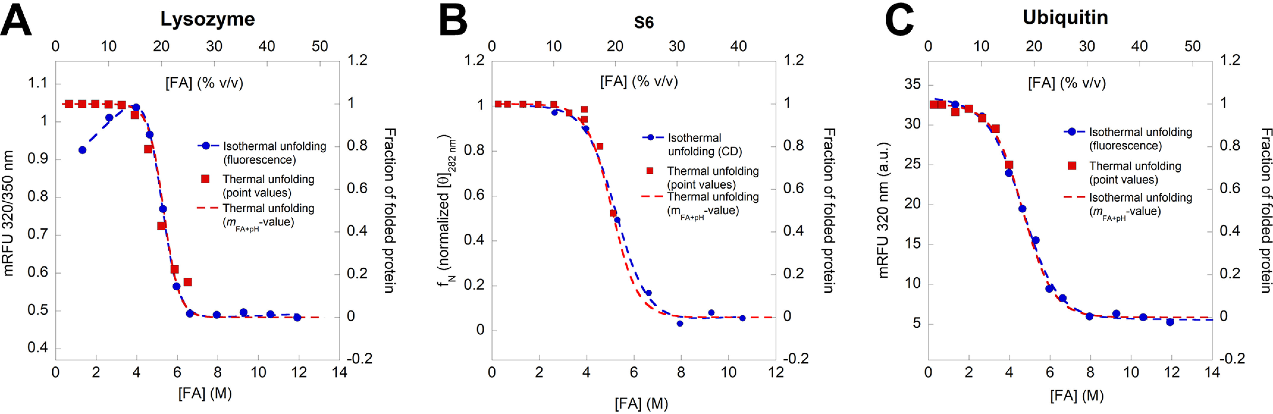
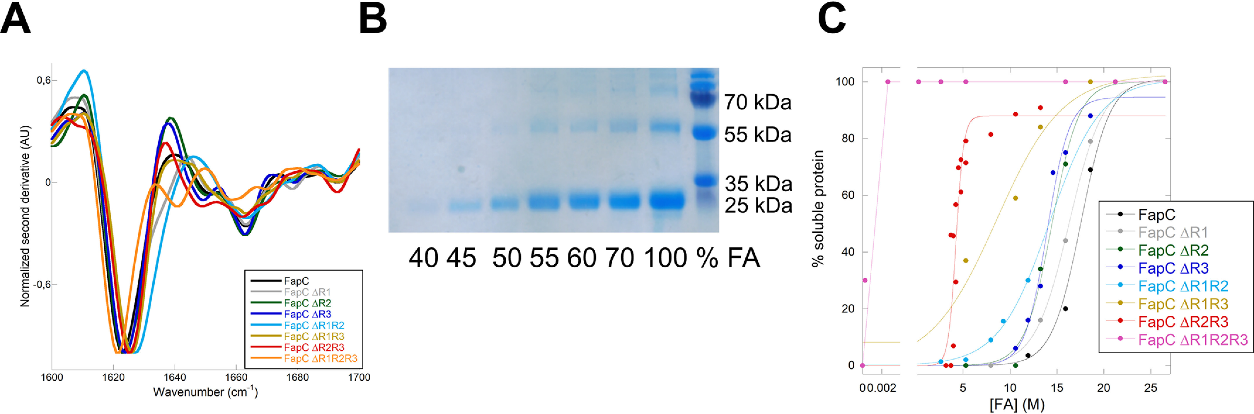
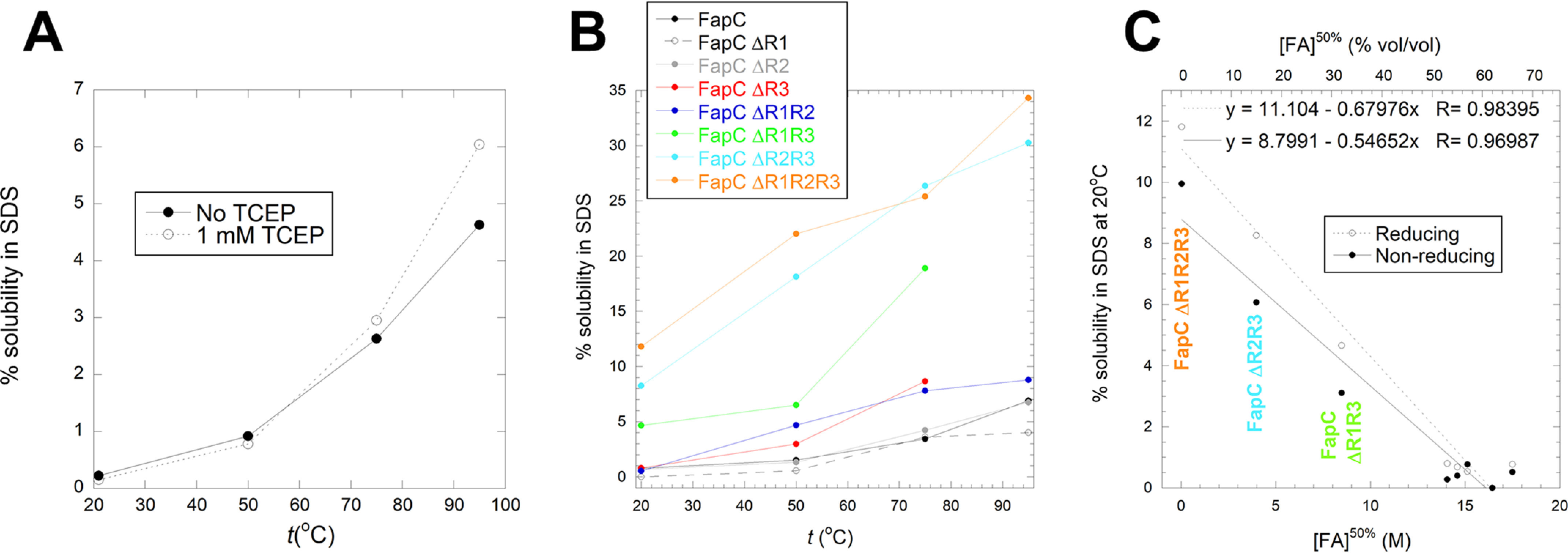
Bacterial functional amyloids are evolutionarily optimized to aggregate, so much so that the extreme robustness of functional amyloid makes it very difficult to examine their structure-function relationships in a detailed manner. Previous work has shown that functional amyloids are resistant to conventional chemical denaturants, but they dissolve in formic acid (FA) at high concentrations. However, systematic investigation requires a quantitative analysis of FA's ability to denature proteins. Amyloid formed by Pseudomonas sp. protein FapC provides an excellent model to investigate FA denaturation. It contains three imperfect repeats, and stepwise removal of these repeats slows fibrillation and increases fragmentation during aggregation. However, the link to stability is unclear. We first calibrated FA denaturation using three small, globular, and acid-resistant proteins. This revealed a linear relationship between the concentration of FA and the free energy of unfolding with a slope of mFA+pH (the combined contribution of FA and FA-induced lowering of pH), as well as a robust correlation between protein size and mFA+pH We then measured the solubilization of fibrils formed from different FapC variants with varying numbers of repeats as a function of the concentration of FA. This revealed a decline in the number of residues driving amyloid formation upon deleting at least two repeats. The midpoint of denaturation declined with the removal of repeats. Complete removal of all repeats led to fibrils that were solubilized at FA concentrations 2-3 orders of magnitude lower than the repeat-containing variants, showing that at least one repeat is required for the stability of functional amyloid.

Keywords: FapC; Pseudomonas; fibril; formic acid; functional amyloid; heat capacity; m-values; protein denaturation; protein stability; thermodynamics.


Conflict of interest statement

Conflict of interest—The authors declare that they have no conflicts of interest with the contents of this article.

Figures

Figure 1.
Figure 1.
The fapA–F operon and the FapC protein. The fapA–F operon encodes the six Fap proteins, FapA–F. The Pseudomonas sp. strain UK4 FapC protein consists of a 24-aa N-terminal signal peptide (yellow) and three imperfect repeats (R1–R3, red) separated by two linker regions (L1–L2, light gray). Start and end amino acid positions for the different regions are shown in the upper corners, and the length is shown below.
Figure 2.
Figure 2.
Thermal unfolding curves of lysozyme (A), S6 (B), and ubiquitin (C) in 10%, v/v, FA and in HCl solutions with the corresponding pH. In 10% FA all three proteins show significantly reduced thermal stability while maintaining a reversible two-state unfolding (Fig. S2). The sigmoidal curves were fitted using Equation 1.
Figure 3.
Figure 3.
Melting temperature of lysozyme (A), S6 (B), and ubiquitin (C). The FA concentration is represented both as molar concentration (M) and volume percentage (v/v%). Each melting temperature, tm, is obtained by thermal unfolding of the proteins at a given concentration of FA or HCl followed by fitting the sigmoidal unfolding curves of Equation 1 (Fig. 2). tm values for all three proteins decrease significantly at high FA concentrations compared with HCl solutions of the corresponding pH.
Figure 4.
Figure 4.
Determination of the specific heat capacity of denaturation (ΔCp) of lysozyme in FA (A) and HCl (B), S6 in FA (C) and ubiquitin in FA (D) by plotting δHTm versus tm. ΔCp of S6 and ubiquitin in HCl could not be determined because of insignificant difference in measured tm values at different concentrations of HCl.
Figure 5.
Figure 5.
Correlation between the change in the free energy of unfolding ΔΔGD-N and FA concentration (blue), HCl (red), and HCl-subtracted effect of FA (green) of lysozyme (A), S6 (B), and ubiquitin (C). D, the m values found in panels AC are plotted versus the number of residues of the model proteins. These FA-based m values can be compared to m values for the same three proteins in GdmCl, resulting in a slope that is 3–4 times more steep for GdmCl reflecting FA's comparatively lower strength as a denaturant.
Figure 6.
Figure 6.
Unfolding of lysozyme (A), S6 (B), and ubiquitin (C), measured by intrinsic aromatic fluorescence (lysozyme and ubiquitin) and near-UV CD (S6). Blue points are measured equilibrium values, whereas red points represent the predicted fraction of folded protein at 25 °C based on thermal unfolding values. The red stippled curves are best fits of Equation 4 to the red points. These show a very good correspondence with the measured isothermal equilibrium data set.
Figure 7.
Figure 7.
All FapC proteins can form amyloid fibrils. A, FapC variants were desalted from 8 M GdmCl into MQ water and immediately fibrillated before FTIR analysis was performed on the formed fibrils. The normalized second derivative shown here validates the amyloid nature of the different fibrils. B, representative gel of the supernatants after treatment with FA (here shown for FapC ΔR3 protein). The band around 25 kDa is FapC solubilized by FA. C, percentage of total protein present in supernatant after FA treatment as a function of FA concentration. Gel bands were quantified with ImageJ, normalized to the 100% FA band, and fitted to Equation 8. There is a marked decrease in stability when the number of repeats is reduced.
Figure 8.
Figure 8.
Solubilization of FapC fibrils in SDS. A, solubility of WT FapC in SDS at 20–95°C under reducing (1 mm TCEP) or nonreducing conditions. Solubility was quantified as in Fig. 7C. B, solubility under reducing conditions in SDS at 20–95°C for all 8 FapC variants. C, correlation between solubility in SDS at room temperature and the midpoint of denaturation in FA, shown for all 8 FapC variants. The three most destabilized variants are indicated with the same colors as those in panel B.

References

    1. Pham C. L., Kwan A. H., and Sunde M. (2014) Functional amyloid: widespread in Nature, diverse in purpose. Essays Biochem. 56, 207–219 10.1042/bse0560207 - DOI - PubMed
    1. Dueholm M., Nielsen P. H., Chapman M. R., and Otzen D. E. (2012) Functional amyloids in Bacteria. in Amyloid Fibrils and Prefibrillar Aggregates (Otzen D. E., ed.), pp. 411–438, Wiley-VCH Verlag GmbH, Berlin, Germany
    1. Olsen A., Jonsson A., and Normark S. (1989) Fibronectin binding mediated by a novel class of surface organelles on Escherichia coli. Nature 338, 652–655 10.1038/338652a0 - DOI - PubMed
    1. Collinson S. K., Emody L., Muller K. H., Trust T. J., and Kay W. W. (1991) Purification and characterization of thin, aggregative fimbriae from Salmonella enteritidis. J. Bacteriol. 173, 4773–4781 10.1128/jb.173.15.4773-4781.1991 - DOI - PMC - PubMed
    1. Dueholm M. S., Albertsen M., Otzen D., and Nielsen P. H. (2012) Curli functional amyloid systems are phylogenetically widespread and display large diversity in operon and protein structure. PLoS ONE 7, e51274 10.1371/journal.pone.0051274 - DOI - PMC - PubMed

Publication types

Associated data