. 2020 Aug 11;117(32):19599-19603.
doi: 10.1073/pnas.2001776117. Epub 2020 Jul 27.

Elevated energy requirement of cone photoreceptors

Affiliations

Elevated energy requirement of cone photoreceptors

Norianne T Ingram et al. Proc Natl Acad Sci U S A. .

Abstract

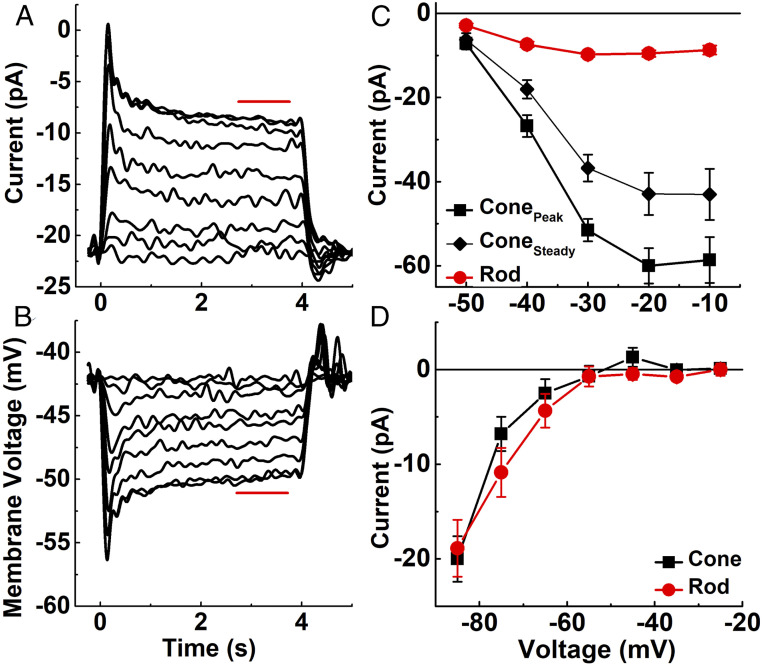
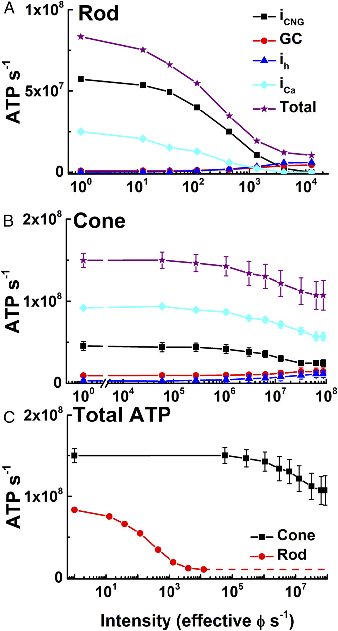
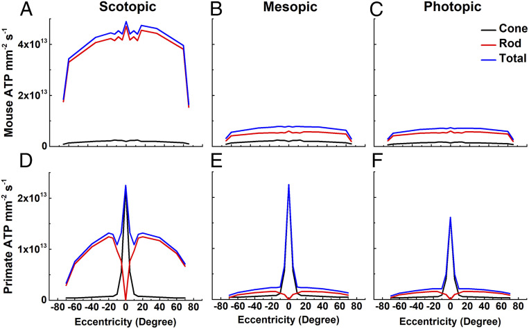
We have used recent measurements of mammalian cone light responses and voltage-gated currents to calculate cone ATP utilization and compare it to that of rods. The largest expenditure of ATP results from ion transport, particularly from removal of Na+ entering outer segment light-dependent channels and inner segment hyperpolarization-activated cyclic nucleotide-gated channels, and from ATP-dependent pumping of Ca2+ entering voltage-gated channels at the synaptic terminal. Single cones expend nearly twice as much energy as single rods in darkness, largely because they make more synapses with second-order retinal cells and thus must extrude more Ca2+ In daylight, cone ATP utilization per cell remains high because cones never remain saturated and must continue to export Na+ and synaptic Ca2+ even in bright illumination. In mouse and human retina, rods greatly outnumber cones and consume more energy overall even in background light. In primates, however, the high density of cones in the fovea produces a pronounced peak of ATP utilization, which becomes particularly prominent in daylight and may make this part of the retina especially sensitive to changes in energy availability.

Keywords: degeneration; fovea; metabolism; photoreceptor; retina.


Conflict of interest statement

The authors declare no competing interest.

Figures

Fig. 1.
Fig. 1.
Light responses and voltage-gated currents of mouse cones. (A and B) Cones from Cx36−/− retinas were illuminated with 4 s of steady 405-nm light at following intensities (in effective photons s−1 at the λmax value of either the M or S cone pigment): 4.4 × 10⁴, 2.1 × 10⁵, 8.1 × 10⁵, 2.3 × 10⁶, 4.8 × 10⁶, 9.6 × 10⁶, 2.4 × 10⁷, 4.8 × 10⁷, 6.6 × 10⁷. (A) Mean current responses from four cones held at a membrane potential of −50 mV. (B) Mean voltage responses from seven cones to the same light-intensity steps as in A. Red bars indicate region of responses averaged to calculate steady-state response. (C) Amplitude of Ca2+ current to depolarizing voltage steps in darkness, measured in the presence of 200 μm niflumic acid in the external perfusion solution to block Ca2+-activated Cl currents (4). Photoreceptors were held at −70 mV and then stepped for 0.75 s to depolarizing potentials (−50 to 0 mV in 10-mV increments). Data show peak current and current measured just before termination of the voltage step (“steady”) plotted against holding potential. Symbols are mean ± SEM values for 14 cones and 9 rods. The horizontal line indicates zero current. (D) Mean ih current from 7 cones and 13 rods recorded as in Ingram et al. (4) by holding Vm at −30 mV and stepping for 400 ms to hyperpolarizing potentials (−25 mV to −85 mV in 10-mV increments). We included 25 mM tetraethylammonium and 10 μm isradipine in the external perfusion solution to block K+ and Ca2+ currents. The horizontal line indicates zero current.
Fig. 2.
Fig. 2.
ATP consumption in rods and cones in steady light. (A and B) Net ATP consumption for rods (A) and cones (B) as a function of light intensity in photons s−1, effective at the λmax of the rod or cone photopigments (SI Appendix). ATP consumption was then calculated from physiological measurements in Fig. 1 (SI Appendix), due to CNG channels (iCNG, black squares), HCN channels (ih, blue triangles), Ca2+ influx at synaptic terminals (iCa, cyan diamonds), guanylyl cyclase (GC, red circles), and sum of all four (total, purple stars). (C) Total ATP consumed for rods (red) and cones (black). Data are mean ± SEM.
Fig. 3.
Fig. 3.
ATP consumption across retinal eccentricities. We used total ATP consumption for rods and cones from Fig. 2 together with measurements of the density of photoreceptors as a function of retinal eccentricity (mouse, 32; human, 36) to calculate the metabolic demand of the outer retina in mouse (AC) and human (DF) in three ambient light intensities: scotopic (darkness; 0 effective ϕ cell−1s−1) (A and D); mesopic (12,600 effective ϕ rod−1s−1 or 12,500 effective ϕ cone−1s−1) (B and E); and photopic (8.8 × 10⁷ effective ϕ rod−1s−1 or 8.7 × 10⁷ effective ϕ cone−1s−1) (C and F). The same values were used for cone currents, even though foveal cone outer segments are three times longer than mouse cones (38, 39) and likely have larger circulating currents. No attempt was made to take the difference in the number of synaptic ribbons in the pedicles between foveal and peripheral cones into consideration (40); these factors would only increase the difference in ATP consumption between rods and cones.

References

    1. Linsenmeier R. A., Braun R. D., Oxygen distribution and consumption in the cat retina during normoxia and hypoxemia. J. Gen. Physiol. 99, 177–197 (1992). - PMC - PubMed
    1. Okawa H., Sampath A. P., Laughlin S. B., Fain G. L., ATP consumption by mammalian rod photoreceptors in darkness and in light. Curr. Biol. 18, 1917–1921 (2008). - PMC - PubMed
    1. Dawis S. M. et al. ., Adenosine triphosphate utilization rates and metabolic pool sizes in intact cells measured by transfer of 18O from water. Biophys. J. 55, 79–99 (1989). - PMC - PubMed
    1. Ingram N. T., Sampath A. P., Fain G. L., Membrane conductances of mouse cone photoreceptors. J. Gen. Physiol. 152, e201912520 (2020). - PMC - PubMed
    1. Attwell D., Laughlin S. B., An energy budget for signaling in the grey matter of the brain. J. Cereb. Blood Flow Metab. 21, 1133–1145 (2001). - PubMed

Publication types

MeSH terms