. 2020 Aug 11;117(32):19321-19327.
doi: 10.1073/pnas.1921856117. Epub 2020 Jul 27.

Hedgehog signaling is necessary and sufficient to mediate craniofacial plasticity in teleosts

Affiliations

Hedgehog signaling is necessary and sufficient to mediate craniofacial plasticity in teleosts

Dina Navon et al. Proc Natl Acad Sci U S A. .

Abstract

Phenotypic plasticity, the ability of a single genotype to produce multiple phenotypes under different environmental conditions, is critical for the origins and maintenance of biodiversity; however, the genetic mechanisms underlying plasticity as well as how variation in those mechanisms can drive evolutionary change remain poorly understood. Here, we examine the cichlid feeding apparatus, an icon of both prodigious evolutionary divergence and adaptive phenotypic plasticity. We first provide a tissue-level mechanism for plasticity in craniofacial shape by measuring rates of bone deposition within functionally salient elements of the feeding apparatus in fishes forced to employ alternate foraging modes. We show that levels and patterns of phenotypic plasticity are distinct among closely related cichlid species, underscoring the evolutionary potential of this trait. Next, we demonstrate that hedgehog (Hh) signaling, which has been implicated in the evolutionary divergence of cichlid feeding architecture, is associated with environmentally induced rates of bone deposition. Finally, to demonstrate that Hh levels are the cause of the plastic response and not simply the consequence of producing more bone, we use transgenic zebrafish in which Hh levels could be experimentally manipulated under different foraging conditions. Notably, we find that the ability to modulate bone deposition rates in different environments is dampened when Hh levels are reduced, whereas the sensitivity of bone deposition to different mechanical demands increases with elevated Hh levels. These data advance a mechanistic understanding of phenotypic plasticity in the teleost feeding apparatus and in doing so contribute key insights into the origins of adaptive morphological radiations.

Keywords: craniofacial; ecodevo; flexible stem; hedgehog signaling; phenotypic plasticity.


Conflict of interest statement

The authors declare no competing interest.

Figures

Fig. 1.
Fig. 1.
A four-bar linkage chain is shown describing the kinematics of lower jaw opening in the cichlid skull (29). The three red bars represent movable links, while the single black bar is a fixed link. The system is driven by the levator opercula muscle (not shown), which originates on the skull and inserts along the dorsal aspect of the opercle bone (OP, gray). With the contraction of this muscle, the OP (input link) swings posteriodorsally, pulling the retroarticular process ([RA], output link) of the lower jaw (purple) through the interopercle (IOP) bone (blue, coupler link), which results in lower jaw depression via rotation around the mandible–quadrate joint, located just dorsal to the RA. Variation in the geometry of this linkage will influence the efficiency of the system (e.g., output per unit input) and is associated with cichlid species adapted to different diets (14, 20). Notably, the shapes of 2/3 moveable links (i.e., the IOP and RA links) in this system are associated with and responsive to Hh signaling levels (13, 14).
Fig. 2.
Fig. 2.
Predictions are shown for the possible roles of Hh signaling on bone growth and plasticity. The null hypothesis is that bone growth is not plastic, and manipulation of Hh levels has no effect on bone growth (A). Based on previous work from many laboratories, we do not expect this to be the case. Hh signaling is known to play important roles in bone development (15), and the cichlid jaw has been shown to be plastic (27). Another possible outcome is that Hh levels influence bone growth but that the relative effect is similar across environments (B). Finally, we may find that Hh levels influence plasticity such that the effect of Hh levels on bone growth depends on the environment (C). For the experiments in this paper, E1 and E2 represent alternate feeding regimes.
Fig. 3.
Fig. 3.
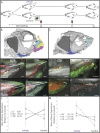
Rates of bone deposition are shown for different cichlid species and zebrafish genotypes. (A) Schematic of the experimental design: Fish families were split and trained on alternate benthic/pelagic diets for 1 to 2 wk, at time 0 (T0) both groups were injected with a red fluorochrome (alizarin), at T1 (i.e., 1 mo later), a second green fluorochrome (calcein) was injected, and at T2 (i.e., 1 wk later) animals were killed and prepared for bone imaging. (B and C) µCT reconstructions of a representative cichlid (B) and zebrafish (C) highlighting the bones that were analyzed, including the premaxilla (yellow), maxilla (teal), mandible (pink), IOP (blue), and OP (dark gray). Black arrows in B and C indicate the locations of matrix deposition measurements. Black boxes indicate the anterior portion of the IOP shown in DK and quantified in L and M. Differences in bone deposition in TRC reared in benthic versus pelagic foraging treatments are illustrated by D and E, respectively. Examples from MZ are shown in F and G, WT zebrafish are shown in H and I, and zebrafish carrying the Hh ++ transgene Tg(hsp70l:shha-EGFP) are shown in J and K. White bars indicate the measurement of matrix deposition between T0 and T1. Gray scale bars equal 50 μm for all panels. (L and M) Reaction norms showing the strength and direction of plasticity in the IOP for the three cichlid species (L) and the three zebrafish genotypes (M). The Hh – transgene is Tg(hsp70l:gli2DR-EGFP). Symbols represent means. Numbers next to symbols represent sample sizes for each experimental replicate. Error bars are 95% CIs.

References

    1. Bradshaw A. D., Evolutionary significance of phenotypic plasticity in plants. Adv. Genet. 13, 115–155 (1965).
    1. Schlichting C. D., Pigliucci M., Phenotypic Evolution. A Reaction Norm Perspective, (Sinauer Associates, Sunderland, MA, 1998).
    1. Pigliucci M., Evolution of phenotypic plasticity: Where are we going now? Trends Ecol. Evol. 20, 481–486 (2005). - PubMed
    1. Luo L., Actin cytoskeleton regulation in neuronal morphogenesis and structural plasticity. Annu. Rev. Cell Dev. Biol. 18, 601–635 (2002). - PubMed
    1. Flück M., Hoppeler H., “Molecular basis of skeletal muscle plasticity-from gene to form and function” in Reviews of Physiology, Biochemistry and Pharmacology, (Springer, Berlin, Germany, 2003), Vol. 146, pp. 159–216. - PubMed

Publication types