. 2020 Sep 18;88(10):e00372-20.
doi: 10.1128/IAI.00372-20. Print 2020 Sep 18.

Leukocidins and the Nuclease Nuc Prevent Neutrophil-Mediated Killing of Staphylococcus aureus Biofilms

Affiliations

Leukocidins and the Nuclease Nuc Prevent Neutrophil-Mediated Killing of Staphylococcus aureus Biofilms

Mohini Bhattacharya et al. Infect Immun. .

Abstract

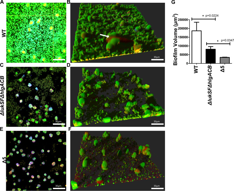
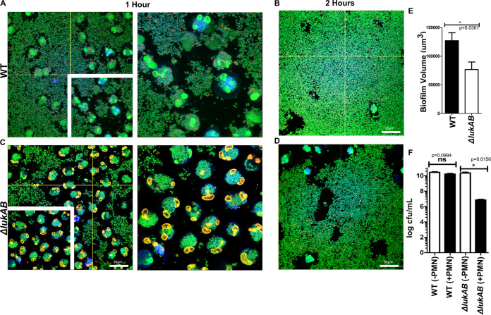
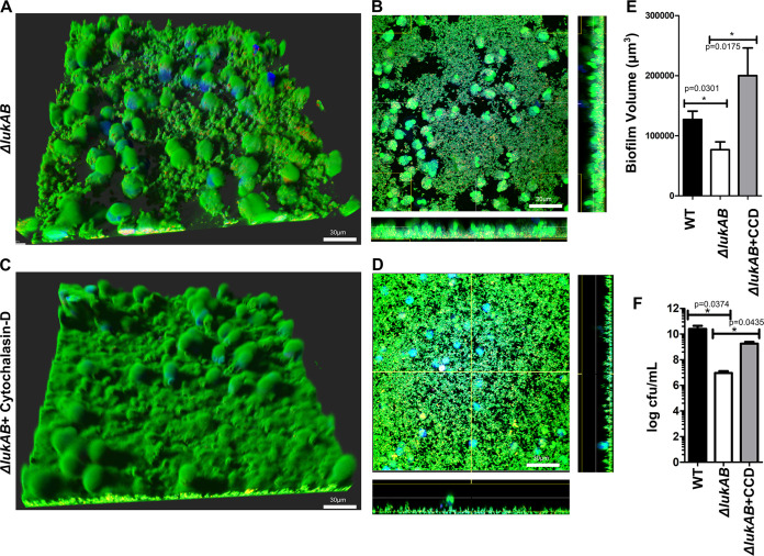
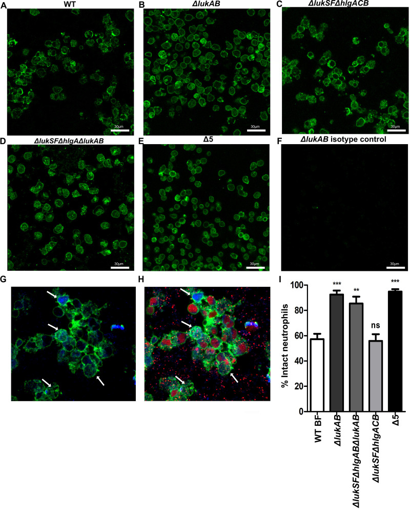
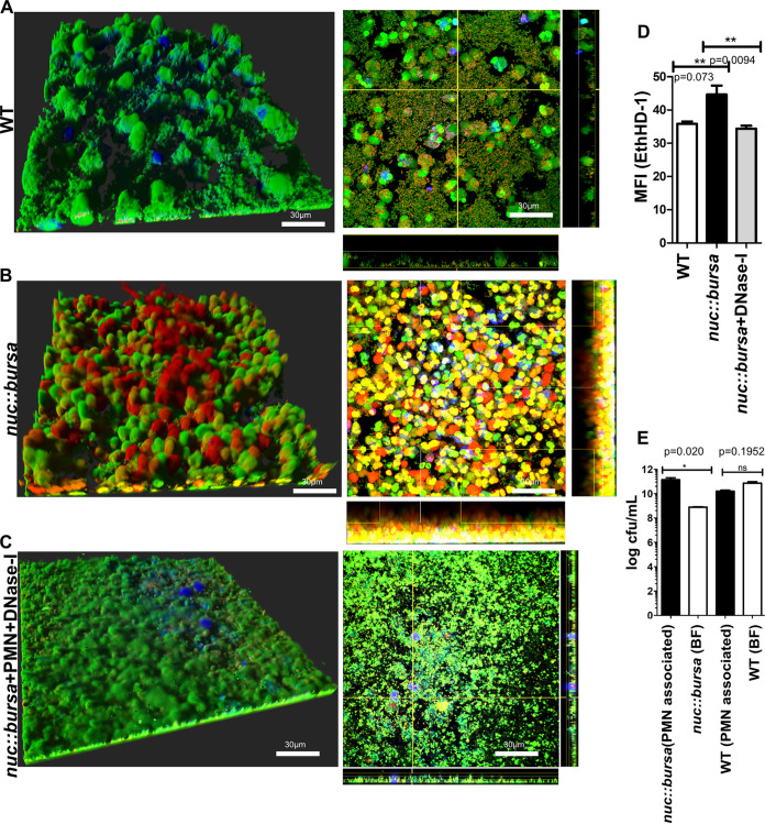
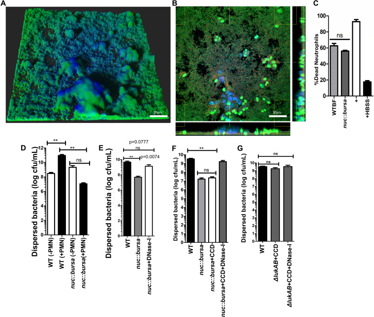
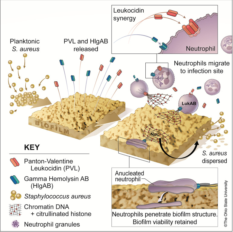
Bacterial biofilms are linked with chronic infections and have properties distinct from those of planktonic, single-celled bacteria. The virulence mechanisms associated with Staphylococcus aureus biofilms are becoming better understood. Human neutrophils are critical for the innate immune response to S. aureus infection. Here, we describe two virulence strategies that converge to promote the ability of S. aureus biofilms to evade killing by neutrophils. Specifically, we show that while neutrophils exposed to S. aureus biofilms produce extracellular traps (NETs) and phagocytose bacteria, both mechanisms are inefficient in clearance of the biofilm biomass. This is attributed to the leukocidin LukAB, which promotes S. aureus survival during phagocytosis. We also show that the persistence of biofilm bacteria trapped in NETs is facilitated by S. aureus nuclease (Nuc)-mediated degradation of NET DNA. This study describes key aspects of the interaction between primary human neutrophils and S. aureus biofilms and provides insight into how S. aureus evades the neutrophil response to cause persistent infections.

Keywords: NETs; S. aureus; biofilms; neutrophil; phagocytosis.


Figures

FIG 1
FIG 1
Leukocidins are required for survival of biofilms exposed to neutrophils. Wild-type USA300 biofilms were incubated with Cell Tracker Blue-labeled primary human neutrophils for 2 h, stained with Syto-9 (viable bacterial cells; green) and ethidium homodimer-1 (DNA of dead mammalian cells; red), and imaged using confocal laser scanning microscopy. (A) Image of a section taken close to the base of the biofilm. (B) Three-dimensional image showing a wild-type USA300 biofilm after a 2-h incubation with primary human neutrophils. The arrow indicates DNA of a dying neutrophil. (C and D) Experiments similar to those for panels A and B, performed with biofilms of a ΔlukSF ΔhlgACB USA300 strain. (E and F) Experiments similar to those for panels A and B, performed with biofilms of a Δ5 USA300 strain, lacking all leukocidin proteins. (G) Volume quantification of WT, ΔlukSF ΔhlgACB, and Δ5 USA300 biofilms after treatment with neutrophils for 2 h. Biofilms were grown in μ-slides and captured at a ×600 magnification. Images represent the majority population phenotype seen in six independent experiments performed in triplicate. Student t tests were performed for pairwise comparisons.
FIG 2
FIG 2
LukAB contributes to bacterial survival when biofilms are in contact with neutrophils. (A and B) Wild-type USA300 biofilms were incubated with Cell Tracker Blue-labeled primary human neutrophils for 1 (A) and 2 (B) h, stained with Syto-9 (viable bacteria), and imaged using confocal laser scanning microscopy. Representative images of sections taken close to the base of biofilms. Insets show a ×1.2 optical zoom of the area outlined in white. (C and D) Experiments similar to those described for panels A and B, performed with an isogenic ΔlukAB USA300 strain. (E) Biofilm biomass measured at 1 h after incubation of wild-type (WT) and ΔlukAB USA300 biofilms with neutrophils for 1 h. (F) Total CFU per milliliter of biofilms from indicated strains, incubated with (+PMN) and without (−PMN) neutrophils for 1 h. Biofilms were grown in μ-slides and captured at a ×600 total magnification. Results are averages of six independent experiments performed in triplicate, with standard errors of the means (SEM). Images and measurements of volume were taken using Imaris software version X 6.4. Enumerations of CFU per milliliter were done independently, using biofilms grown in silicone tubing. Student t tests were performed for pairwise comparisons.
FIG 3
FIG 3
LukAB facilitates survival of biofilm bacteria in a phagocytosis-dependent manner. (A) Biofilms from a ΔlukAB USA300 strain incubated with Cell Tracker Blue-labeled primary human neutrophils for 1 h and stained with Syto-9 (live bacteria) and ethidium homodimer-1 (dead/dying neutrophils). (B) Representative image of a section taken close to the base of the biofilm. (C and D) Cell Tracker Blue-labeled primary human neutrophils were incubated with cytochalasin D for 1 h before incubation with ΔlukAB USA300 biofilms for 1 h and stained as described for panels A and B. (E) Biofilm biomass measured at 1 h after incubation with neutrophils for the ΔlukAB USA300 strain treated with cytochalasin D (+CCD). (F) Numbers of CFU per milliliter, calculated for biomass retrieved from biofilms of the ΔlukAB USA300 strain without and with cytochalasin D (+CCD). Images were captured at a total magnification of ×600, and measurements were taken using Imaris software version X 6.4. Results are averages from six independent experiments performed in triplicate, with SEM. Enumerations of CFU per milliliter were done independently, using biofilms grown in silicone tubing. Student t tests were performed for pairwise comparisons.
FIG 4
FIG 4
PVL, HlgAB, and LukAB are required for biofilms to evade neutrophil killing. (A) Neutrophils were incubated with wild type biofilms and imaged after 1 h. Staining of neutrophils and bacteria was done as described for Fig. 1. (B) Representative image of section taken close to the base of wild-type biofilms and imaged as described for panel A. (C and D) Experiments performed similar to those for panel A, with an isogenic ΔlukSF ΔhlgA ΔlukAB mutant of USA300. (E and F) Experiments performed similar to those for panel A, with an isogenic mutant lacking all 5 leukocidins (Δ5 USA300). (G) Biofilm volume quantified after a 1-h incubation of biofilms from the indicated strains with neutrophils. (H) Measurements of total bacteria (CFU per milliliter) isolated from biofilms of indicated strains, after a 1-h incubation with neutrophils. Results are averages from six independent experiments performed in triplicate, with SEM. Enumerations of CFU per milliliter were done independently, using biofilms grown in silicone tubing. Multiple comparisons were done using one-way analysis of variance and Tukey’s post hoc analysis where appropriate. **, P < 0.01; ***, P < 0.001; ns, not significant. Images were taken using Imaris software version X 6.4. MFI calculations were performed using ImageJ.
FIG 5
FIG 5
LukAB and not NETosis induces neutrophil lysis. (A to E) Biofilms of the indicated USA300 strains were exposed to neutrophils for 1 h. Neutrophils were then labeled with an anti-CD45 antibody and stained with an Alexa Fluor 488 secondary antibody (green). (F) An anti-IgG antibody was used as an isotype control. (G and H) Wild-type biofilms incubated with Cell Tracker Blue-labeled primary human neutrophils (blue) for 1 h, stained with ethidium homodimer-1 (red, dead/dying neutrophils) and an FITC-labeled anti-CD45 antibody (green). Arrows indicate cells that retained staining with Cell Tracker Blue and anti-CD45 antibody but not ethidium homodimer-1. (I) Percent intact neutrophils undergoing NETosis, calculated for the indicated strains by counting the number of neutrophils that stained with anti-CD45+ Cell Tracker Blue as a percentage of the total number of cells per field (anti-CD45 plus Cell Tracker Blue plus EthHD-1), after a 1-h incubation with the respective strains, for 12 fields per strain, from 6 independent experimental replicates. Results are averages from six independent experiments. Images were taken at a total magnification of ×600. Digital zoom (×1.2) was applied to images in G and H, using Imaris software version X 6.4.
FIG 6
FIG 6
Nuc facilitates survival of biofilm S. aureus from NETosis-mediated killing. (A and B) Cell Tracker Blue-labeled neutrophils were incubated with biofilms of the wild type (WT) or an isogenic nuc::bursa USA300 strain for 1 h and stained as described for Fig. 1. Sections taken close to the base of the biofilm are shown in the panels on the right. (C) Images similar to those in A andB, with nuc::bursa USA300 biofilms that were treated with DNase I for 1 h after incubation with neutrophils. (D) Mean fluorescence intensity measurements of ethidium homodimer-1 staining, comparing wild-type (WT) biofilms treated with neutrophils to biofilms of nuc::bursa strains or nuc::bursa strains plus DNase I, similarly treated. (E) Numbers of CFU per milliliter calculated for biofilms grown in silicone tubing and treated with neutrophils for 1 h. Neutrophil-associated bacteria were enumerated and compared to biofilm-associated extracellular populations (BF) for wild-type and nuc::bursa USA300 strains. Results are averages from six independent experiments performed in triplicate, with SEM. Enumerations of CFU per milliliter were done independently, using biofilms grown in silicone tubing (as described in Materials and Methods). Student t tests were performed for pairwise comparisons. Multiple comparisons were done using one-way analysis of variance and a Tukey’s post hoc analysis where appropriate. **, P < 0.01; ns, not significant. Images were taken using Imaris software version X 6.4. MFI calculations were performed using ImageJ.
FIG 7
FIG 7
Nuc-mediated breakdown of NET DNA contributes to biofilm bacterial dispersal. (A) nuc::bursa USA300 biofilms were treated with neutrophils for 1 h and subsequently with wild-type (WT) biofilm supernatant for an additional hour. Staining was done as described for Fig. 1. (B) Cross section of biofilm from panel A, incubated with neutrophils taken at the bottom of the biofilm. (C) Neutrophil killing activity of spent media collected from wild-type and nuc::bursa USA300 biofilms. Neutrophils were incubated with spent media for 30 min. LIVE-DEAD measurements show a ratio of Syto-9 (live) to propidium iodide fluorescence calculated as a percentage against a positive control (neutrophils plus 0.1% SDS). Neutrophils treated with Hanks balanced salt solution (HBSS) were used as a negative control. (D) Comparisons of dispersed bacteria before (−PMN) and after (+PMN) treatment of wild-type (WT) or nuc::bursa USA300 biofilms with neutrophils for 1 h. (E) Enumerations similar to those in panel D, comparing neutrophil-treated wild-type biofilms with nuc::bursa biofilms with and without DNase I treatment subsequent to incubation with neutrophils. (F) Counts (CFU per milliliter) of dispersed bacterial populations collected after a 60-min incubation of wild-type and nuc::bursa USA300 biofilms with primary human neutrophils, before and after treatment with cytochalasin D with and without DNase I. (G) Enumerations similar to those in panel F, performed with neutrophils treated with WT and ΔlukAB USA300 biofilms. Measurements of CFU per milliliter were done using biofilms grown in silicone tubing. Results are averages from six independent experiments performed in triplicate, with SEM. **, P < 0.01, using one-way analysis of variance and Tukey’s post hoc analysis; ns, not significant.
FIG 8
FIG 8
Summary of results. The activity of leukocidins PVL and gamma hemolysin AB (HlgAB) released from biofilms results in the induction of neutrophil extracellular traps (NETosis). While anuclear, neutrophils are still capable of penetrating the biofilm structure and phagocytosing bacteria; the antimicrobial activity of NETs is insufficient for clearing S. aureus biofilms. The leukocidin LukAB allows survival of bacteria during phagocytosis, and NET DNA is broken down by the nuclease (Nuc), resulting in dispersal of bacteria and thus perpetuating chronic infection. (Copyright The Ohio State University.)

References

    1. Lowy FD. 1998. Staphylococcus aureus Infections. N Engl J Med 339:520–532. doi:10.1056/NEJM199808203390806. - DOI - PubMed
    1. Chapman JR, Balasubramanian D, Tam K, Askenazi M, Copin R, Shopsin B, Torres VJ, Ueberheide BM. 2017. Using quantitative spectrometry to understand the influence of genetics and nutritional perturbations on the virulence potential of Staphylococcus aureus. Mol Cell Proteomics 16:S15–S28. doi:10.1074/mcp.O116.065581. - DOI - PMC - PubMed
    1. Periasamy S, Joo H-S, Duong AC, Bach T-H, Tan VY, Chatterjee SS, Cheung GYC, Otto M. 2012. How Staphylococcus aureus biofilms develop their characteristic structure. Proc Natl Acad Sci U S A 109:1281–1286. doi:10.1073/pnas.1115006109. - DOI - PMC - PubMed
    1. Costerton JW, Geesey GG, Cheng KJ. 1978. How bacteria stick. Sci Am 238:86–95. doi:10.1038/scientificamerican0178-86. - DOI - PubMed
    1. Ammons MCB, Tripet BP, Carlson RP, Kirker KR, Gross MA, Stanisich JJ, Copié V. 2014. Quantitative NMR metabolite profiling of methicillin-resistant and methicillin-susceptible Staphylococcus aureus discriminates between biofilm and planktonic phenotypes. J Proteome Res 3:2973–2985. doi:10.1021/pr500120c. - DOI - PMC - PubMed

Publication types

MeSH terms

LinkOut - more resources