Review
. 2020 Jul 27;11(7):584.
doi: 10.1038/s41419-020-02803-4.

Bioactive DNA from extracellular vesicles and particles

Affiliations
Review

Bioactive DNA from extracellular vesicles and particles

Ethan Z Malkin et al. Cell Death Dis. .

Abstract

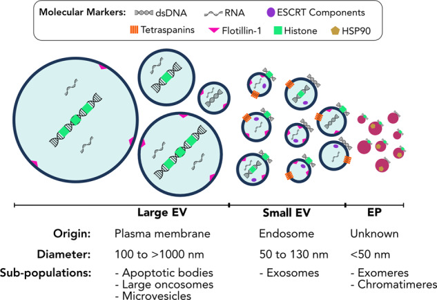
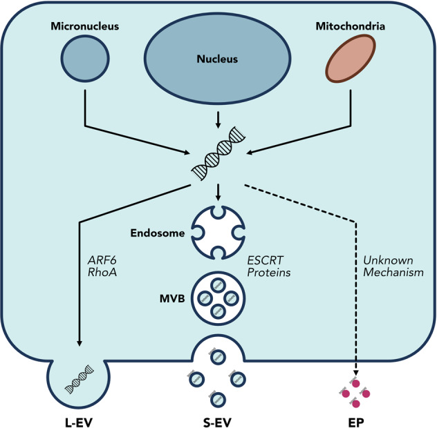
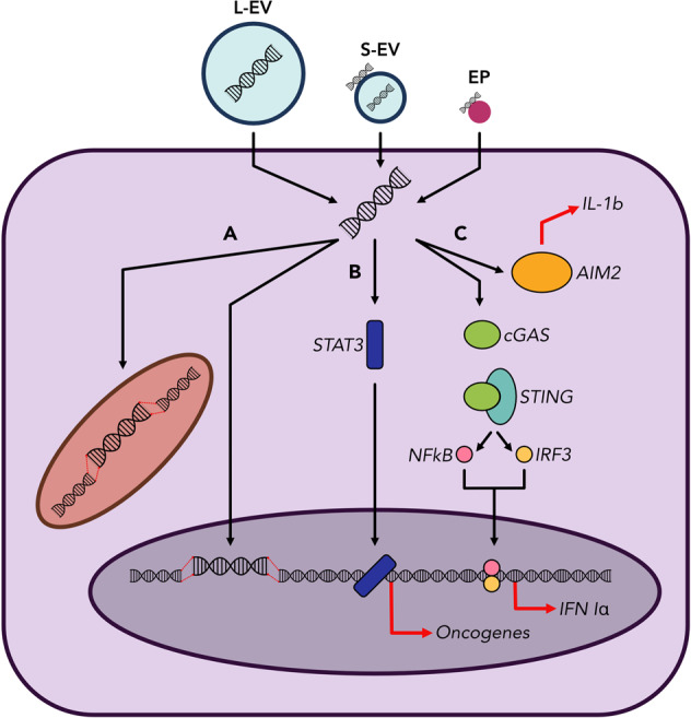
Extracellular vesicles (EVs) and particles (EPs) have recently emerged as active carriers of molecular biomarkers and mediators of intercellular communication. While most investigations have focused exclusively on the protein, lipid and RNA constituents of these extracellular entities, EV/EP DNA remains poorly understood, despite DNA being found in association with virtually all EV/EP populations. The functional potential of EV/EP DNA has been proposed in a number of pathological states, including malignancies and autoimmune diseases. Moreover, the effectiveness of cell-free DNA as the biomarker of choice in emerging liquid biopsy applications highlights the role that EV/EP DNA may play as a novel disease biomarker. In this review, we provide a comprehensive overview of EV/EP DNA studies conducted to date, with a particular focus on the roles of EV/EP DNA as a functional mediator and molecular biomarker in various pathologic states. We also review what is currently known about the origins, structure, localisation and distribution of EV/EP DNA, highlighting current controversies as well as opportunities for future investigation.


Conflict of interest statement

S.V.B. provides consultation for Bristol-Myers Squibb, receives institutional research support from Nektar Therapeutics, is a co-inventor on a patent relating to mutation-based ctDNA detection technology that has been licensed to Roche Molecular Diagnostics and on patent applications relating to methylation-based ctDNA detection technology, and is co-founder and provides consultation for DNAMx, Inc.

Figures

Fig. 1
Fig. 1. Extracellular vesicles and particles comprise heterogenous populations.
Large extracellular vesicles (L-EVs) are characterised by their formation from the plasma membrane, their predominantly large size, and the long fragment length of their luminal DNA. Small extracellular vesicles (S-EVs) are primarily characterised by their endosomal origins, surface protein markers, and smaller size, though overlap in size does exist between larger S-EVs and smaller L-EVs. Extracellular particles (EPs) are small protein-nucleic-acid complexes that are not membrane-bound; the mechanisms of EP biogenesis are unknown. Within each of these groups, multiple subpopulations have been described. However, the full extent of EV/EP subpopulation heterogeneity has yet to be realised, as exemplified by the differences in size and molecular markers both between and within different EV/EP populations. ESCRT endosomal sorting complexes required for transport, HSP90 heat shock protein 90.
Fig. 2
Fig. 2. DNA loading mechanisms are unique for different EV/EP populations.
DNA is released into the cytosol from the nucleus and micronuclei in response to genomic instability and DNA damage; mitochondria can also release DNA into the cytosol. The precise mechanisms by which DNA is loaded into EVs are unknown, but ARF6 and RhoA have been implicated in L-EV DNA loading, while the ESCRT family of proteins are involved in S-EV DNA loading. The mechanisms of DNA loading onto EPs remains unknown. Moreover, the means by which DNA associates with the surface membrane of S-EVs is not fully understood. Consequently, EV/EP DNA loading mechanisms and structure represent areas that require further study.
Fig. 3
Fig. 3. EV/EP DNA directly influences physiological function by acting on recipient cells.
EV/EPs deliver DNA to the recipient cell cytosol. a Horizontal gene transfer. EV/EP DNA can translocate to the recipient cell nucleus or mitochondria, where it is integrated into the host genome. Subsequent transcription of this DNA heavily influences recipient cell function, and phenotypic changes depend on the genotype of cells from which the EV/EP DNA was derived. b Activation of oncogenic pathways. EV/EP DNA can activate or cause the up-regulation of various intracellular signalling proteins, such as STAT3, causing translocation to the nucleus and over-expression of oncogenes that drive a pro-tumourigenic phenotype in the recipient cell. c Activation of inflammatory pathways. EV/EP DNA can trigger various cytosolic DNA receptors, including AIM2 (which subsequently produces interleukins) and cGAS. Activation of cGAS causes downstream release of Type I Interferons that induce inflammatory responses unique to the disease context.

References

    1. Thierry, A. R., El Messaoudi, S., Gahan, P. B., Anker, P. & Stroun, M. Origins, structures, and functions of circulating DNA in oncology. Cancer Metastasis Rev.35, 347–376 (2016). - PMC - PubMed
    1. Wan, J. C. M. et al. Liquid biopsies come of age: towards implementation of circulating tumour DNA. Nat. Rev. Cancer17, 223–238 (2017). - PubMed
    1. Cescon, D. W., Bratman, S. V., Chan, S. M. & Siu, L. L. Circulating tumor DNA and liquid biopsy in oncology. Nat. Cancer1, 276–290 (2020). - PubMed
    1. Fuchs, T. A. et al. Extracellular DNA traps promote thrombosis. Proc. Natl Acad. Sci. USA107, 15880–15885 (2010). - PMC - PubMed
    1. Hawes, M. C., Wen, F. & Elquza, E. Extracellular DNA: a bridge to cancer. Cancer Res.75, 4260–4264 (2015). - PubMed

Publication types

Grants and funding