Review
. 2020 Jul 27;12(1):21.
doi: 10.1038/s41368-020-00089-y.

Computational technology for nasal cartilage-related clinical research and application

Affiliations
Review

Computational technology for nasal cartilage-related clinical research and application

Bing Shi et al. Int J Oral Sci. .

Abstract

Surgeons need to understand the effects of the nasal cartilage on facial morphology, the function of both soft tissues and hard tissues and nasal function when performing nasal surgery. In nasal cartilage-related surgery, the main goals for clinical research should include clarification of surgical goals, rationalization of surgical methods, precision and personalization of surgical design and preparation and improved convenience of doctor-patient communication. Computational technology has become an effective way to achieve these goals. Advances in three-dimensional (3D) imaging technology will promote nasal cartilage-related applications, including research on computational modelling technology, computational simulation technology, virtual surgery planning and 3D printing technology. These technologies are destined to revolutionize nasal surgery further. In this review, we summarize the advantages, latest findings and application progress of various computational technologies used in clinical nasal cartilage-related work and research. The application prospects of each technique are also discussed.


Conflict of interest statement

The authors declare no competing interests.

Figures

Fig. 1
Fig. 1
Flowchart for starting a nasal cartilage-related study based on computational technology. Arrows indicate the workflow of starting a nasal cartilage-related study based on computational technology. Black arrows indicate how to perform the study step by step; red arrows demonstrate how to make the decisions to choose the appropriate methods. The flowchart also demonstrates the structure of this review
Fig. 2
Fig. 2
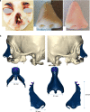
Nasal cartilage obtained from micro-CT scan. The three different views of the alar cartilage were obtained from micro-CT
Fig. 3
Fig. 3
Nasal cartilage and facial muscles obtained from MRI scans. ac Micro-MRI scan of the nasolabial region of an induced infant with unilateral complete cleft lip., The red arrow indicates the position of the cartilage. AC alar cartilage, ULC upper lateral cartilage, NS nasal septum cartilage. d 3D model reconstruction of the nasolabial region based on micro-MRI, including soft tissue and cartilage. e Facial muscles obtained from MRI
Fig. 4
Fig. 4
Different nasal cartilage reconstruction models used in different studies. a Patient-specific nasal cartilage reconstruction based on patient imaging data.b Reconstructed soft tissue model based on patient imaging data and reconstructed cartilage based on soft tissue morphology.c Simplified model of nasal cartilage
Fig. 5
Fig. 5
Finite element analyses of primary unilateral cleft lip rhinoplasty. a Two opposite forces at both sides of the cleft simulate the closure of a cleft lip. b, c Two paths illustrating the surface deformation and stress of the soft tissue. dg The directions of force loading for the finite element simulation and schematic diagrams of four common suspension sutures during primary cleft lip rhinoplasty
Fig. 6
Fig. 6
Finite element analyses of secondary unilateral cleft lip rhinoplasty. a Typical secondary unilateral cleft lip nasal deformity. b 3D reconstruction model of the patient-specific nose. cf Schematic diagrams of two sutures in secondary cleft lip rhinoplasty and the directions of force loading during the finite element simulation
Fig. 7
Fig. 7
Computational fluid dynamics for handling nasal surgery. a CFD for analysing a patient-specific airway. b Streamlines and velocity contours show the post-surgical airflow changes for different patients. The changes in airflow through the nasal valve area can be clearly idenitified
Fig. 8
Fig. 8
Application of 3D printing in nasal cartilage-related surgery. a 3D printing of a patient-specific nasal prosthesis.b Patient-specific design and 3D printing of a meshed titanium nasal prosthesis

References

    1. Lavernia L, Brown WE, Wong BJF, Hu JC, Athanasiou KA. Toward tissue-engineering of nasal cartilages. Acta Biomater. 2019;88:42–56. - PubMed
    1. Pelttari K, Mumme M, Barbero A, Martin I. Nasal chondrocytes as a neural crest-derived cell source for regenerative medicine. Curr. Opin. Biotechnol. 2017;47:1–6. - PubMed
    1. Bhumiratana S, et al. Tissue-engineered autologous grafts for facial bone reconstruction. Sci. Transl. Med. 2016;8:343ra383. - PMC - PubMed
    1. Huang H, et al. Recapitulation of unilateral cleft lip nasal deformity on normal nasal structure: a finite element model analysis. J. Craniofac. Surg. 2018;29:2220–2225. - PubMed
    1. Huang H, Luo X, Cheng X, Shi B, Li J. Biomechanical simulation of correcting primary unilateral cleft lip nasal deformity. PLoS ONE. 2018;13:e0199964. - PMC - PubMed