. 2020 Oct;5(10):1285-1298.
doi: 10.1038/s41564-020-0758-1. Epub 2020 Jul 27.

Structures of capsid and capsid-associated tegument complex inside the Epstein-Barr virus

Affiliations

Structures of capsid and capsid-associated tegument complex inside the Epstein-Barr virus

Wei Liu et al. Nat Microbiol. 2020 Oct.

Abstract

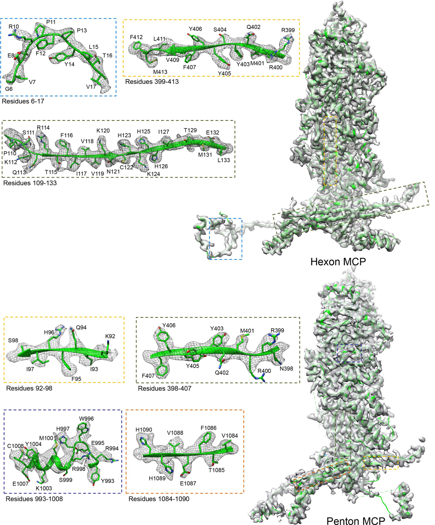
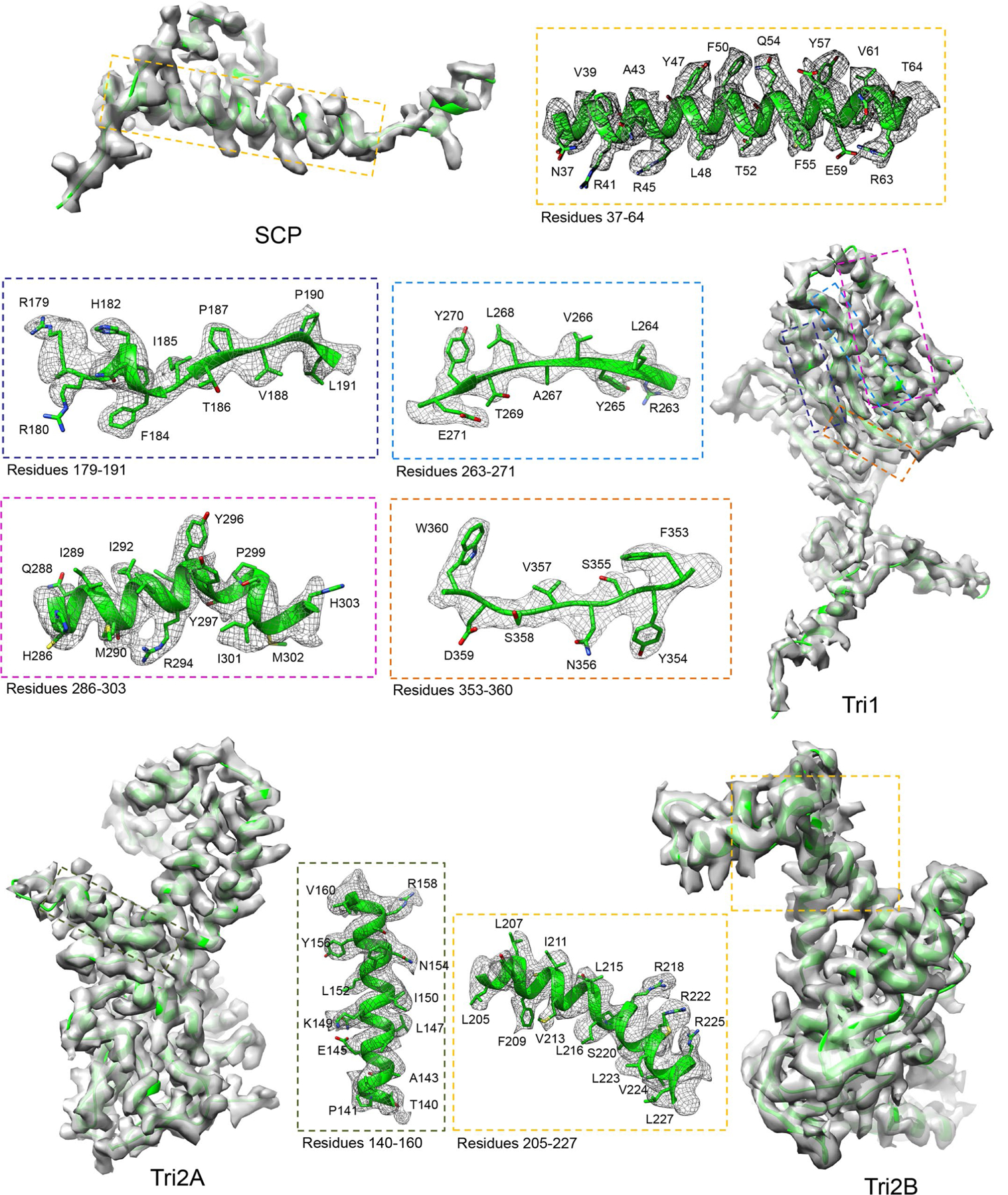
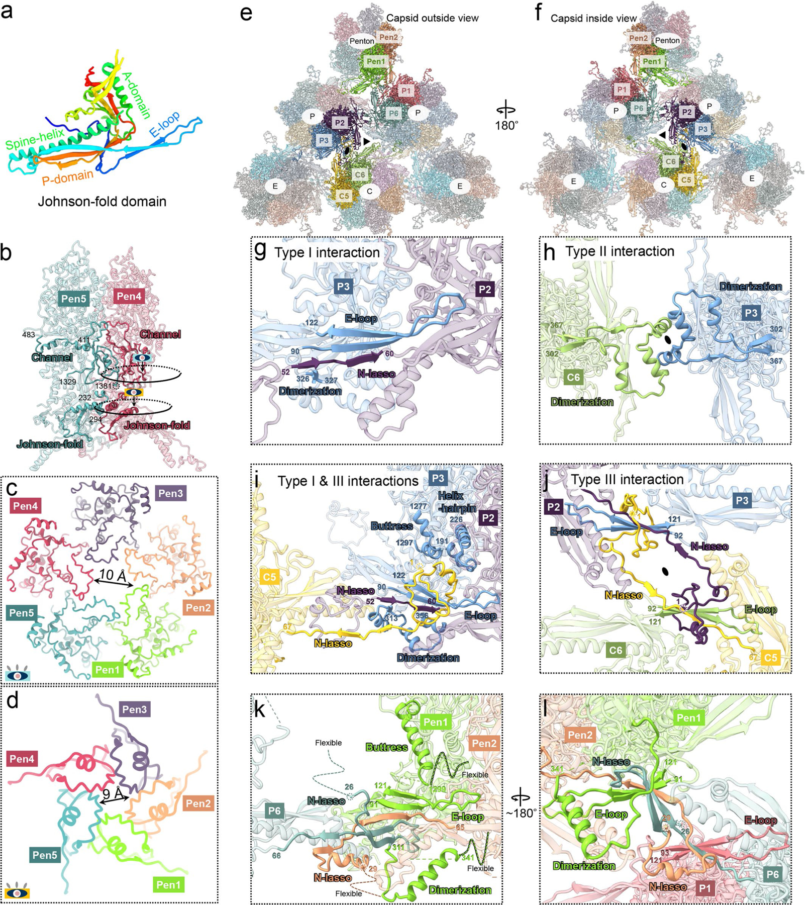
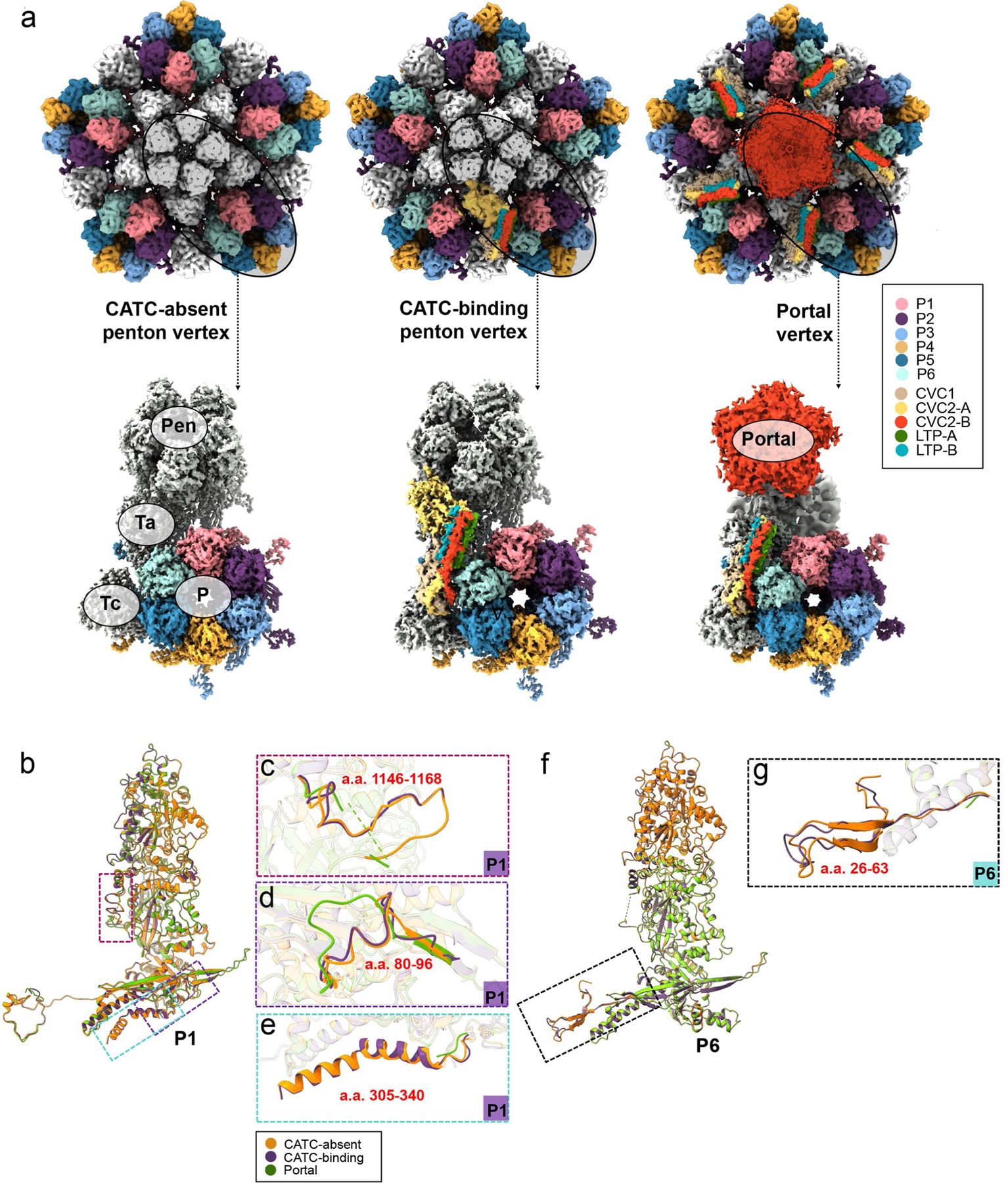
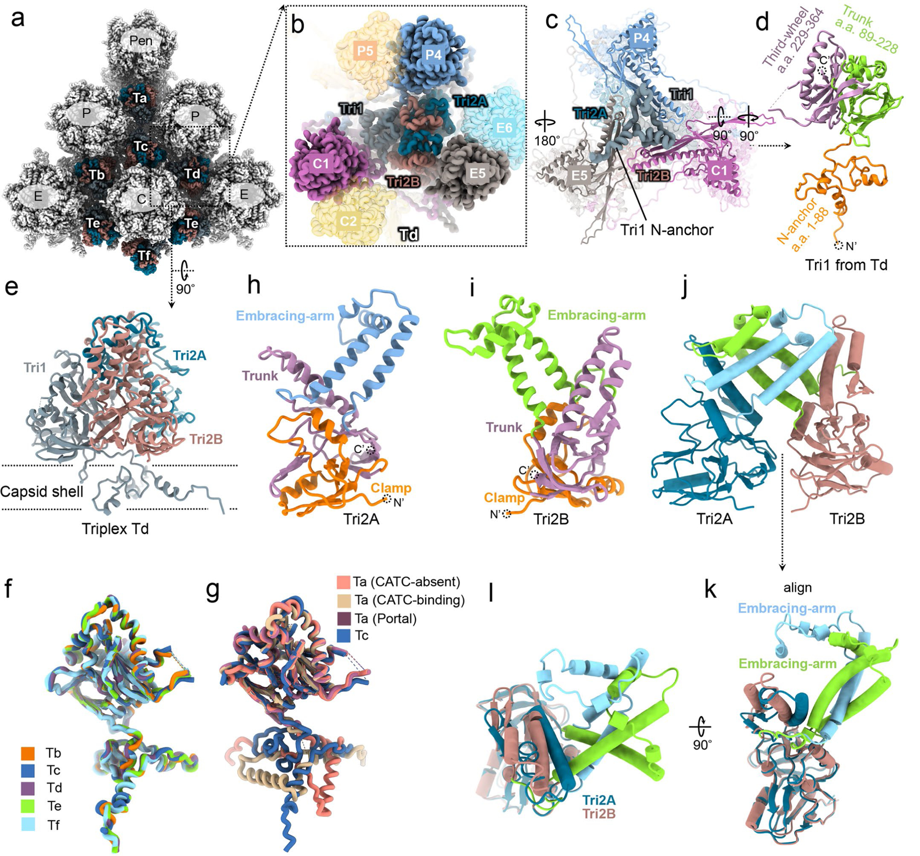
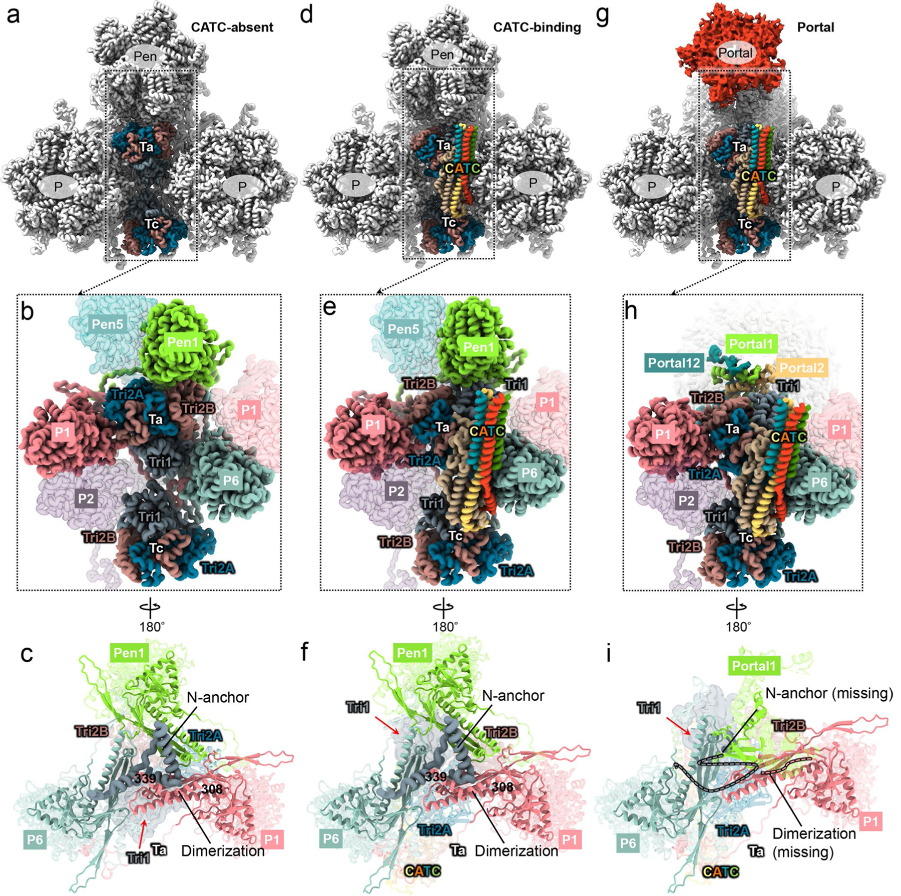
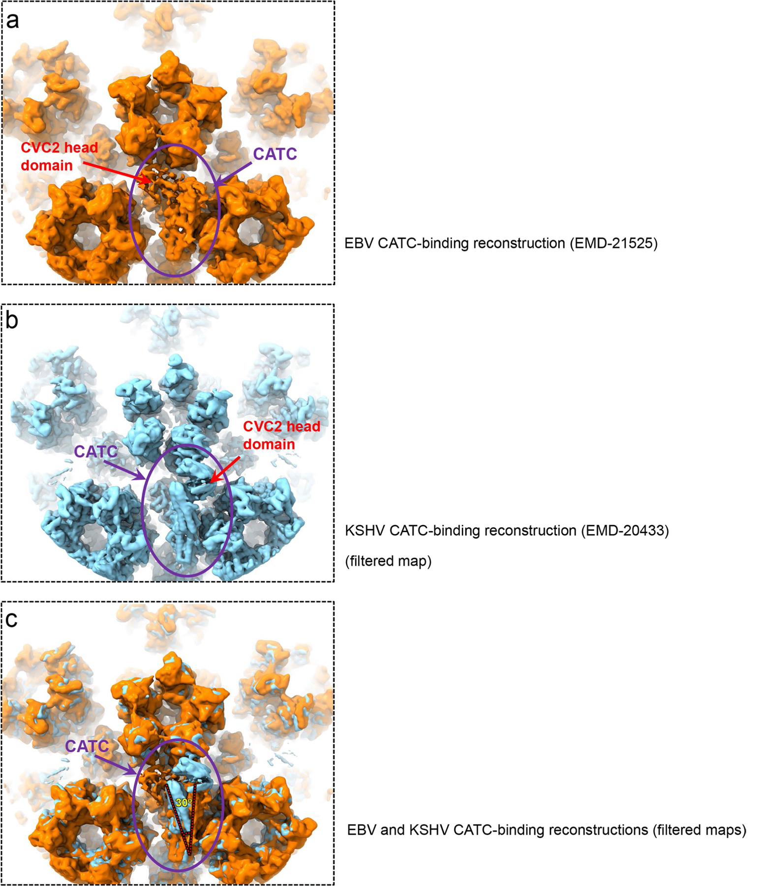
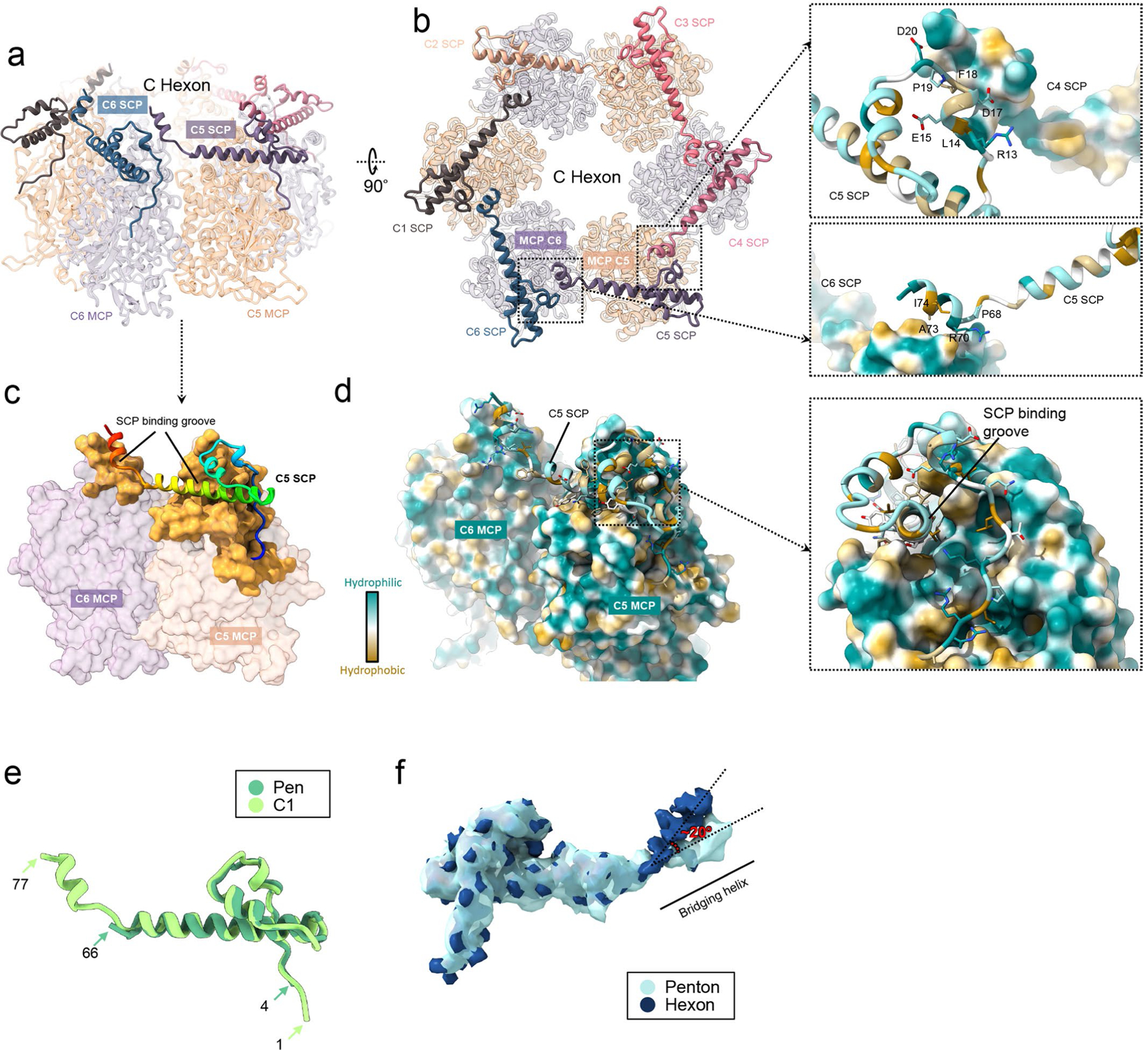
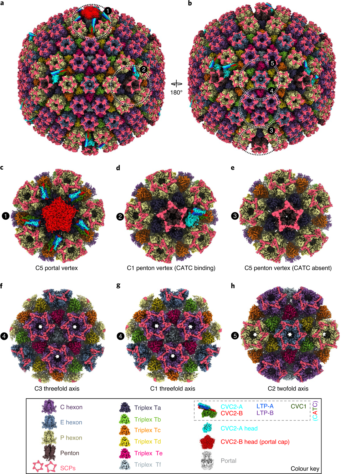
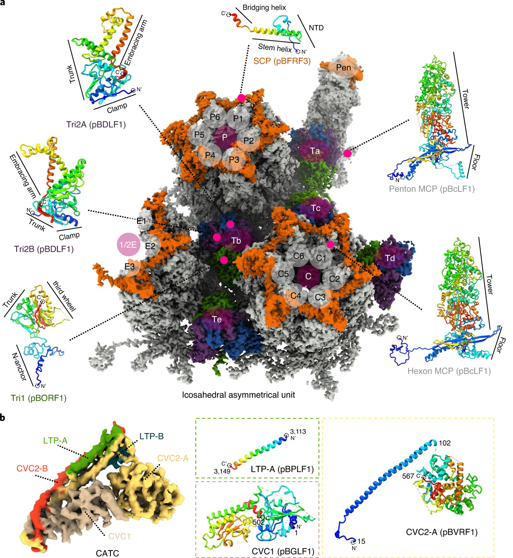
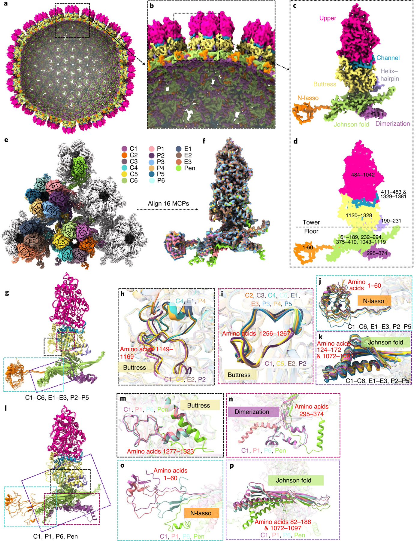
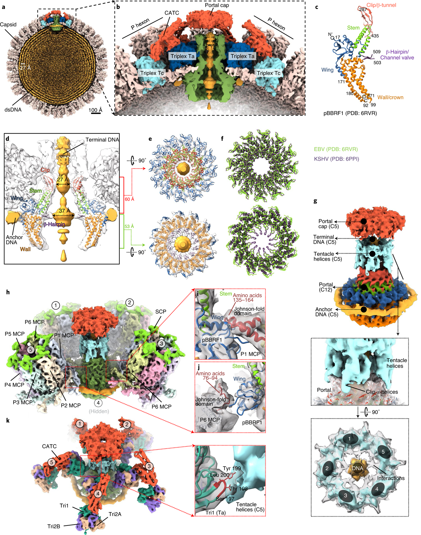
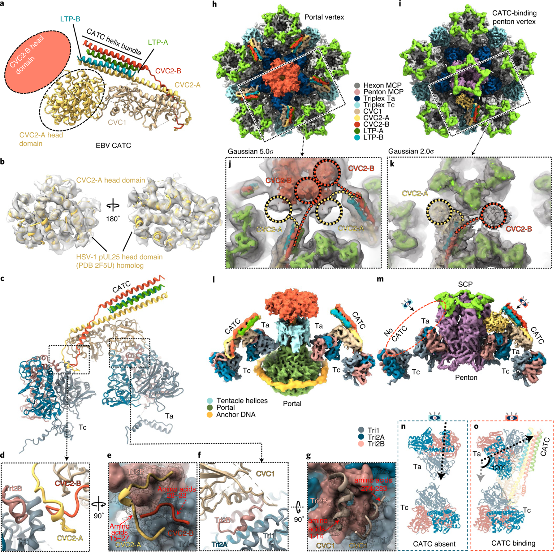
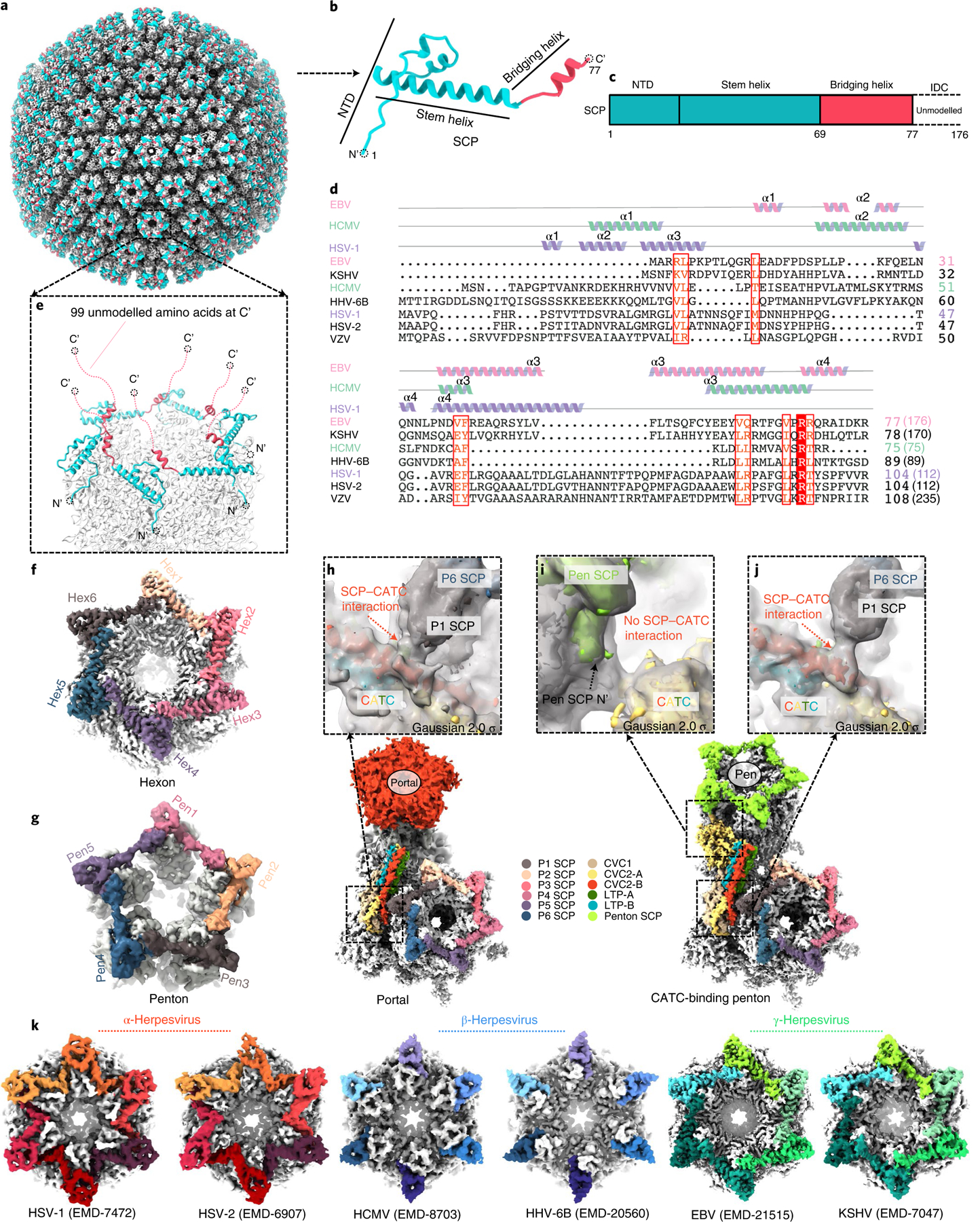
As the first discovered human cancer virus, Epstein-Barr virus (EBV) causes Burkitt's lymphoma and nasopharyngeal carcinoma. Isolating virions for determining high-resolution structures has been hindered by latency-a hallmark of EBV infection-and atomic structures are thus available only for recombinantly expressed EBV proteins. In the present study, by symmetry relaxation and subparticle reconstruction, we have determined near-atomic-resolution structures of the EBV capsid with an asymmetrically attached DNA-translocating portal and capsid-associated tegument complexes from cryogenic electron microscopy images of just 2,048 EBV virions obtained by chemical induction. The resulting atomic models reveal structural plasticity among the 20 conformers of the major capsid protein, 2 conformers of the small capsid protein (SCP), 4 conformers of the triplex monomer proteins and 2 conformers of the triplex dimer proteins. Plasticity reaches the greatest level at the capsid-tegument interfaces involving SCP and capsid-associated tegument complexes (CATC): SCPs crown pentons/hexons and mediate tegument protein binding, and CATCs bind and rotate all five periportal triplexes, but notably only about one peri-penton triplex. These results offer insights into the EBV capsid assembly and a mechanism for recruiting cell-regulating factors into the tegument compartment as 'cargoes', and should inform future anti-EBV strategies.


Conflict of interest statement

Competing interests

The authors declare no competing interests.

Figures

Extended Data Fig. 1 |
Extended Data Fig. 1 |. Density maps (gray) and atomic models (ribbon) of a P4 hexon MCP and a penton MCP segmented from the C2 2-fold and the C5 CATC-absent 5-fold sub-particle reconstructions.
Boxed regions are enlarged in boxes with edges colored correspondingly, with density shown as gray mesh and atomic models as ribbon/sticks.
Extended Data Fig. 2 |
Extended Data Fig. 2 |. Density maps (gray) and atomic models (ribbon) of an SCP, a tri1, a tri2a, and a tri2B monomer.
An SCP (for example, e1 SCP, was docked and segmented out from the 3.0-Å C6 hexon sub-particle reconstruction) is superposed with the atomic model (ribbon). Density maps (gray) of the Tri1, Tri2A, and Tri2B (segmented out from triplex Tb of the C3 3-fold sub-particle reconstruction) at 3.4 Å are superposed with their atomic models (ribbon). Boxed regions are enlarged in boxes with edges colored correspondingly, with density shown as gray mesh and atomic models as ribbon/sticks.
Extended Data Fig. 3 |
Extended Data Fig. 3 |. MCP interactions.
a, Atomic model of the Johnson-fold domain of MCP shown in rainbow-colored ribbon. b-d, MCP-MCP interactions in penton (b) and constrictions in penton channel (c, d). The colored eye symbols in (b) indicate the view directions in (c) and (d). e, f, Part of the MCP network viewed from outside (e) and inside (f) the capsid. g-j, Three types of network interactions among hexon MCPs. Type I interactions (g) are hydrogen bonds in an intra-capsomeric augmentation of β-strands from adjacent MCPs (for example, P2 and P3) in the same capsomer. Type II interactions h, inter-capsomeric interactions among a pair of MCPs (for example, P3 and C6), join two dimerization domains. Type III interactions i, j, characterized by the lassoing action of the N-lasso domain (for example, P3, C5, and C6) among three MCPs, build on and fortify type I interactions (j). k, l, Penton MCP interactions with hexon MCP subunits P1 and P6. Note that penton MCP lacks type II and III interactions and the N-lasso domain of P6 hexon MCP differs from those in other hexon MCPs.
Extended Data Fig. 4 |
Extended Data Fig. 4 |. Plasticity of MCP structures and MCP interactions with CATC.
a-g, Plasticity of MCPs related to interactions with portal or CATC. Shown in (a) are the three types of vertices—CATC-absent penton vertex, CATC-binding penton vertex, and portal vertex—and how MCP subunits P1 and P6 are variably engaged in CATC or portal interactions. Corresponding MCPs were extracted from the three types of vertices and aligned. The superpositions of the three aligned P1 MCPs (b, with zoomed-in areas shown in c-e) and of the three aligned P6 MCPs f, with zoomed-in areas shown in g, show structural plasticity.
Extended Data Fig. 5 |
Extended Data Fig. 5 |. Triplex structures and plasticity of the tri1 N-anchor domains.
a, Distribution of triplexes Ta, Tb, Tc, Td, Te, and Tf among the penton and three types of hexons (C, e, and P). b-e, enlarged view of a triplex Td with three adjacent hexon MCP subunits (C1, e5, and P4) from outside (b) and inside (c). The inside view (c) shows that N-anchor domain of Td Tri1 lines along the three valleys of the MCP inner floor. Also shown are side views of Tri1 monomer (d) and triplex Td (e). f, Superpositions of Tri1 monomers from different triplexes except Ta. g, Superpositions of Tri1 monomers from triplex Tc, peri-penton triplex Ta before and after CATC binding, and peri-portal Ta. h-j, Tri2A (h) and Tri2B (i) shown as ribbons side by side, or together as pipe-and-plank (j). k, l, Two orthogonal views of the superposition of the aligned Tri2A and Tri2B, showing nearly identical clamp and trunk domains, but different embracing arm domain.
Extended Data Fig. 6 |
Extended Data Fig. 6 |. Interactions between triplexes and MCP subunits at three types of vertex sub-particle reconstructions.
a–c, Interactions in CATC-absent penton vertex sub-particle reconstruction. d–f, Interactions in CATC-binding penton vertex sub-particle reconstruction. g–i, Interactions in portal vertex sub-particle reconstruction. Overviews (a, d, g) and close-up views (b, e, h) of the triplexe Ta and Tc region observed from outside the capsid. In the close-up views, the locations of the three Ta subunits (Tri1, Tri2A and Tri2B) are rotated counter-clockwise by 120° in both the CATC-binding penton vertex (e) and the portal vertex (h), as compared to those in the CATC-absent penton vertex (b). By contrast, there is no rotation in triplex Tc. From inside the capsid, the N-anchor domain of Tri1 is secured by the dimerization domain of P1 MCP in the penton vertex (c) and remains unrotated after CATC binding (f). Neither Tri1 N-anchor domain nor the P1 MCP dimerization domain is resolved (i), suggesting flexibility of both.
Extended Data Fig. 7 |
Extended Data Fig. 7 |. Fitting of the atomic model of the recombinant portal protein.
a, b, The recent published portal structure9 was docked in our in situ structure of the portal (semi-transparent gray) as either a dodecamer viewed in two orthogonal directions (a) or a monomer (b). c, Five insets show good fittings of five domains of the portal protein.
Extended Data Fig. 8 |
Extended Data Fig. 8 |. Plasticity of CATC attachment on the capsid.
a, Global view of the C5 whole virus reconstruction showing consensus-patterned (that is, averaged) occupancy of CATCs at penton vertices. b, c, Same as in (a) but with only CATC densities (Gaussian-filtered [2σ], CVC2 head domains are removed for clarity) displayed at two progressively lower thresholds. The two thresholds (0.01 and 0.005) were chosen interactively in UCSF Chimera such that at the first threshold one CATC are visible at the portal-proximal positions (b), and at the second threshold two CATCs are visible at the portal-distal positions (c). d, Histogram of number of CATC per penton vertex.
Extended Data Fig. 9 |
Extended Data Fig. 9 |. Structural differences of CATCs in EBV and KSHV.
a-b, Low-pass filtered sub-particle reconstructions of eBV CATC-binding penton vertex (a) and of KSHV CATC-binding penton vertex (eMD-20433) (b). c, Superposition of (a) and (b) showing that eBV and KSHV CATC structures differ in two aspects: First, CVC2 head domain in KSHV heads right, as opposite to that in eBV, which heads left; Second, their helix bundles have an ~30º angle difference.
Extended Data Fig. 10 |
Extended Data Fig. 10 |. Interactions of SCP with hexon MCP.
a, b SCP binds the upper domain of C hexon MCPs and six copies of SCP form a flower-shaped ring crowning and stabilize the hexon mainly by hydrophilic (right panels) interactions. c, d SCP interacts with two adjacent MCPs on their upper domains by inserting its stem helix into the SCP binding grooves (gold) (c) mainly by hydrophobic interactions (d). e, f Superposition of representative hexon SCP (for example, C1) and a penton SCP’s atomic models (e) and density maps (f) reveals the plasticity of SCP protein in eBV.
Fig. 1 |
Fig. 1 |. Subparticle reconstructions and architecture of the EBV capsid with portal and CATCs.
a,b, Shaded-surface representations of the eBV C5 whole-virus reconstruction, revealing the DNA-translocating portal vertex (a) and variable attachments of CATCs (a,b). b is the back view of a. ch, Reconstructions for subparticles exemplified by the circled areas in a and b, including C5 reconstruction of the portal vertex (c), C1 reconstruction of the CATC-binding penton vertex (d), C5 reconstruction of the CATC-absent penton vertex (e), C3 (f) and C1 (g) reconstructions of the threefold axis region, and C2 reconstruction of the twofold axis region (h). Colour keys of structural components are at the bottom.
Fig. 2 |
Fig. 2 |. Atomic models of representative EBV capsid and CATC subunits.
a, Density map for an icosahedral asymmetrical unit segmented from the three main-axis subparticle reconstructions and coloured by protein types: MCP (grey), Tri1 (green), Tri2A (blue), Tri2B (purple) and SCP (orange). b, Density map for CATC segmented from the C1 CATC-binding penton vertex subparticle reconstruction and coloured by protein subunits. In both a and b, representative atomic models of protein subunits are shown next to the density map as ribbons rainbow coloured from blue (N terminus) to red (C terminus).
Fig. 3 |
Fig. 3 |. Plasticity of the MCP structures.
ad, Cut-away (a) and zoomed-in (b) views of the C5 whole-virus reconstruction with only MCP subunits shown, coloured by domains defined in c and d. Ranges of amino-acid residues in each domain are numbered in d. ep, Structural plasticity of the 16 MCP subunits at quasi-equivalent positions within an icosahedral asymmetrical unit (coloured in e). The superposition of 16 aligned MCPs (f) shows only small variations among the subunits C1–C6, e1–e3 and P2–P5 (g, with zoomed-in areas in hk), but greater structural variations for subunits P1, P6 and Pen compared with subunit C1 (l, with zoomed-in areas in mp).
Fig. 4 |
Fig. 4 |. Capsid accommodation of the DNA-translocating portal complex and periportal CATCs.
a,b, Clipped (a) and zoomed-in (b) views of the C5 whole-virus reconstruction, showing packaged dsDNA within the capsid with neighbouring dsDNA duplexes spaced ~27 Å apart (a) and structural components around the portal vertex (b). c, Atomic model of the recombinant portal protein shown as a monomer coloured by domains. df, Clipped view of the portal vertex region showing the fitted atomic models (ribbon) of two opposing subunits (d) and the two constrictions along the DNA-translocating channel (e). The superposition (f) of eBV and KSHV portal atomic models reveals similarities along these constrictions. gl, Composite map of eBV portal region, showing interactions of the portal complex and DNA, and the MCP and Tri1. The C12 portal subparticle reconstruction was placed into the C5 portal vertex subparticle reconstruction by referencing HSV-1 and KSHV C1 portal vertex structures18,19, showing DNA, tentacle helices and portal cap structures surrounding the fitted atomic model of the portal complex (g). Five surrounding P hexons (h) interact with the wing domain of the portal protein through amino-acid segments 135–164 (i) and 76–94 (j) of P1 MCP and P6 MCP, respectively. Both segments are located within the Johnson-fold domain of the MCP. Likewise, surrounding the structure shown in g are five Ta triplexes (k), the Tri1 subunit of which interacts at residues 198 and 199 with the tentacle helices (l).
Fig. 5 |
Fig. 5 |. CATC and its interactions with triplexes ta and tc.
ag, Atomic model of a peri-penton CATC showing hypothetical placement of the head domains of CVC2-A and CVC2-B conformers (a,b) and CATC interactions with triplexes Ta and Tc (cg). Helices resolved in the density map (semi-transparent grey in b) of the CVC2-A head domain match those of the homologous HSV-1 pUL25 atomic model (ribbons in b). CATC interactions with triplex Ta and Tc are shown in c and detailed in d, e, f and g, respectively. ho, CATC accommodation at the portal and penton vertices. Subparticle reconstructions of the portal vertex (h) and the CATC-binding penton vertex (i) with insets showing low-pass-filtered density maps (semi-transparent grey). In the penton vertex subparticle reconstruction, the CATC helix bundle is near two densities (circled) that are probably head domains of CVC2-A (yellow circle) and CVC2-B (red circle) (k). In the portal vertex reconstruction, the CATC helix bundle is connected to the portal cap (j), suggesting that the CVC2 head domains emanating from CATC contribute to the portal cap. l,m, Side views of the portal vertex (l) and CATC-binding penton vertex (m). n,o, Comparison of triplex Ta external orientation relative to triplex Tc in the absence (n) and presence (o) of CATC at the penton vertex, showing that the CATC binding rotates Ta apical domains for 120° counterclockwise.
Fig. 6 |
Fig. 6 |. Plasticity of the SCP structure and implications for tegument protein recruitment.
ag, Structure of the eBV SCP in the penton and hexon. The SCP has a helix-rich N-terminal half (b) that sits on top of both the penton and the hexon of the capsid (a), bridging adjacent MCP subunits (eg) and the flexible C-terminal half emanating into the tegument layer (e). c, Schematic diagram of the domain organization of SCP. Structure and sequence alignments (d) indicate that eBV SCP differs from known SCP structures. Lengths of SCP sequences are indicated in parentheses. ICD, intrinsically disordered C-terminal domain; VZV, varicella-zoster virus; HHV, human herpesvirus. fg, Representative eBV hexon (f) and penton (g). hj, Interactions between CATC and hexon SCP near the portal vertex (h) and the penton vertex (i and j). k, Comparison of interactions between SCP (colour) and MCP (grey) in the hexons of three subfamilies of herpesviruses.

References

    1. Epstein MA, Achong BG & Barr YM Virus particles in cultured lymphoblasts from Burkitt’s lymphoma. Lancet 283, 702–703 (1964). - PubMed
    1. Salahuddin SZ et al. Isolation of a new virus, HBLV, in patients with lymphoproliferative disorders. Science 234, 596–601 (1986). - PubMed
    1. Andersson JP Clinical aspects on Epstein–Barr virus infection. Scand. J. Infect. Dis. Suppl 80, 94–104 (1991). - PubMed
    1. Fugl A & Andersen CL Epstein–Barr virus and its association with disease—a review of relevance to general practice. BMC Fam. Pract 20, 62 (2019). - PMC - PubMed
    1. Bochkarev A et al. Crystal structure of the DNA-binding domain of the Epstein–Barr virus origin-binding protein EBNA1. Cell 83, 39–46 (1995). - PubMed

Publication types