. 2020 Aug;22(8):960-972.
doi: 10.1038/s41556-020-0550-8. Epub 2020 Jul 27.

Biophysical properties of AKAP95 protein condensates regulate splicing and tumorigenesis

Affiliations

Biophysical properties of AKAP95 protein condensates regulate splicing and tumorigenesis

Wei Li et al. Nat Cell Biol. 2020 Aug.

Abstract

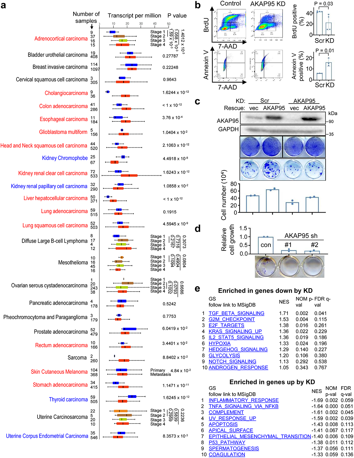
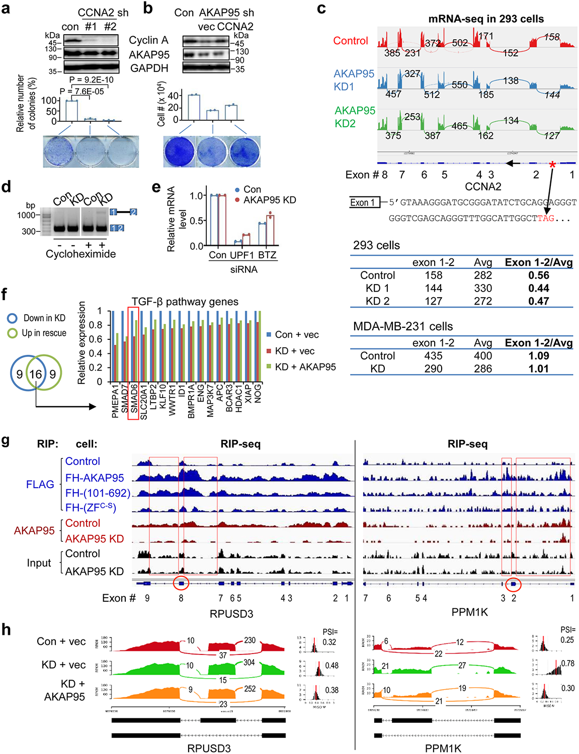
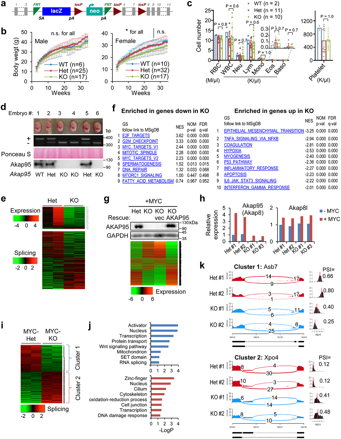
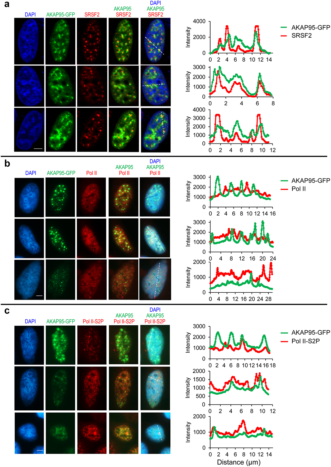
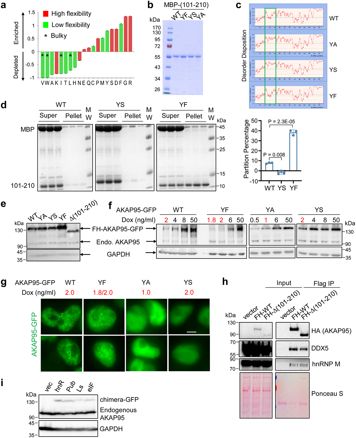
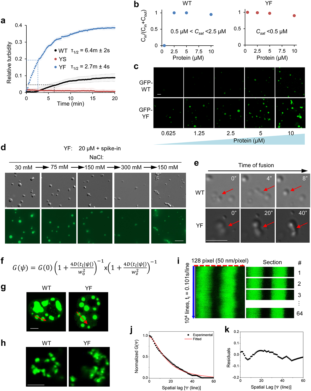
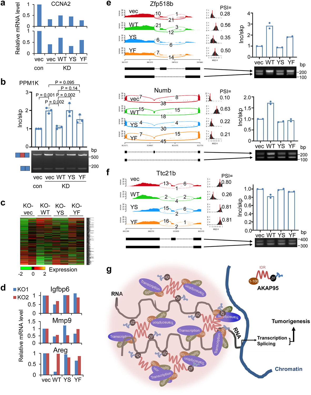
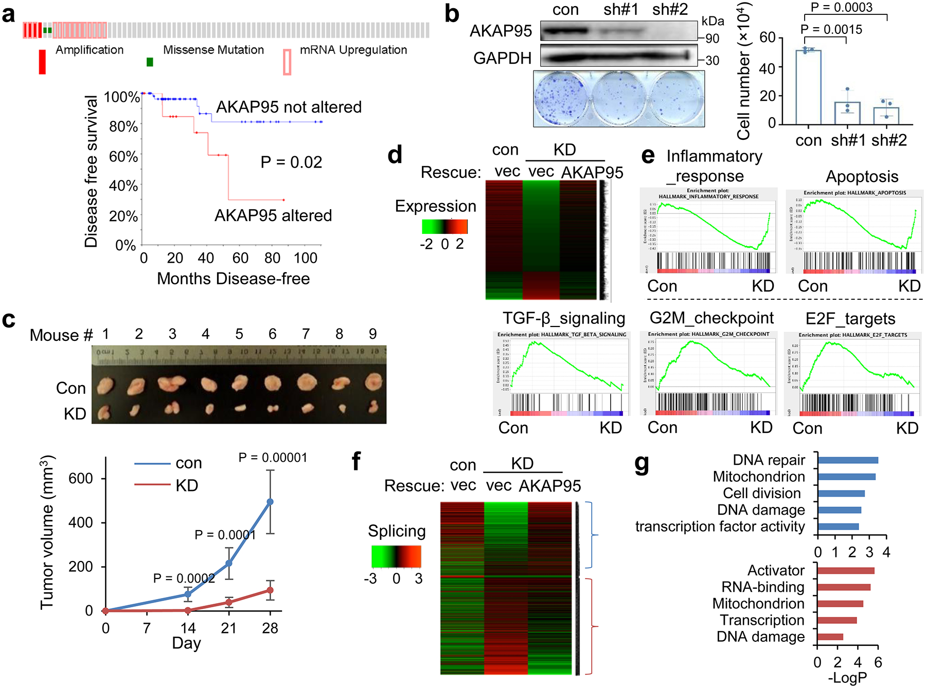
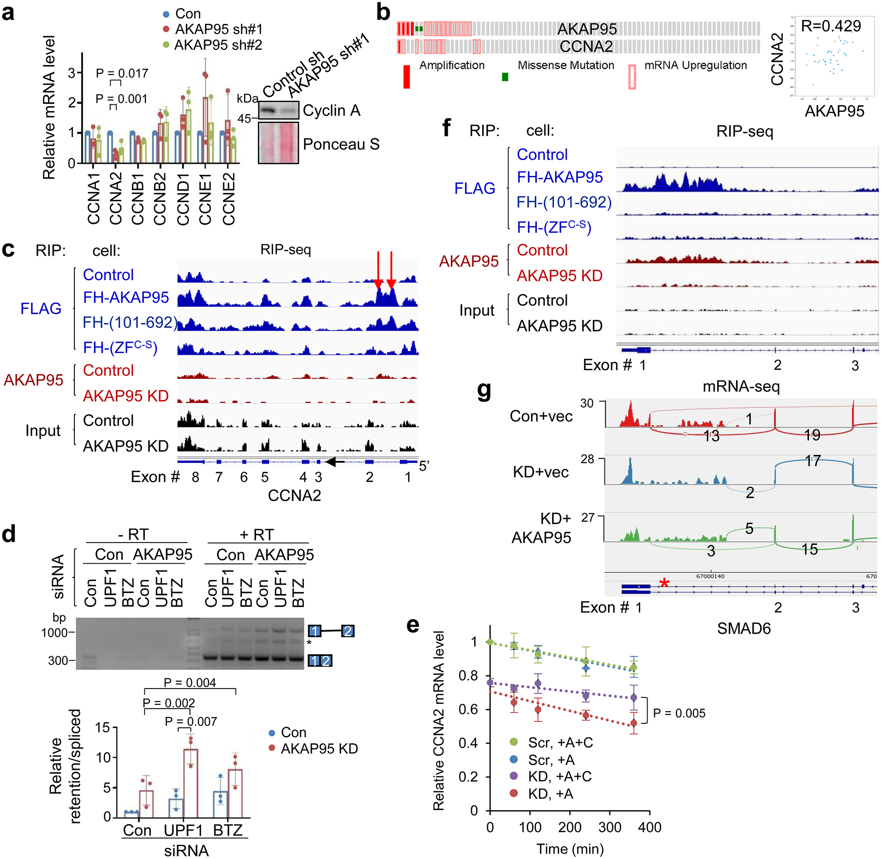
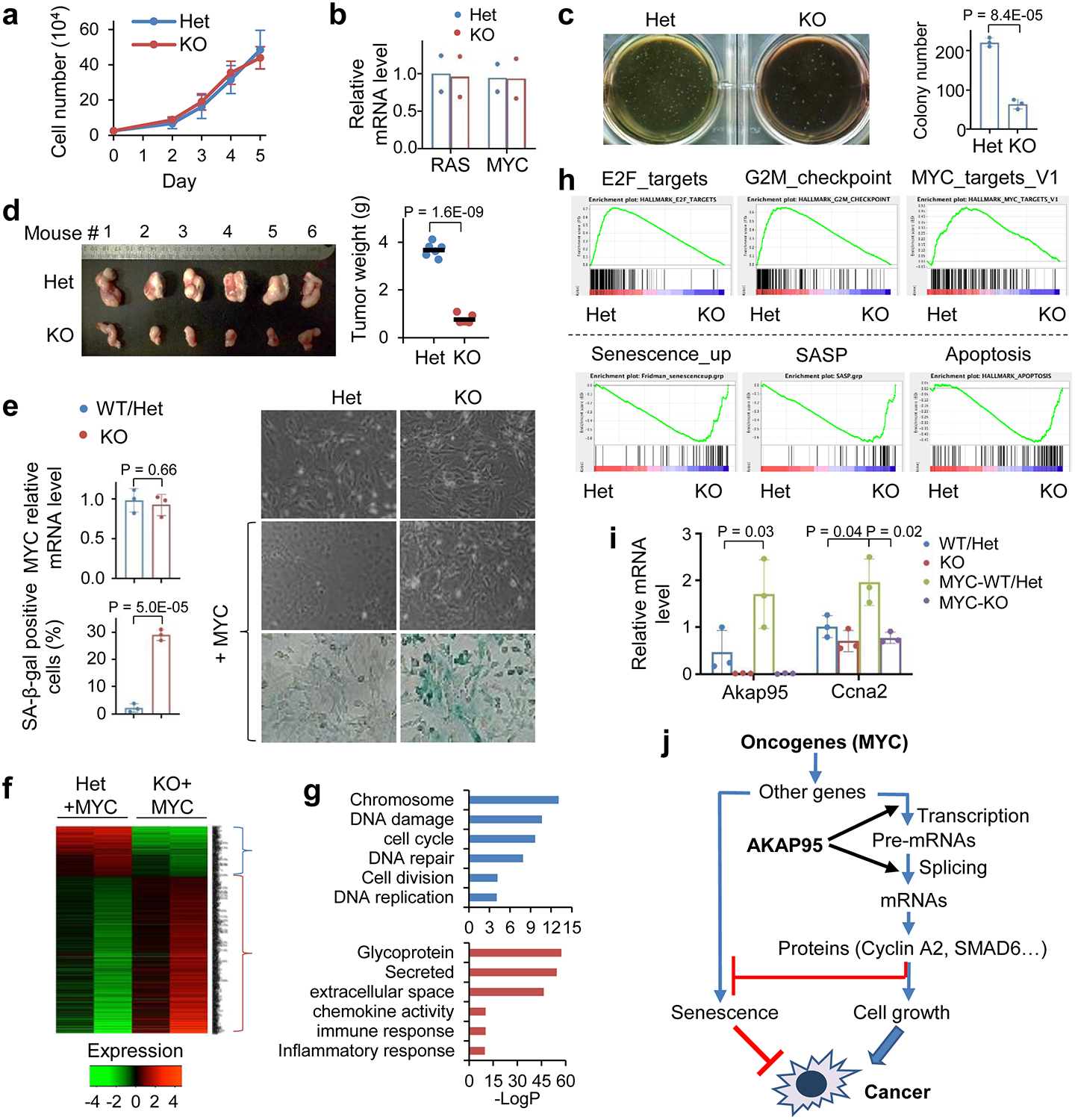
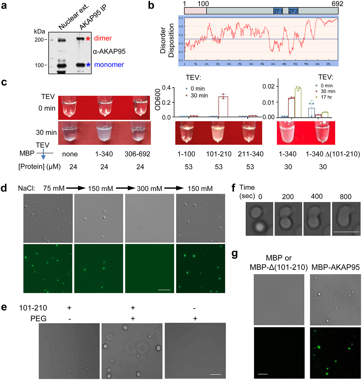
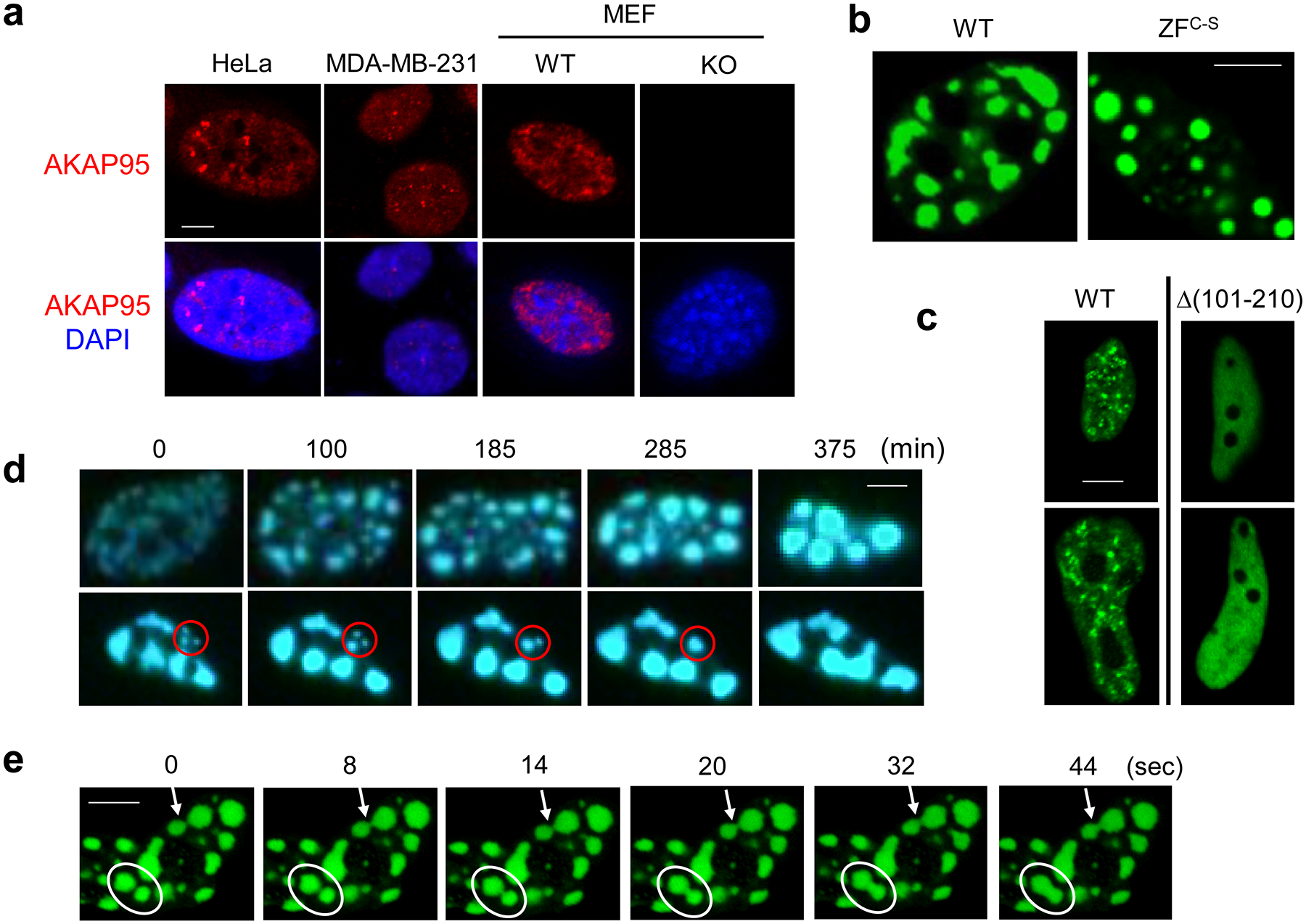
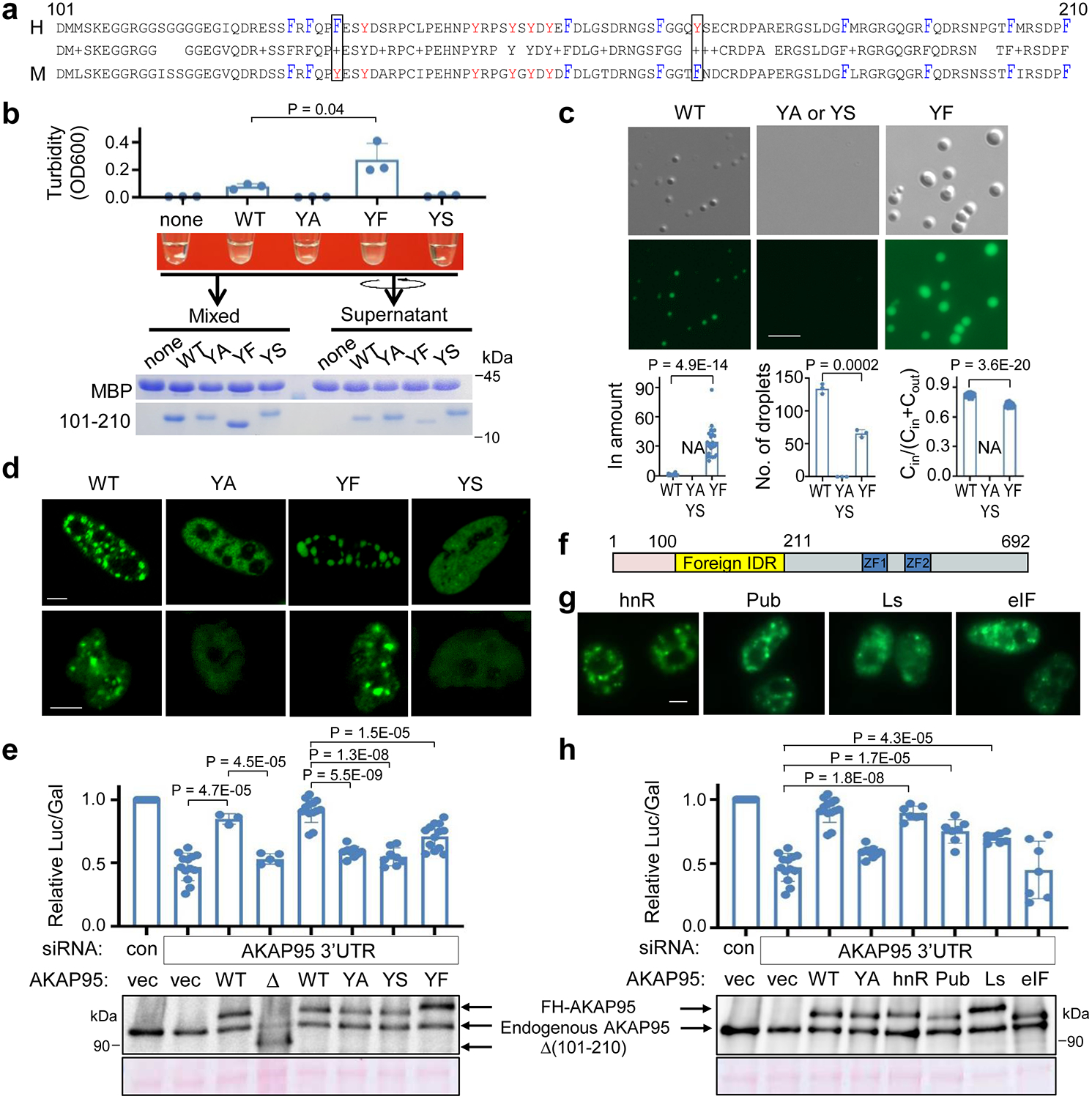
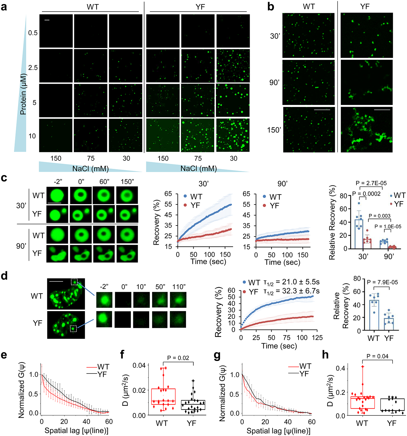
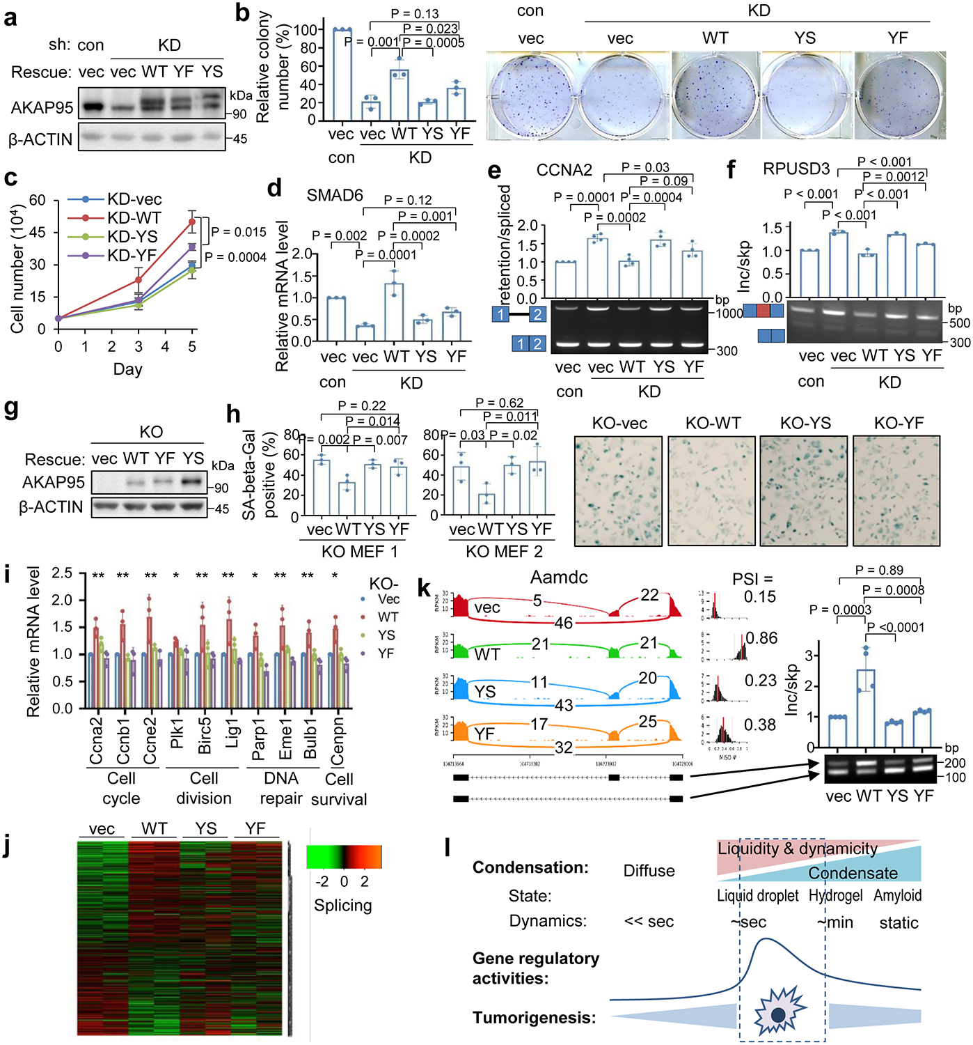
It remains unknown if biophysical or material properties of biomolecular condensates regulate cancer. Here we show that AKAP95, a nuclear protein that regulates transcription and RNA splicing, plays an important role in tumorigenesis by supporting cancer cell growth and suppressing oncogene-induced senescence. AKAP95 forms phase-separated and liquid-like condensates in vitro and in nucleus. Mutations of key residues to different amino acids perturb AKAP95 condensation in opposite directions. Importantly, the activity of AKAP95 in splice regulation is abolished by disruption of condensation, significantly impaired by hardening of condensates, and regained by substituting its condensation-mediating region with other condensation-mediating regions from irrelevant proteins. Moreover, the abilities of AKAP95 in regulating gene expression and supporting tumorigenesis require AKAP95 to form condensates with proper liquidity and dynamicity. These results link phase separation to tumorigenesis and uncover an important role of appropriate biophysical properties of protein condensates in gene regulation and cancer.


Conflict of interest statement

Competing interests

The authors declare no competing interests.

Figures

Extended Data Fig. 1
Extended Data Fig. 1. AKAP95 regulates cancer cell growth and gene expression
a, RNA-Seq–derived gene expression levels from TCGA were analyzed by UALCAN portal. Box plot analysis shows relative expression of AKAP95 in 28 types of cancer (red box) versus normal (blue box) samples, unless indicated for different cancer stages or tumor grades for certain cancer types. Cancer type in red font has significantly higher AKAP95 expression in cancer than in normal (or in later stage than in earlier stage). Cancer type in blue font has significantly lower AKAP95 expression in cancer than in normal samples (or in later stage than in earlier stage). Numbers on the left of the plot stand for the number of samples. Data are median (line), 25–75th percentiles (box) and minimum-maximum values recorded (whiskers). b, Assays for cell proliferation by BrdU incorporation (top) and apoptosis by Annexin V staining (bottom) for control and AKAP95-KD MDA-MB-231 cells. Images of flow cytometry results are shown (left), and percentages of cells positive for BrdU or Annexin V are shown as mean ± SD from n = 3 independent KD assays. c, MDA-MB-231 cells were infected to express scramble (control) or AKAP95 shRNA #1 (KD) and indicated constructs. Top, immunoblotting of total cell lysates. Middle, images of these cells seeded at high (5 ×10⁴ cells/well in 6-well plate, top) and low (400 cells/well in 24-well plate, bottom) densities and stained with crystal violet. Bottom, cell numbers (seeded at high densities) as mean from 2 independent experiments. d, MCF7 cells were infected to express scramble control shRNA or two AKAP95 shRNAs. Bottom, images of indicated cell colonies stained with crystal violet. Top, cell numbers were quantified and presented in the bar graph as mean from 2 independent experiments. e, Top 10 gene sets enriched in genes down- (top, n = 951 genes) and up- (bottom, n = 294 genes) regulated by AKAP95 KD in MDA-MB-231 cells. NES, normalized enrichment score. P values by two-sided Student’s t-test for a-d and modified Fisher’s exact test for e. Uncropped blots and statistical source data are provided as in source data extended data fig 1.
Extended Data Fig. 2
Extended Data Fig. 2. AKAP95 directly regulates splicing of key transcripts for cancer
a,b, MDA-MB-231 cells virally expressing control or indicated shRNAs (a) or shRNA combined with indicated constructs (b) were subject to immunoblotting of total cell lysates (top) and colony formation assay. Middle, colony numbers as mean ± SD from n = 3 (a) or mean from 2 (b) independent experiments. Bottom, images of cells stained with crystal violet. c, Top, mRNA-seq profiles for CCNA2 in control or AKAP95 KD 293 cells. The numbers of exon junction reads are indicated. The red asterisk at the gene diagram indicates a stop codon 57 bp downstream of exon 1 in the intron. The number of reads for the junction of exons 1 and 2, and for the average neighboring exons, and their ratios are in the tables below for indicated cells. d, Total RNAs were used for RT-PCR for intron 1 region in control and AKAP95-KD MDA-MB-231 cells treated with or without cycloheximide for 6 hours. Repeated 3 times. e, Relative mRNA levels of UPF in UPF1-KD samples and BTZ in BTZ-KD samples, respectively, each relative to the control samples, as determined by RT-qPCR and normalized to GAPDH. Mean is from 2 independent experiments. f, Relative expression level of TGF-β pathway genes based on RNA-seq reads from control and AKAP95-KD MDA-MB-231 cells expressing vector or AKAP95. Venn diagram shows numbers of TGF-β pathway genes (from GSEA) downregulated by AKAP95 KD and upregulated by rescue with AKAP95 expression, and the relative expression of the 16 overlapped genes in both categories are plotted. One representative analysis from 2 repeats. g, RIP-seq profiles showing AKAP95 binding to RPUSD3 and PPM1K pre-mRNAs. Track information is the same as in Fig. 2c. Red circles indicate the alternatively included exons (corresponding to the middle exon in the gene diagrams in (h), and red boxes show AKAP95 binding at the introns flanking these exons. h, Sashimi plots showing that the alternative splicing of RPUSD3 and PPM1K pre-mRNAs was affected by AKAP95 KD and rescued by restored expression of AKAP95. The numbers of exon junction reads and PSI are indicated. P values by two-sided Student’s t-test for a and b. Uncropped blots and statistical source data are provided as in source data extended data fig 2.
Extended Data Fig. 3
Extended Data Fig. 3. AKAP95 is required for transformation and overcoming oncogene-induced cellular senescence
a, Schematic of the “Knockout-first” Akap95 allele (null without further recombination). b, Body weights of mice with indicated Akap95 genotypes. Not significant (n.s.) between any two groups at any time for male, or after week 26 for female. *between Het and KO before week 26 for female. c, Peripheral blood profiles of 8-week mice of indicated genotype. n in b, c refers to the number of mice analyzed. d, MEFs were derived from mouse embryos. Images of six embryos from the same litter were shown at the top, followed with genotyping results, Ponceau S staining, and immunoblotting of MEFs. e, Top, heatmap showing relative expression levels of genes and clustered by changes in un-transduced KO MEFs from 2 embryos each. It includes 203 and 20 genes down- or up-regulated in KO, respectively. Bottom, heatmap showing relative alternative splicing of genes clustered by PSI changes. It includes 285 and 332 alternative splicing events with decreased or increased PSI in KO, respectively. Also see Supplementary Table 2f and 7. f, Top 10 gene sets enriched in genes down- (left, n = 265 genes) and up- (right, n = 742 genes) regulated in the MYC-transduced KO versus Het MEFs. g, Rescue of the gene expression profile by introduction of human AKAP95 into the MYC-transduced KO MEFs as shown by immunoblotting and heatmap for relative expression of down- or up-regulated genes in the indicated cells. Also see Supplementary Table 2b. Repeated 2 times. h, Relative expression of Akap95 and Akap8l in un-transduced (-MYC) and MYC-transduced (+MYC) MEFs from n = 2 Het and two KO embryos, as determined by normalized RNA-seq reads. i, Heatmap showing relative alternative splicing of genes clustered by PSI changes in MYC-transduced MEFs from 2 embryos each. It includes 216 and 252 alternative splicing events with decreased or increased PSI in KO versus Het MEFs, respectively. Also see Supplementary Table 2c. j, Gene ontology analysis for the indicated gene clusters from the heatmap in i. Blue (n = 216 genes) and red (n = 252 genes) show functions significantly enriched in genes with PSI increase or decrease by KO, respectively. k, Sashimi plots showing alternative splicing changes for each gene cluster from the heatmap using two examples, Asb7 for cluster 1, and Xpo4 for cluster 2. ns, not significant, *P < 0.05, by two-sided Student’s t-test for b, one-way ANOVA for c and modified Fisher’s exact test for f, j. Uncropped blots and statistical source data are provided as in source data extended data fig 3.
Extended Data Fig. 4
Extended Data Fig. 4. AKAP95 phase separation in vitro
a, 293T cells were transfected with either empty vector (vec), or indicated AKAP95 construct with FLAG-HA-tag. Following α-Flag IP, the pulldown proteins were boiled and resolved by SDS-PAGE and detected by immunoblotting with α-HA. Blue and red asterisks indicate monomer and dimer, respectively. b, Identification of 1–100 as a probable prion subsequence on AKAP95. By the PLAAC program, using homo sapiens as background and core length of 30. c, Purified MBP and MBP fused to AKAP95 truncations as indicated or full-length AKAP95 (1–692) were resolved on SDS-PAGE and stained with Coomassie blue. d, MBP fused to AKAP95 truncations as indicated or full-length AKAP95 were resolved on SDS-PAGE and stained with Coomassie blue following treatment with TEV protease. Note that the cleaved MBP serves as a better indicator for cleavage efficiency as staining signal various for protein fragments of different sequences and sizes. e, Another event of fusion of two droplets formed by 50 μM MBP-AKAP95 (101–210) in 30 mM NaCl and 10% of PEG6000 after treatment with TEV protease for 30 min. Scale bar, 5 μm. Also see Supplementary Video 1. f, Quantification of nuclear AKAP95 concentration by anti-AKAP95 Western blot. Total lysates from indicated number of MDA-MB-231 (M231) and flp-TREx 293 cells (f293, un-induced and dox-induced for FH-AKAP95 expression) were loaded, along with indicated ng of purified MBP-AKAP95. AKAP95 signal of un-induced f293 is similar to that of 25 ng of MBP-AKAP95. All experiments were repeated 2 times. Uncropped blots are provided as in source data extended data fig 4.
Extended Data Fig. 5
Extended Data Fig. 5. AKAP95 partially localizes in nuclear speckles and with actively transcribing Pol II
Fluorescence microscopy images of HeLa cells transiently expressing AKAP95-GFP. Nucleus DNA was stained by DAPI, and specific proteins were stained with antibodies for SRSF2 (a), Pol II (b), and Pol II-S2P (c). The assays were repeated 10 times for each staining and show similar trend. Right, quantification of the signal intensity of indicated molecules across the dotted lines shown in the images. Quantification by Image J. Scale bar, 5 μm for all. Statistical source data are provided as in source data extended data fig 5.
Extended Data Fig. 6
Extended Data Fig. 6. AKAP95 condensation requires Tyrosine in 101–210 and regulates splicing
a, Amino acid enrichment for AKAP95 (101–210). By Composition Profiler, using SwissProt 51 Dataset as background. b, Purified MBP fused to AKAP95 (101–210) WT and mutants were resolved on SDS-PAGE and stained with Coomassie blue. Repeated 2 times. c, Disorder plot of AKAP95 WT or mutants with indicated mutations in 101–210. d, MBP-AKAP95 (101–210) WT, YS, and YF, all at 35 μM and in 30 mM NaCl, were treated with TEV protease for 2 hrs in 3 independent assays, and subjected to centrifugation. The supernatant and pellets (resuspended in the same volume as the supernatant) were resolved by SDS-PAGE followed with coomassie blue staining. MBP signal in the pellet reflects residual supernatant fraction, and its percentage [MBP pellet/(supernatant + pellet)] was subtracted from the (101–210) pellet percentage. Such normalized (101–210) pellet percentages are plotted as Partition Percentage as mean ± SD of n = 3 independent experiments. It is most likely that all supernatants may also have substantial portion of condensates. Moreover, the size cutoff of condensates is also arbitrary, as protein assemblies may take a continuum of size distribution71. e, Immunoblotting by α-AKAP95 (top) or GAPDH (bottom) of total lysates from Flp-In T-Rex 293 cell lines induced to express full-length AKAP95 WT or indicated mutants fused to GFP. Repeated 3 times. f,g, Indicated full-length AKAP95 WT or mutants fused to GFP were induced by various concentrations of doxycycline in Flp-In T-Rex 293 cell lines. Immunoblotting of total cell lysates with indicated antibodies (f). The doxycycline concentrations in red font activated the transgene at the near endogenous level, and were selected for treating cells and fluorescence microscopy assays of fixed cells in (g). Scale bar, 5 μm. Repeated 2 times. h, 293T cells transiently expressing indicated constructs with FLAG-HA-tag were used for α-FLAG immunoprecipitation and immunoblotting with indicated antibodies and Ponceau S staining. Repeated 2 times. i, Immunoblotting of 293T cells transfected with empty vector or indicated FLAG-HA-tagged AKAP95 chimeras fused to GFP. Bottom, by anti-GAPDH. Top, by anti-AKAP95 (Bethyl Laboratories, A301–062A, recognizes an epitope in a region between residue 575 and 625 of human AKAP95). Repeated 2 times. P values by two-sided Student’s t-test for d. Uncropped blots and statistical source data are provided as in source data extended data fig 6.
Extended Data Fig. 7
Extended Data Fig. 7. YF mutation alters material properties of AKAP95 condensates
a, OD450 at different time after TEV protease treatment of 50 μM MBP-AKAP95 (101–210) WT, YS, and YF in 150 mM NaCl, as mean ± SD of readings after subtracting that of MBP at each time (constant at 0.07–0.08) from n = 3 independent assays. Dashed lines show half of the maximum turbidity and time (τ1/2) to reach it. b, Ratio of protein concentration inside the droplets over sum of inside and outside for (101–210) WT and YF at increasing protein concentrations and in 30 mM NaCl, as mean ± SD from n = 6 randomly picked droplets each, in one representative assay from 3 repeats based on Fig. 7a. c, Confocal microscopy images of GFP-AKAP95 (101–210) WT and YF at increasing protein concentrations, all in 150 mM NaCl and 10% of PEG6000 after TEV protease treatment for 20 min. Repeated 2 times with similar results. d, DIC and fluorescence microscopy images for 20 μM MBP-AKAP95 (101–210) YF spiked with Oregon-green-labeled same protein (molar ratio 10:1), after TEV protease treatment for 30 min. Changes in NaCl concentration is indicated. Images were taken 5 min after salt adjustment. Repeated 2 times with similar results. e, Different extent of droplet fusion (arrows) by AKAP95 (101–210) WT and YF, both at 50 μM and in 30 mM NaCl and 10% of PEG6000 after TEV protease treatment for 30 min. A similar trend was observed in 5 fusion events (or attempted fusion for YF) for each. Also see Supplementary Videos 4–6. f, Equation for Line Raster Scan Image Correlation fitting autocorrelation G(Ψ), which depends on G(0)=γ/N (γ: beam profile, N = number of mobile particles), diffusion coefficient D (μm2/s), line scan time tl, pixel size Ψ, radial beam waist w0 and axial beam waist wz. The radial waist w0 (0.218 μm) was calibrated with sub-diffraction beads (0.1 μm) diluted solution as reported before72. The axial waist wz was considered equal to 3*w0. g, Fluorescence confocal microscope image of HeLa cell expressing full-length AKAP95 WT or YF fused to GFP. Arrow indicates the scanned region for in vivo Line RICS. h, Fluorescence confocal microscope image of GFP-AKAP95 (101–210) WT and YF in 150 mM NaCl and 10% of PEG6000 after TEV protease treatment for 20 min, for in vitro Line RICS experiments. i, Line Fluorescence carpet formed by ~104 lines (each line is composed by 128 pixels, 50 nm/pixel) acquired with 0.101 s line scan time and 32.8 μs/pixel. Line RICS autocorrelation curves were computed on 64 sections of 128 lines followed by averaging all the curves. The sectioning of the line carpet permitted to avoid the effect of the movement of the condensates on the measurement of the autocorrelation curves. (λex=488 nm). j, Line RICS autocorrelation curves for experimental data and fitted. k, Residuals of the fitting. Experiments in g-i were repeated 3 times with similar results. Scale bar, 2 μm for c and h, 5 μm for d, e, and g.
Extended Data Fig. 8
Extended Data Fig. 8. Regulation of tumorigenesis and gene expression by AKAP95 requires its condensation with appropriate material properties
a,b, MDA-MB-231 cells were virally infected to stably express scramble (control) or AKAP95 shRNA #1 (KD) and the indicated constructs including empty vector (vec) and FLAG-HA-tagged full-length AKAP95 WT or mutants. a, Relative CCNA2 mRNA level as determined by RT-qPCR and normalized to GAPDH, and plotted for each of the 2 biological repeats individually. b, RT-PCR for ratios for exon-included over -skipped PPM1K transcript, as mean ± SD from n = 3 biological repeats. c-f, MYC-transduced Akap95 KO MEFs were transduced with vector or constructs expressing HA-tagged full-length AKAP95 WT or mutants. c, Heatmap showing relative expression levels of genes changed in MYC-transduced KO MEFs (from 2 embryos each) stably expressing indicated rescue constructs. Also see Supplementary Table 2d. d, Relative mRNA levels of indicated SASP genes as determined by RNA-seq reads from 2 biological repeats (KO1 and KO2). e,f, Sashimi plots showing example genes for which the alternative exon inclusion was promoted (e) or suppressed (f) by introduction of AKAP95 WT, but not as effectively by YS or YF, and RT-PCR for the inclusion of the alternative exon, as mean from 2 embryos each. g, A model for how AKAP95 condensates may regulate gene expression for tumorigenesis. P values by one-way ANOVA followed by Tukey’s post hoc test. Uncropped blots and statistical source data are provided as in source data extended data fig 8.
Fig. 1.
Fig. 1.. AKAP95 regulates cancer cell growth and gene expression.
a, Overexpression of AKAP95 in breast cancer tissues of 82 TNBC patient samples. From cBioPortal. Top, each box is a patient sample. Bottom, disease-free survival curves of patients with or without AKAP95 alterations. n = 17 and 65 patient samples for AKAP95 altered and not altered, respectively. b, Growth assay for MDA-MB-231 cells expressing control or two AKAP95 shRNAs. Left, immunoblotting of total cell lysates and images of cell colonies stained with crystal violet. Right, numbers of cells in growth assays as mean ± SD from n = 3 independent experiments. c, Tumors from xenograft of control or AKAP95-KD MDA-MB-231 cells in immune-deficient mice. Tumor volumes at the indicated days post transplantation are plotted as mean ± SD (n = 9 mice). d,e, RNA-seq analysis in MDA-MB-231 cells expressing control or AKAP95 shRNA #1 and the indicated vector or AKAP95-expressing construct. One representative analysis from 2 repeats. d, Heatmap showing relative expression levels of genes down- or up-regulated in the indicated cells. It includes 951 and 294 genes down- and up-regulated in KD compared to control cells, respectively. Also see Supplementary Table 1a. e, GSEA for gene expression profiles of control and AKAP95-KD cells. Plots above and below the broken line show gene sets significantly enriched in up- and down-regulated genes by AKAP95 KD, respectively. f, Heatmap showing relative alternative splicing and clustered by changes in percent-spliced-in (PSI) values in the indicated cells. It includes 807 and 1275 alternative splicing events with decreased or increased PSI in KD cells, respectively. Also see Supplementary Table 1b. g, Gene ontology analysis for the indicated clusters from the heatmap in f. Blue (n = 807 genes) and red (n = 1275 genes) show functions significantly enriched in genes with PSI increase or decrease by AKAP95 KD, respectively. P values by log-rank test for a, Student’s t-test for b and d, and modified Fisher’s exact test for g. All two-sided. Uncropped blots and statistical source data are provided in source data fig 1.
Fig. 2.
Fig. 2.. AKAP95 directly regulates splicing of key transcripts for cancer.
a, CCNA2 expression in MDA-MB-231 cells upon AKAP95 KD. Left, relative mRNA levels of indicated cyclins were determined by RT-qPCR and normalized to GAPDH, and presented as mean ± SD from n = 3 biological repeats. Right, immunoblotting for Cyclin A1/A2. b, Co-overexpression of AKAP95 and CCNA2 in breast cancer tissues of TNBC patients. Left, each box represents a patient. Right, correlation of their mRNA levels in the TNBC patients with indicated Pearson correlation coefficient. From cBioPortal. n = 82 patient samples. c,f, RNA immunoprecipitation-sequencing (RIP-seq) profiles for CCNA2 (c) and SMAD6 (f) based on our previous work. Blue, anti-FLAG RIP-seq in control or 293 cells expressing the FLAG-HA-tagged AKAP95 WT or mutants. Red, anti-AKAP95 RIP-seq in control or AKAP95-KD 293 cells. Black, profiles of total input RNAs. All profiles have the same Y-axis scale. Arrows indicate AKAP95-binding sites at intron 1. One representative RIP-seq analysis from 2 repeats. d, Total RNAs were used for RT-PCR, in the absence (- RT) or presence (+ RT) of reverse transcriptase, for CCNA2 intron 1 in MDA-MB-231 cells with indicated siRNAs. Top, PCR products on agarose gel. Asterisk, an unknown amplification product. Repeated 3 times. Bottom, relative ratios of the signal for the intron 1- retaining transcript over the intron 1-spliced transcript, as mean ± SD from n = 3 independent experiments. e, Assay for CCNA2 mRNA stability. Control (Scr) and AKAP95-KD MDA-MB-231 cells were treated starting from 0 min with Actinomycin D (+A, to block RNA synthesis) and cycloheximide (+C, to block NMD) or not as indicated. Total RNA at indicated times were used for RT-PCR and normalized to ACTB, as mean ± SD from n = 3 biological repeats. g, mRNA-seq profiles for SMAD6 in MDA-MB-231 cells expressing control or AKAP95 shRNA #1 (KD) and vector or AKAP95-expressing construct. Asterisk, a stop codon. P values by two-sided Student’s t-test for a and e and one-way ANOVA followed by Tukey’s post hoc test for d. Uncropped blots and statistical source data are provided as in source data fig 2.
Fig. 3.
Fig. 3.. AKAP95 is dispensable for normal cell growth but required for transformation and suppressing oncogene-induced senescence.
a, Growth of MEFs from Akap95+/- (Het) and Akap95−/− (KO) embryos (n = 6 embryos each). b, Relative HRAS and MYC mRNA levels by RT-qPCR and normalized to Actb, as mean from HRASG12V and MYC transduced MEFs (2 embryos each). c, HRAS-MYC-transduced MEFs in colony formation assay. Colony numbers as mean ± SD from n = 6 experiments using MEFs of 2 embryos each. d, Six mice received HRAS-MYC-transduced Het and KO MEFs on each flank. Tumor weights (week 4) are plotted. Each dot represents a tumor. e-i, MYC-transduced MEFs from 3 KO and 3 Akap95-expressing (1 WT, 2 Het) embryos. e, Right, images of cells before and after MYC transduction. Images of SA-beta-galactosidase activity assay are at bottom. Relative MYC mRNA levels after transduction were determined by RT-qPCR and normalized to Actb (left top). Percentage of SA-beta-gal-positive cells are plotted (left bottom). Both as mean ± SD from MEFs (n = 3 embryos each). f, Heatmap showing relative expression of genes and clustered by changes in KO MEFs (2 embryos each separately analyzed), with 265 and 742 genes down- or up-regulated in KO, respectively. Also see Supplementary Table 2a. g, Gene ontology analysis for the indicated gene clusters from the heatmap in f. Blue (n = 265 genes) and red (n = 742 genes) show functions significantly enriched in down- and up-regulated genes, respectively. h, GSEA plots above and below dashed line show gene sets significantly enriched in genes down- and up-regulated in the MYC-transduced KO compared to Het MEFs, respectively. i, Relative Akap95 and Ccna2 mRNA levels before and after MYC transduction by RT-qPCR and normalized to Actb, as mean ± SD from MEFs from n = 3 embryos each. j, Diagram summarizing regulation of tumorigenesis by AKAP95 through gene expression control. P values by two-sided Student’s t-test for all except one-way ANOVA followed by Tukey’s post hoc test for i, and modified Fisher’s exact test for g. Statistical source data are provided as in source data fig 3.
Fig. 4.
Fig. 4.. AKAP95 undergoes phase separation with liquid-like properties in vitro.
a, Immunoblotting for AKAP95 in HeLa cell nuclear extract and AKAP95 immunoprecipitation from the extract. Samples were boiled in the presence of DTT and resolved by SDS-PAGE. b, Disorder plot of human AKAP95. c, Turbidity by pictures and OD600 of MBP (none) and MBP fused to AKAP95 truncations at indicated concentrations all in 30 mM NaCl before and after TEV protease treatment for indicated time. OD600 is plotted as mean ± SD from n = 3 biological repeats. d, DIC (top) and fluorescence microscopy (bottom) images for 20 μM MBP-AKAP95 (101–210) and spiked with Oregon-green-labeled same protein (molar ratio 10:1) after TEV protease treatment for 30 min. Changes in NaCl concentration is indicated. Images were taken 5 min after salt adjustment. e, Phase contrast images of 50 μM MBP-AKAP95 (101–210) in 30 mM NaCl in the absence and presence of 10% of PEG6000 after TEV protease treatment for 30 min. f, Fusion of two droplets formed by 50 μM MBP-AKAP95 (101–210) in 30 mM NaCl and 10% of PEG6000 after TEV protease treatment for 30 min. Also see Video 1. g, DIC and fluorescence microscopy images of 6.25 μM MBP, MBP fused to Δ(101–210) or full-length AKAP95 in 150 mM NaCl, spiked with Oregon-green-labeled AKAP95 (101–210) at a molar ratio of 150:1 after TEV protease treatment for 30 min. Note that the lack of any condensates in the DIC images showed the inability of Δ(101–210) in condensation. Experiments in a, d, e-g were repeated 4 times. Scale bar, 5 μm for all. Uncropped blots and statistical source data are provided as in source data fig 4.
Fig. 5.
Fig. 5.. AKAP95 forms dynamic foci in cell nucleus.
a, Immunostaining of endogenous AKAP95 (red) and DNA (DAPI, blue) in indicated cancer cell lines and primary MEFs from WT and Akap95 KO embryos. b, Confocal microscopy images of AKAP95 WT or ZFC-S fused to GFP in nuclei following transfection into HeLa cells. c, Fluorescence microscopy images of HeLa cells transiently expressing AKAP95 WT or Δ(101–210 fused to GFP. d, HeLa cells were transfected with AKAP95-GFP, and two nuclei were imaged at different time points. Time 0 was 24 hr after transfection. Note the growth and merge of the foci, especially those in the red circle. e, Rapid fusion of AKAP95 (ZFC-S)-GFP foci in a HeLa cell nucleus. The white oval and arrow show two different fusion events. These images are from Video 2. All experiments were Repeated 4 times. Scale bar, 5 μm for all.
Fig. 6.
Fig. 6.. AKAP95 condensation requires Tyrosine in 101–210 and regulates splicing.
a, Alignment of human and mouse AKAP95 (101–210). Middle row shows identical residues (letter) and conservative mutations (“+”). Tyr, red; Phe, blue and tall. Box, Tyr and Phe swapping. b, MBP alone (none) or MBP-AKAP95 (101–210) WT or mutants all at 50 μM and in 30 mM NaCl after TEV protease treatment for 30 min. Turbidity of each reaction was shown, and by OD600 as mean ± SD from n = 3 (for YA, YS) or 4 (the rest) independent assays. Samples taken after mixing and from supernatant after centrifugation were resolved by SDS-PAGE followed by coomassie blue staining. c, DIC and fluorescence microscopy images for 10 μM Oregon-green-labeled MBP-AKAP95 (101–210) WT and mutants in 30 mM NaCl after TEV protease treatment for 30 min. Plots from left to right show relative protein amount in droplet, number of droplets in a field, and ratio of protein concentration inside droplets over sum of inside and outside droplets, respectively, as mean ± SD from n = 24 randomly picked droplets, except for number of droplets from n = 3 randomly picked fields, in one representative assay from 5 repeats. NA, not applicable. d, Fluorescence microscopy images of HeLa cells (top) and Flp-In T-Rex 293 cell lines expressing (bottom) GFP fusions with full-length AKAP95 WT or mutants. Repeated 4 times. e,h, HEK293 cells co-transfected with indicated siRNAs and plasmids were subject to splice reporter assay (top) and immunoblotting with α-AKAP95 (bottom). Δ = Δ(101–210). Mean ± SD from n = 8 [except 5 for Δ(101–210) and 13 for YF and 2nd WT] independent transfections are plotted in e and 7 independent transfections in h. f, Schematic of AKAP95 chimeras. g, Fluorescence microscopy images of 293T cells transfected with indicated AKAP95 chimeras fused to GFP. Repeated 3 times. P values by two-sided Student’s t-test for b and one-way ANOVA followed by Tukey’s post hoc test for e and h. Scale bar, 5 μm for all. Uncropped blots and statistical source data are provided as in source data fig 6.
Fig. 7.
Fig. 7.. YF mutation enhances AKAP95 condensation propensity and renders condensates more solid-like state.
a, Fluorescence microscopy images of Oregon-green-labeled AKAP95 (101–210) WT and YF at indicated protein and NaCl concentrations after MBP cleavage for 30 min. Repeated 3 times. b, Fluorescence microscopy images of 50 μM AKAP95 (101–210) WT and YF in 30 mM NaCl both spiked with Oregon-green-labeled (101–210) WT (molar ratio 150:1) after MBP cleavage for 30’ and imaged immediately (30’) or after incubation for 60 (90’) or 120 (150’) more minutes. Repeated 3 times. c, FRAP of 10 μM GFP-AKAP95 (101–210) WT and YF after 30 min of MBP cleavage in 150 mM NaCl. FRAP was performed immediately (30’) or after incubation for 60 min more (90’). Left, fluorescence microscopy images of droplets at indicated times. Middle, FRAP recovery curves as mean ± SD. Right, mean ± SD of recovery at the final time. n = 7 independent measurements. d, FRAP of Full-length AKAP95 WT and YF fused to GFP in HeLa cell nuclei. Left, fluorescence microscopy images of foci. The photobleached focus was boxed and amplified for indicated time points. Middle, FRAP recovery curves as mean ± SD. Right, mean ± SD of recovery (relative to minimal level) at the final time. n = 7 independent measurements. e,f, Diffusion of full length AKAP95 WT and YF fused to GFP in HeLa cell nuclei, showing Line RICS normalized autocorrelation curves G(Ψ) as function of Spatial Lag (Ψ) (e) and diffusion coefficients (f), as mean ± SD (n = 20 or 22 independent measurements for WT, YF, respectively). g,h, Diffusion coefficients of purified GFP-AKAP95 (101–210) WT and YF, showing Line RICS normalized autocorrelation curves G(Ψ) (g) and diffusion coefficients (h), as mean ± SD (n = 22 or 13 independent measurements for WT, YF, respectively). Scale bars, 10 (a, b), 2 (c), 5 (d) μm. P values by two-sided Student’s t-test (c, d) or Mann-Whitney U test (f, h). For box-and-whisker plots, data are median (line), 25–75th percentiles (box) and minimum-maximum values recorded (whiskers). Statistical source data are provided as in source data fig 7.
Fig. 8.
Fig. 8.. Regulation of tumorigenesis and gene expression by AKAP95 requires its condensation with proper biophysical properties.
a-f, MDA-MB-231 transduced with control or AKAP95 shRNA #1 (KD) and vector or FLAG-HA-tagged full-length AKAP95 WT or mutants. n = 3 biological repeats for a-f, except n = 4 biological repeats for e. a, Immunoblotting of total cell lysates. b, Colony formation assays. Left, colony numbers as mean ± SD. Right, images of cells stained with crystal violet. c, Growth of cultured cells, as mean ± SD. d, Relative SMAD6 mRNA level were determined by RT-qPCR and normalized to GAPDH, as mean ± SD. e,f, RT-PCR for ratios for intron 1-retained over -spliced CCNA2 (e) and exon-included over -skipped RPUSD3 transcripts, as mean ± SD. g-k, MYC-transduced Akap95 KO MEFs transduced with vector or HA-tagged full-length AKAP95 WT or mutants. g, Immunoblotting of total cell lysates. Repeated 3 times. h, Left, percentage of SA-beta-gal-positive cells as mean ± SD (n = 3 different images of MEFs from two embryos). Right, images from KO MEF 1. i, Relative mRNA levels of indicated genes with related functions at bottom by RT-qPCR and normalized to Gapdh, as mean ± SD (n = 3 independent experiments). * or ** between vec and WT, WT and YS, WT and YF, except for Plk1, for which * only between vec and WT, WT and YF. *P<0.05, **P<0.01. j, Heatmap showing relative alternative splicing with PSI changes in MYC-transduced KO MEFs expressing indicated constructs (2 embryos each). Also see Supplementary Table 2e. k, Sashimi plot showing Aamdc alternative splicing that was rescued by introduction of AKAP95 WT, but not but the mutant, and RT-PCR for the inclusion of the alternative exon as mean ± SD (n = 4 biological replicates pooled from 2 embryos each). l, Diagram showing impact of material properties of AKAP95 WT and mutants on gene regulation and tumorigenesis. P values by two-sided Student’s t-test for c and one-way ANOVA followed by Tukey’s post hoc test for all other analyses. Uncropped blots and statistical source data are provided as in source data fig 8.

Comment in

References

    1. Courchaine EM, Lu A & Neugebauer KM Droplet organelles? The EMBO journal 35, 1603–1612 (2016). - PMC - PubMed
    1. Hyman AA, Weber CA & Julicher F Liquid-liquid phase separation in biology. Annual review of cell and developmental biology 30, 39–58 (2014). - PubMed
    1. Shin Y & Brangwynne CP Liquid phase condensation in cell physiology and disease. Science (New York, N.Y.) 357 (2017). - PubMed
    1. Banani SF, Lee HO, Hyman AA & Rosen MK Biomolecular condensates: organizers of cellular biochemistry. Nature reviews. Molecular cell biology 18, 285–298 (2017). - PMC - PubMed
    1. Boeynaems S et al. Protein Phase Separation: A New Phase in Cell Biology. Trends in cell biology 28, 420–435 (2018). - PMC - PubMed

Publication types

MeSH terms