. 2020 Jul 3:8:690.
doi: 10.3389/fbioe.2020.00690. eCollection 2020.

Lipid-Based Nanovesicles for Simultaneous Intracellular Delivery of Hydrophobic, Hydrophilic, and Amphiphilic Species

Affiliations

Lipid-Based Nanovesicles for Simultaneous Intracellular Delivery of Hydrophobic, Hydrophilic, and Amphiphilic Species

Antonella Zacheo et al. Front Bioeng Biotechnol. .

Abstract

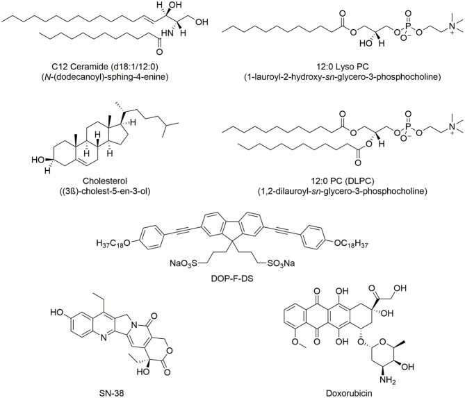
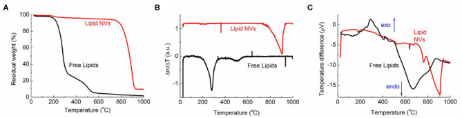
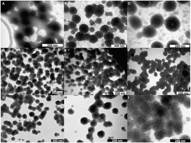
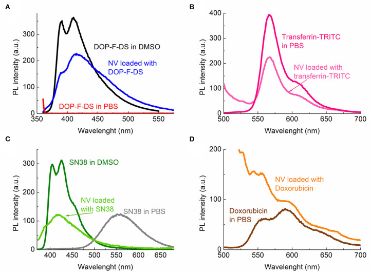
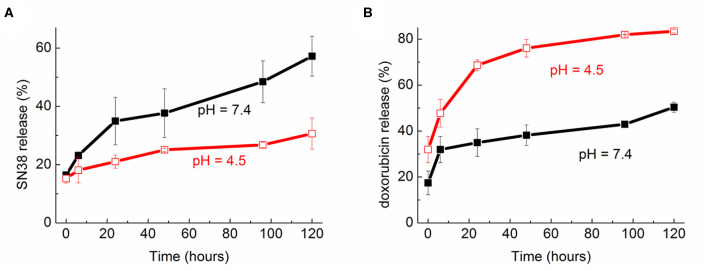
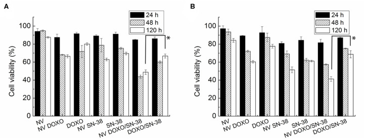
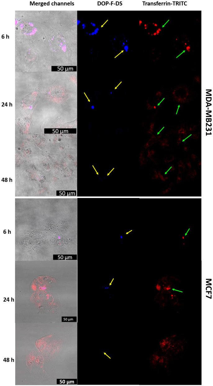
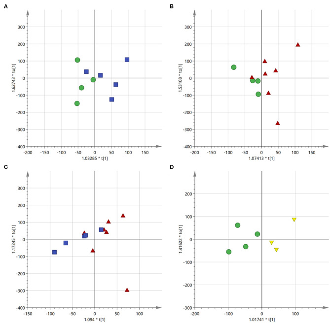
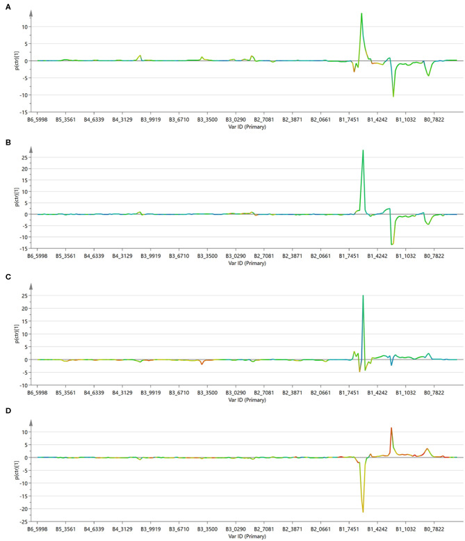
Lipid nanovesicles (NVs) are the first nanoformulation that entered the clinical use in oncology for the treatment of solid tumors. They are indeed versatile systems which can be loaded with either hydrophobic or hydrophilic molecules, for both imaging and drug delivery, and with high biocompatibility, and limited immunogenicity. In the present work, NVs with a lipid composition resembling that of natural vesicles were prepared using the ultrasonication method. The NVs were successfully loaded with fluorophores molecules (DOP-F-DS and a fluorescent protein), inorganic nanoparticles (quantum dots and magnetic nanoparticles), and anti-cancer drugs (SN-38 and doxorubicin). The encapsulation of such different molecules showed the versatility of the developed systems. The size of the vesicles varied from 100 up to 300 nm depending on the type of loaded species, which were accommodated either into the lipid bilayer or into the aqueous core according to their hydrophobic or hydrophilic nature. Viability assays were performed on cellular models of breast cancer (MCF-7 and MDA-MB-231). Results showed that NVs with encapsulated both drugs simultaneously led to a significant reduction of the cellular activity (up to 22%) compared to the free drugs or to the NVs encapsulated with only one drug. Lipidomic analysis suggested that the mechanism of action of the drugs is the same, whether they are free or encapsulated, but administration of the drugs by means of nanovesicles is more efficient in inducing cellular damage, likely because of a quicker internalization and a sustained release. This study confirms the versatility and the potential of lipid NVs for cancer treatment, as well as the validity of the ultrasound preparation method for their preparation.

Keywords: SN-38; breast cancer; doxorubicin; lipidomic analysis; nanoparticle; nanovesicle.


Figures

Figure 1
Figure 1
Chemical structure of the lipids used in the synthesis of the NVs and the encapsulated organic molecules, (i.e., the DOP-F-DS fluorophore and the two drugs used in cellular study).
Figure 2
Figure 2
TGA and DSC data for NVs (red curve) and free lipids (black curve). (A) Percentage weight, (B) first derivative curves, and (C) DSC data.
Figure 3
Figure 3
TEM images of nanovesicles either empty (A) or loaded with (B) QDs, (C) MNPs, (D) DOP-F-DS, (E) transferrin-TRITC, (F) DOP-F-DS, and transferrin-TRITC, (G) doxorubicin, (H) SN-38, and (I) both drugs.
Figure 4
Figure 4
Photoluminescence spectra of the fluorescent species either free or encapsulated: (A) DOP-F-DS; (B) transferrin-TRITC; (C) SN-38; and (D) doxorubicin.
Figure 5
Figure 5
Release profile over time (up to 120 h) of (A) SN-38 and (B) doxorubicin from the nanovesicles at two pH (7.4 and 4.5).
Figure 6
Figure 6
MTT viability assay of (A) MDA-MB-231 and (B) MCF-7 cells administered for 24, 48, and 120 h with empty nanovesicles, nanovesicles loaded with DOXO, free DOXO, nanovesicles loaded with SN-38, free SN-38, nanovesicles loaded with both drugs, and both free drugs, respectively. The viability of the cells incubated with the loaded nanovesicles was compared with that of the cells incubated with free drugs, at 120 h in both cell lines. Statistical analysis was performed via t-test considering it significant for *p < 0.05.
Figure 7
Figure 7
CLSM images of MDA-MB-231 and MCF-7 cells incubated with liposomes loaded with transferrin-TRITC and DOP-F-DS for 6, 24, and 48 h. The left column refers to the fluorescence channels merged with the bright field, while the central and right columns refer to the blue and red fluorescence channels, respectively. The scale bars correspond to 50 μm.
Figure 8
Figure 8
Score scatter plot for the first component of the OPLS-DA models. (A) Control cells (green circles) vs. free drugs (blue squares); (B) control cells (green circles) vs. drugs encapsulated into the NVs (red triangles); (C) cells treated with free drugs (blue squares) vs. those treated with the encapsulated drugs (red triangles); (D) control cells (green circles) vs. empty NVs (yellow triangles).
Figure 9
Figure 9
S-line plot for the first component of the OPLS-DA models. (A) Control cells vs. free drugs; (B) control cells vs. drugs encapsulated into the NVs; (C) cells treated with free drugs vs. those treated with the encapsulated drugs; (D) control cells vs. empty NVs.

Similar articles

Cited by

References

    1. Al-Jamal W. T., Al-Jamal K. T., Tian B., Lacerda L., Bomans P. H., Frederik P. M., et al. . (2008). Lipid–quantum dot bilayer vesicles enhance tumor cell uptake and retention in vitro and in vivo. ACS Nano 2, 408–418. 10.1021/nn700176a - DOI - PubMed
    1. Antimisiaris S. G., Mourtas S., Marazioti A. (2018). Exosomes and exosome-inspired vesicles for targeted drug delivery. Pharmaceutics 10:218. 10.3390/pharmaceutics10040218 - DOI - PMC - PubMed
    1. Armitage E. G., Southam A. D. (2016). Monitoring cancer prognosis, diagnosis and treatment efficacy using metabolomics and lipidomics. Metabolomics 12:146. 10.1007/s11306-016-1093-7 - DOI - PMC - PubMed
    1. Barenholz Y. (2012). Doxil(R)–the first FDA-approved nano-drug: lessons learned. J. Control. Release 160, 117–134. 10.1016/j.jconrel.2012.03.020 - DOI - PubMed
    1. Bothun G. D., Rabideau A. E., Stoner M. A. (2009). Hepatoma cell uptake of cationic multifluorescent quantum dot liposomes. J. Phys. Chem. B 113, 7725–7728. 10.1021/jp9017458 - DOI - PubMed