. 2020 Jul 3:8:709.
doi: 10.3389/fbioe.2020.00709. eCollection 2020.

Conductive Hydroxyethyl Cellulose/Soy Protein Isolate/Polyaniline Conduits for Enhancing Peripheral Nerve Regeneration via Electrical Stimulation

Affiliations

Conductive Hydroxyethyl Cellulose/Soy Protein Isolate/Polyaniline Conduits for Enhancing Peripheral Nerve Regeneration via Electrical Stimulation

Ping Wu et al. Front Bioeng Biotechnol. .

Abstract

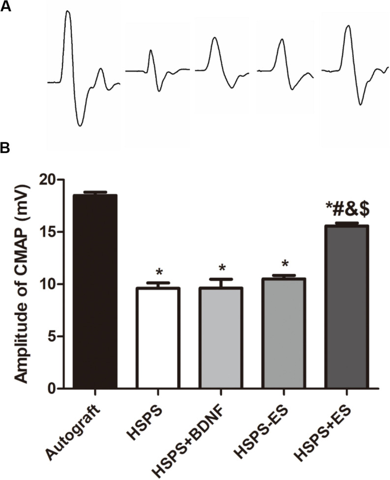
Nerve regeneration remains a challenge to the treatment of peripheral nerve injury. Electrical stimulation (ES) is an assistant treatment to enhance recovery from peripheral nerve injury. A conductive nerve guide conduit was prepared from hydroxyethyl cellulose (HEC)/soy protein isolate (SPI)/PANI sponge (HSPS) and then the HSPS conduits were used to repair 10 mm sciatic nerve injury in rat model with or without ES, using HSPS+brain-derived neurotrophic factor (BDNF) and autografts as controls. The nerve repairing capacities were evaluated by animal experiments of behavioristics, electrophysiology, toluidine blue staining, and transmission electron microscopy (TEM) in the regenerated nerves. The results revealed that the nerve regeneration efficiency of HSPS conduits with ES (HSPS+ES) group was the best among the conduit groups but slightly lower than that of autografts group. HSPS+ES group even exhibited notably increased in the BDNF expression of regenerated nerve tissues, which was also confirmed through in vitro experiments that exogenous BDNF could promote Schwann cells proliferation and MBP protein expression. As a result, this work provided a strategy to repair nerve defect using conductive HSPS as nerve guide conduit and using ES as an extrinsic physical cue to promote the expression of endogenous BDNF.

Keywords: electrical stimulation; hydroxyethyl cellulose; peripheral nerve injury; polyaniline; soy protein isolate.


Figures

FIGURE 1
FIGURE 1
(A) General observation images of HEC/SPI conduit. (B) General observation images of HSPS conduit. (C) Longitudinal view SEM images of the HSPS conduit. (D) Transverse cross-section view SEM images of the HSPS conduit. (E) General observation images of HSPS-ES conduit with electrodes. (F) Schematic diagram of HSPS+ES electrical stimulation in animals.
FIGURE 2
FIGURE 2
In vitro cell experiments. (A) Schwann cells S100 IF identification, scale bar = 100 μm. (B) BDNF promotes the Schwann cells proliferation. (C) BDNF promotes the Schwann cells PCNA. (D) MBP protein relative expression levels. *P < 0.05 compared with the 0 ng/mL group.
FIGURE 3
FIGURE 3
Immunofluorescence image of BDNF proteins in the nerve of HSPS-ES group (A) and HSPS+ES group (B).
FIGURE 4
FIGURE 4
The sciatic functional index (SFI) in each group. (A) Images of footprints the SD rats and (B) statistical results of SFI values in each group at 3 months after surgery. *P < 0.05 compared with the Autograft group, #P < 0.05 compared with the HSPS group, &P < 0.05 compared with the HSPS+BDNF group, [dollar]P < 0.05 compared with the HSPS-ES group.
FIGURE 5
FIGURE 5
The results of electrophysiological test. (A) Representative CMAPs recordings on the injured side for each group. (B) The statistical results of peak amplitude of CMAPs. *P < 0.05 compared with the Autograft group, #P < 0.05 compared with the HSPS group, &P < 0.05 compared with the HSPS+BDNF group, [dollar]P < 0.05 compared with the HSPS-ES group.
FIGURE 6
FIGURE 6
Images of HE-stained longitudinal sections of the regenerated nerve in each group at 3 months after surgery, the red arrows represented the diameter of the regenerated nerves.
FIGURE 7
FIGURE 7
Images of toluidine blue stained regenerated nerve cross sections and the statistical density of myelinated nerve fibers in regenerated nerves for each group, scale bar = 20 μm. *P < 0.05 compared with the Autograft group, #P < 0.05 compared with the HSPS group, &P < 0.05 compared with the HSPS+BDNF group, [dollar]P < 0.05 compared with the HSPS-ES group.
FIGURE 8
FIGURE 8
Images of toluidine blue stained regenerated nerve cross sections and the statistical density of myelinated nerve fibers in regenerated nerves for each group. (A) TEM images of the middle portion of regenerated nerve and statistical analysis of (B) axon diameter, (C) thickness of myelin sheath, and (D) G-ratio. *P < 0.05 compared with the Autograft group, #P < 0.05 compared with the HSPS group, &P < 0.05 compared with the HSPS+BDNF group, [dollar]P < 0.05 compared with the HSPS-ES group.
FIGURE 9
FIGURE 9
The toxicity assessment of the HSPS conduits in vivo.

Similar articles

Cited by

References

    1. Arteshi Y., Aghanejad A., Davaran S., Omidi Y. (2018). Biocompatible and electroconductive polyaniline-based biomaterials for electrical stimulation. Eur. Polymer J. 108 150–170. 10.1016/j.eurpolymj.2018.08.036 - DOI
    1. Carvalho C. R., Oliveira J. M., Reis R. L. (2019). Modern trends for peripheral nerve repair and regeneration: beyond the hollow nerve guidance conduit. Front. Bioeng. Biotechnol. 7:337. 10.3389/fbioe.2019.00337 - DOI - PMC - PubMed
    1. Charthad J., Chang T. C., Liu Z., Sawaby A., Weber M. J., Baker S., et al. (2018). A mm-sized wireless implantable device for electrical stimulation of peripheral nerves. IEEE Trans. Biomed. Circ. Syst. 12 257–270. 10.1109/TBCAS.2018.2799623 - DOI - PubMed
    1. Chen S., Zhao Y., Yan X., Zhang L., Li G., Yang Y. (2019). PAM/GO/gel/SA composite hydrogel conduit with bioactivity for repairing peripheral nerve injury. J. Biomed. Mater. Res. A 107 1273–1283. 10.1002/jbm.a.36637 - DOI - PubMed
    1. Chen Y., Ke C., Chen C., Lin J. (2018). “Effects of acupuncture on peripheral nerve regeneration,” in Experimental Acupuncturology, ed. Lin J. G. (Singapore: Springer; ), 81–94. 10.1007/978-981-13-0971-7_6 - DOI