

Radiographic bone level around particular laser-treated dental implants: 1 to 6 years multicenter retrospective study
- PMID: 32719900
- PMCID: PMC7385050
- DOI: 10.1186/s40729-020-00230-w
Radiographic bone level around particular laser-treated dental implants: 1 to 6 years multicenter retrospective study
Abstract
Purpose: The aim of the present retrospective study was to evaluate clinical and radiological outcomes, in terms of implant survival rate, marginal bone loss, and peri-implantitis incidence, of a titanium implants with an innovative laser-treated surface.
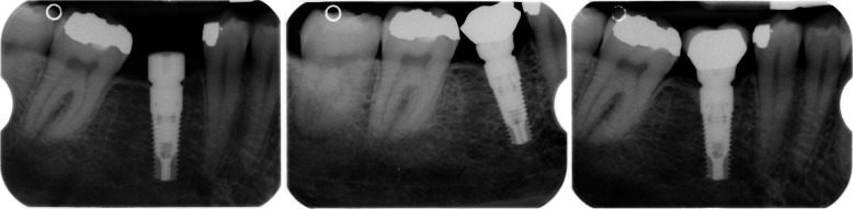
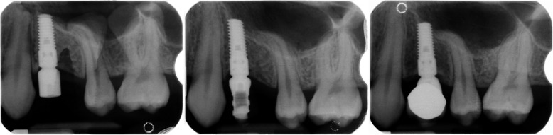
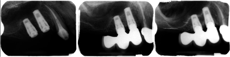
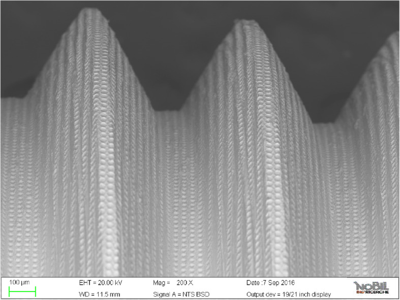
Materials and methods: A total of 502 dental implants were inserted in four dental practices (Udine, Arezzo, Frascati, Roma) between 2008 and 2013. All inserted implants had laser-modified surface characterized by a series of 20-μm-diameter holes (7-10 μm deep) every 10 μm (Synthegra®, Geass srl, Italy). The minimum follow-up period was set at 1 year after the final restoration. Radiographs were taken after implant insertion (T0), at time of loading (T1), and during the follow-up period (last recall, T2). Marginal bone loss and peri-implant disease incidence were recorded.
Results: A total of 502 implants with a maximum follow-up period of 6 years were monitored. The mean differential between T0 and T2 was 0.05 ± 1.08 mm at the mesial aspect and 0.08 ± 1.11 mm at the distal with a mean follow-up period of 35.76 ± 18.05 months. After being in function for 1 to 6 years, implants reported varying behavior: 8.8% of sites did not show any radiographic changes and 38.5% of sites showed bone resorption. The bone appeared to have been growing coronally in 50.7% of the sites measured.
Conclusion: Implants showed a maintenance of marginal bone levels over time, and in many cases, it seems that laser-modified implant surface could promote a bone growth. The low peri-implant disease incidence recorded could be attributed to the laser titanium surface features that seem to prevent bacterial colonization. Future randomized and controlled studies are needed to confirm the results of the present multi-centrical retrospective analysis.
Keywords: Dental implants; Implant survival rate; Laser surface; Osseointegration.
Conflict of interest statement
Claudio Mongardini, Blerina Zeza, Pierluigi Pelagalli, Rodolfo Blasone, Mario Scilla, and Marco Berardini declare that there are no conflicts of interest between them and products listed in the manuscript.
Figures

Similar articles
-
Effect of a laser-ablated micron-scale modification of dental implant collar surface on changes in the vertical and fractal dimensions of peri-implant trabecular bone.Clin Ter. 2020 Sep-Oct;171(5):e385-e392. doi: 10.7417/CT.2020.2245. Clin Ter. 2020. PMID: 32901779
-
Comparative Results of Single Implants With and Without Laser-Microgrooved Collar Placed and Loaded with Different Protocols: A Long-Term (7 to 10 years) Retrospective Multicenter Study.Int J Oral Maxillofac Implants. 2020 Jul/Aug;35(4):841-849. doi: 10.11607/jomi.7605. Int J Oral Maxillofac Implants. 2020. PMID: 32724939
-
One-piece zirconia oral implants: one-year results from a prospective case series. 2. Three-unit fixed dental prosthesis (FDP) reconstruction.J Clin Periodontol. 2013 May;40(5):553-62. doi: 10.1111/jcpe.12093. Epub 2013 Mar 18. J Clin Periodontol. 2013. PMID: 23506654
-
Influence of surgical and prosthetic techniques on marginal bone loss around titanium implants. Part I: immediate loading in fresh extraction sockets.J Prosthodont. 2014 Oct;23(7):521-7. doi: 10.1111/jopr.12153. Epub 2014 Apr 18. J Prosthodont. 2014. PMID: 24750449
-
Can Peri-Implant Marginal Bone Loss Progression and a-MMP-8 Be Considered Indicators of the Subsequent Onset of Peri-Implantitis? A 5-Year Study.Diagnostics (Basel). 2022 Oct 26;12(11):2599. doi: 10.3390/diagnostics12112599. Diagnostics (Basel). 2022. PMID: 36359443 Free PMC article.
Cited by
-
Titanium Surfaces with a Laser-Produced Microchannel Structure Enhance Pre-Osteoblast Proliferation, Maturation, and Extracellular Mineralization In Vitro.Int J Mol Sci. 2024 Mar 16;25(6):3388. doi: 10.3390/ijms25063388. Int J Mol Sci. 2024. PMID: 38542358 Free PMC article.
-
Effectiveness of Photobiomodulation With Low-Level Laser Therapy on the Implant Stability Quotient at Different Time Intervals: A Randomized Clinical Trial.Cureus. 2024 Jan 12;16(1):e52160. doi: 10.7759/cureus.52160. eCollection 2024 Jan. Cureus. 2024. PMID: 38344646 Free PMC article.
References
-
- Adler L, Buhlin K, Jansson L. Survival and complications: A 9- to 15-year retrospective follow-up of dental implant therapy. J Oral Rehabil. 2020;47:67–77. - PubMed
-
- Sinjari B, D'Addazio G, Traini T, Varvara G, Scarano A, Murmura G, Caputi S. A 10-year retrospective comparative human study on screw-retained versus cemented dental implant abutments. J Biol Regul Homeost Agents. 2019;33:787–797. - PubMed
-
- Salman A, Thacker S, Rubin S, Dhingra A, Ioannidou E, Schincaglia GP. Immediate versus delayed loading of mandibular implant-retained overdentures: a 60-month follow-up of a randomized clinical trial. J Clin Periodontol. 2019;46:863–871. - PubMed
-
- Gallardo YNR, da Silva-Olivio IR, Gonzaga L, Sesma N, Martin W. A systematic review of clinical outcomes on patients rehabilitated with complete-arch fixed implant-supported prostheses according to the time of loading. J Prosthodont. 2019 Aug;21. - PubMed
-
- Lemos CAA, Verri FR, Gomes JML, de Souza Batista VE, Cruz RS, Oliveira HFFE, Pellizzer EP. Ceramic versus metal-ceramic implant-supported prostheses: a systematic review and meta-analysis. J Prosthet Dent. 2019;121:879–886.e4. - PubMed
LinkOut - more resources
Full Text Sources
Miscellaneous
