Review
. 2020 Oct;13(5):561-571.
doi: 10.1007/s12178-020-09658-4.

Patch Augmentation in Rotator Cuff Repair

Affiliations
Review

Patch Augmentation in Rotator Cuff Repair

Peter N Chalmers et al. Curr Rev Musculoskelet Med. 2020 Oct.

Abstract

Purpose of the review: Rotator cuff repair has excellent outcomes for many patients but continues to be suboptimal for large, retracted tears, and revision procedures. In these situations, patch augmentation may be considered in order to improve healing. The purpose of this article is to review the history, graft options, indications, surgical technique, outcomes, and complications associated with arthroscopic patch augmentation for rotator cuff repair.

Recent findings: Patch augmentation has been shown in several studies to improve healing rates. After multiple investigations into different materials available for patch augmentation, acellular dermal allograft seems to be the graft with the best scientific support. While multiple techniques have been presented, few studies have compared their performance. While the arthroscopic technique for patch augmentation can be challenging, we present a systematic approach to this procedure with the potential to reliably and predictably perform patch augmentation. This technique is a valuable tool for surgeons that treat rotator cuff pathology.

Keywords: Biologic augmentation; Dermal allograft; Patch augmentation; Rotator cuff repair; Surgical technique.


Conflict of interest statement

Peter Chalmers is a paid consultant for Depuy-Mitek and Arthrex. Robert Tashjian is a paid consultant for Zimmer Biomet, Wright Medical and Depuy-Mitek; has stock in Conextions, INTRAFUSE, Genesis and KATOR; receives intellectual property royalties from Wright Medical, Shoulder Innovations, and Zimmer Biomet; receives publishing royalties from Springer, the Journal of Bone and Joint Surgery and the Journal of the American Association of Orthopaedic Surgeons, and serves on the editorial board for the Journal of the American Association of Orthopaedic Surgeons.

Figures

Fig. 1
Fig. 1
View of massive rotator cuff tear from posterolateral portal
Fig. 2
Fig. 2
Three triple loaded anchors placed at the medial aspect of the anatomic neck of the greater tuberosity
Fig. 3
Fig. 3
View of stitches passed from anchors (2 simple stitches and 1 mattress stitch from the anterior and posterior anchors and 3 simple stitches from the middle anchor)
Fig. 4
Fig. 4
View from the lateral portal after the simple stitches from the anchors are tied leaving the mattress stitches from the anterior and posterior anchors untied. The trailing stitch in the image is one limb from the independent stitches passed through the rotator cuff medial to the anchor stitches
Fig. 5
Fig. 5
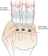
Diagram showing the placement of stitches from the anchors in the rotator cuff along with medial independent stitches in the rotator cuff.
Fig. 6
Fig. 6
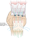
Diagram showing the simple stitches from the anchors tied and the remaining limbs from the mattress anchor stitches as well as one limb of the medial independent stitches in the rotator cuff passed through the graft. Finally, three independent stitches are also passed through the lateral margin of the graft.
Fig. 7
Fig. 7
Lateral view after the graft has been passed in the subacromial space, the medial graft stitches have been tied and cut, the anchor mattress stitches have been tied and their tails have been brought over to 2 lateral row knotless anchors along with three independent stitches passed through the lateral margin of the graft
Fig. 8
Fig. 8
Diagram of final construct with tied medial graft stitches, tied horizontal mattress anchor stitches through the tendon and graft and tails of the tied stitches brought over to two knotless lateral row anchors along with the stitches from the lateral margin of the graft

References

    1. Abdel MP, Salib CG, Mara KC, Pagnano MW, Perry KI, Hanssen AD. Extensor mechanism reconstruction with use of Marlex mesh. J Bone Jt Surg. 2018;100(15):1309–1318. doi: 10.2106/jbjs.17.01165. - DOI - PubMed
    1. Audenaert E, Nuffel VJ, Schepens A, Verhelst M, Verdonk R. Reconstruction of massive rotator cuff lesions with a synthetic interposition graft: a prospective study of 41 patients. Knee Surg Sports Traumatology Arthrosc. 2006;14(4):360–364. doi: 10.1007/s00167-005-0689-7. - DOI - PubMed
    1. Badhe SP, Lawrence TM, Smith FD, Lunn PG. An assessment of porcine dermal xenograft as an augmentation graft in the treatment of extensive rotator cuff tears. J Shoulder Elb Surg. 2008;17(1):S35–S39. doi: 10.1016/j.jse.2007.08.005. - DOI - PubMed
    1. Barber AF, Burns JP, Deutsch A, Labbé MR, Litchfield RB. A prospective, randomized evaluation of acellular human dermal matrix augmentation for arthroscopic rotator cuff repair. Arthrosc J Arthrosc Relat Surg. 2012;28(1):8–15. doi: 10.1016/j.arthro.2011.06.038. - DOI - PubMed
    1. Cai Y-Z, Zhang C, Jin R-L, Shen T, Gu P-C, Lin X-J, et al. Arthroscopic rotator cuff repair with graft augmentation of 3-dimensional biological collagen for moderate to large tears: a randomized controlled study. Am J Sports Medicine. 2018;46(6):1424–1431. doi: 10.1177/0363546518756978. - DOI - PubMed