Review
. 2020 Sep 4;64(2):205-221.
doi: 10.1042/EBC20190074.

CENP-A nucleosome-a chromatin-embedded pedestal for the centromere: lessons learned from structural biology

Affiliations
Review

CENP-A nucleosome-a chromatin-embedded pedestal for the centromere: lessons learned from structural biology

Ahmad Ali-Ahmad et al. Essays Biochem. .

Abstract

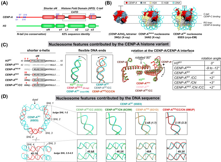
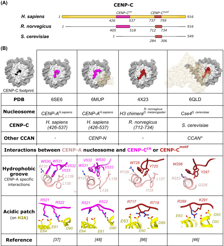
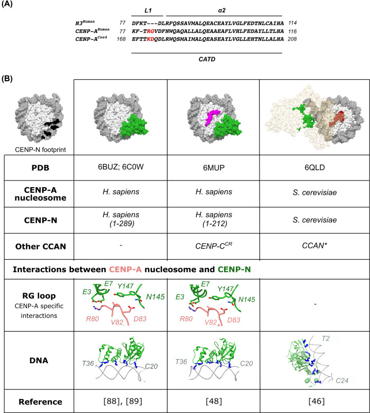
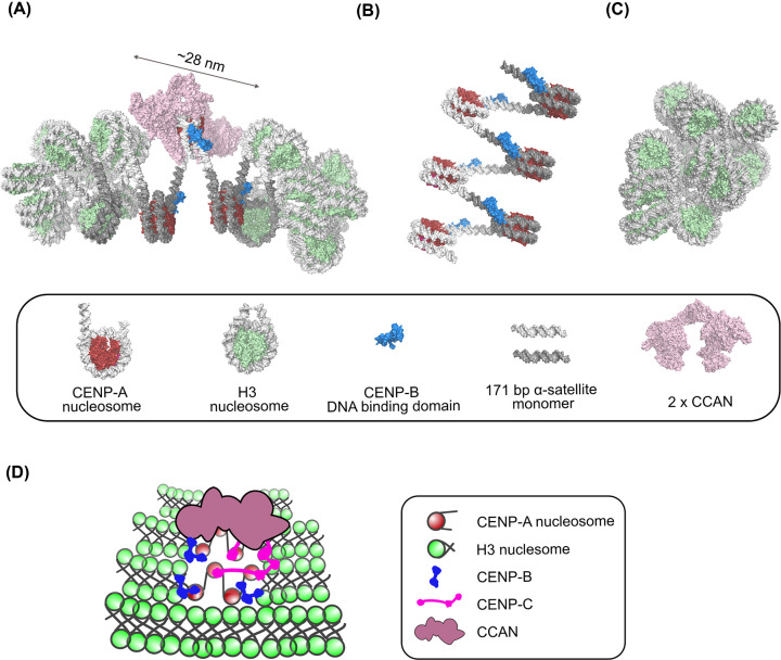
The centromere is a chromosome locus that directs equal segregation of chromosomes during cell division. A nucleosome containing the histone H3 variant CENP-A epigenetically defines the centromere. Here, we summarize findings from recent structural biology studies, including several CryoEM structures, that contributed to elucidate specific features of the CENP-A nucleosome and molecular determinants of its interactions with CENP-C and CENP-N, the only two centromere proteins that directly bind to it. Based on those findings, we propose a role of the CENP-A nucleosome in the organization of centromeric chromatin beyond binding centromeric proteins.

Keywords: CENP-A; centromere; chromatin.


Conflict of interest statement

The authors declare that there are no competing interests associated with the manuscript.

Figures

Figure 1
Figure 1. Structural features of CENP-A nucleosome
(A) Schematic representation of H3 and CENP-A. The post-translational sites on CENP-A are colored in blue and the L1 loop and C-terminal tail, which specifically bind CENP-N and CENP-C respectively, are colored in black. (B) Surface representation of CENP-A-containing (sub)nucleosomal complexes, solved by X-ray crystallography and cryo-EM. CENP-A is in red, H4 is in blue, H2A is in yellow, H2B is in gray and the DNA is in cyan. CENP-A-specific features recognized by CENP-C (C-terminal tail) and CENP-N (RG-loop) are shown in black and boxed on each of the structures. The arrow is showing the position of the four-helix bundle between two CENP-A molecules (rotated 90° in (C), right). PDB ID and the experimental method used are indicated below each structure. (C) Nucleosome features contributed by CENP-A. Left: Multiple sequence alignment, illustrating a shorter α-N helix (boxed sequence) of CENP-A, observed in various structures. Residues interacting with DNA are labeled with one star if only one side of the nucleosome is involved in interactions, and with two stars if residues on both sides are interacting. Middle: overlay of terminal DNA in H3 nucleosome (3LZ0) and CENP-A nucleosome structures obtained by X-ray crystallography (3AN2) or cryo-EM (6SE0). The flexibility of terminal DNA is modulated by the binding of CCAN components (6SE0, 6MUP, 6SEE). Right: Rotation at the four-helix bundle formed between two CENP-A or two H3 molecules (indicated with the arrow in (B) and rotated 90° away from the viewer). The angle between α2 helices (amino acids 86–113) in the canonical nucleosome is taken as a reference. The rotation is most pronounced in the (CENP-A/H4)2 tetramer (3NQJ) and is greatly reduced in the CENP-A nucleosome (6SE0), while it virtually disappears when CENP-C and CENP-N bind the CENP-A nucleosome (6MUP). A table summarizing the measured rotation angles is shown on the right. (D) Nucleosome features contributed by the DNA sequence. Left: the DNA of the CENP-A601 nucleosome (6SE0) is shown in cyan and regions where important DNA path deviation is observed, in comparison with nucleosome wrapped in the α-satellite DNA, are highlighted in red. The distance between DNA gyres is measured between I-36 phosphate and I-41 phosphate. In order to highlight differences in the DNA path around the nucleosome in budge SHL 1–2 and budge SHL 3.5–4.5, on the right, are shown as zoomed views of different overlays. The first four panels show that there are no significant differences in the DNA path between the CENP-A601 nucleosome (cyan; 6SE0) and the CENP-A601 nucleosome, in complex with CENP-C (light green, 6SE6), or with CENP-N (dark green, 6C0W). The overlay of the CENP-A601 nucleosome (cyan; 6SE0) with the CENP-APAS nucleosome (orange; 6O1D) reveals a 2.4-Å deviation in the SHL 1–2 (1.7 Å at the opposite side of the nucleosome) and 2 Å widening of DNA gyres. When the CENP-A601 nucleosome (cyan; 6SE0) is overlaid with the CENP-APAS nucleosome with two CENP-C and two CENP-N molecules (orange; 6O1D), the difference at the SHL 1–2 is 2.4 Å (2.3 Å on the other side of the nucleosome) and widening between DNA gyres increases to 49.3 Å. All nucleosomes were overlaid by aligning the histone cores. Abbreviation: CENP-A601, CENP-A nucleosome on super-positioning 601 sequence. CENP-APAS, CENP-A nucleosome on palindromic α-satellite DNA sequence.
Figure 2
Figure 2. CENP-A nucleosome in complex with CENP-C
(A) A schematic representation of CENP-C from Homo sapiens, Rattus norvegicus and Saccharomyces cerevisiae, indicating nucleosome-binding regions, CENP-CCR and CENP-Cmotif, used in structural studies. (B) A table summarizing information revealed from published 3D-structures of nucleosomes in complex with CENP-C. A surface representation of the CENP-A nucleosome with mapped CENP-C footprint in black is shown on the far left. In the surface representation, histones are shown in light gray, DNA is colored dark gray, CENP-CCR is magenta, the CENP-Cmotif is dark red and other CCAN components shown in transparent beige. The CENP-C residues involved in the interaction with the acidic patch and the CENP-A C-terminal tail are shown as sticks.
Figure 3
Figure 3. CENP-A nucleosome in complex with CENP-N
(A) A multiple sequence alignment of the CATD region of CENP-A (H. sapiens) and CENP-ACse4 (S. cerevisiae) with the corresponding region of H3 (H. sapiens). Insertions in the L1 loop are colored red. (B) A table summarizing information revealed from published 3D-structures of nucleosomes in complex with CENP-N. A surface representation of the CENP-A nucleosome with the mapped CENP-N footprint in black is shown on the far left. In the surface representation, histones are light gray, DNA is dark gray, CENP-N is green, CENP-CCR is magenta, the CENP-Cmotif is dark red, and the other CCAN components are shown in transparent beige. The CENP-N residues interacting with the CENP-A RG loop are shown as sticks. Positively charged residues (arginines and lysines), interacting with the nucleosomal DNA, are shown in blue.
Figure 4
Figure 4. A model of the human CCAN complex in centromeric chromatin
(A) 3D-model of the CCAN/centromeric chromatin complex. The nucleosome modeled harboring CCAN is built using structural information from the H3-CENP-A-H3 tri-nucleosome structure (6L49). CENP-C and CENP-N are modeled on the central nucleosome as in the human CENP-A/CENP-C/CENP-N structure (6MUP). The yeast CCAN complex structure (pink; extracted from 6QLD) deprived of the CENP-ACse4 nucleosome, is docked on the central CENP-A nucleosome by aligning one copy of the CCAN complex on each CENP-N molecule. The DNA binding domain of CENP-B (DBD) (blue; 1HLV) is docked on a degenerated CENP-B box in the α-satellite sequence, as positioned in the CENP-A/CENP-C/CENP-N structure (6MUP). The DNA path was slightly adjusted to accommodate a kink observed in the CENP-B/DNA structure. The 171-bp monomers in the α-satellite sequence are colored light and dark gray (the nucleosome position is based on the CENP-A/CENP-C/CENP-N structure (6MUP)). It is obvious that nucleosomes adjacent to the CCAN loaded CENP-A cannot adopt a H3-H3-H3 packing (6L4A), so two CENP-A nucleosomes with open DNA ends are modelled on each side of the CENP-A, loaded with CCAN to avoid steric clashes. The H3 containing histone core is colored light green, and the CENP-A histone core is colored red. The size of the 2xCCAN:1CENP-A nucleosome complex is indicated. (B) A 3D-model of 6x CENP-A nucleosomes/CENP-B (DBD), based on the open DNA conformation of the CENP-A nucleosome in the H3-CENP-A-H3 tri-nucleosome structure (6L49). Notice the bigger spacing between nucleosomes. (C) A 3D-model of 18x H3 nucleosomes array built based on the H3-H3-H3 tri-nucleosome structure (6L4A). Notice the tight nucleosome packing imposed with DNA crossing over the nucleosome dyad. (D) A schematic diagram of human centromeric chromatin. H3 loaded chromatin is more compact than the CENP-A loaded chromatin. CENP-B binding is enforcing the CENP-A-like (looser) chromatin by introducing a kink close to the nucleosome entry/exit site. Because CENP-B has a dimerization domain, it is also bridging two nucleosomes. CENP-C is structuring the chromatin by binding four CENP-A nucleosomes (dimer with two nucleosome-binding regions) and thus increasing their local concentration. It also interacts with the CCAN.

References

    1. McKinley K.L. and Cheeseman I.M. (2016) The molecular basis for centromere identity and function. Nat. Rev. Mol. Cell Biol. 17, 16–29 10.1038/nrm.2015.5 - DOI - PMC - PubMed
    1. Potapova T. and Gorbsky G.J. (2017) The consequences of chromosome segregation errors in mitosis and meiosis. Biology 6, 12 10.3390/biology6010012 - DOI - PMC - PubMed
    1. Henikoff S., Ahmad K. and Malik H.S. (2001) The centromere paradox: stable inheritance with rapidly evolving DNA. Science 293, 1098–1102 10.1126/science.1062939 - DOI - PubMed
    1. Melters D.P., Bradnam K.R., Young H.A., Telis N., May M.R., Ruby J.G. et al. (2013) Comparative analysis of tandem repeats from hundreds of species reveals unique insights into centromere evolution. Genome Biol. 14, R10 10.1186/gb-2013-14-1-r10 - DOI - PMC - PubMed
    1. Voullaire L.E., Slater H.R., Petrovic V. and Choo K.H. (1993) A functional marker centromere with no detectable alpha-satellite, satellite III, or CENP-B protein: activation of a latent centromere? Am. J. Hum. Genet. 52, 1153–1163 - PMC - PubMed

Publication types