

Myomectomy scar pregnancy: a case report and review of the literature
- PMID: 32720824
- PMCID: PMC7388108
- DOI: 10.1177/0300060520924542
Myomectomy scar pregnancy: a case report and review of the literature
Abstract
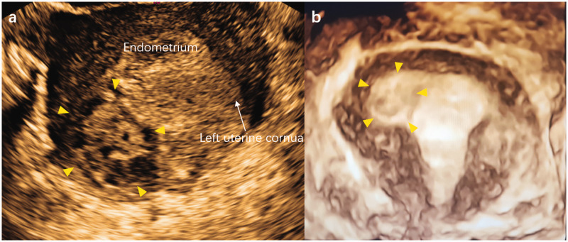
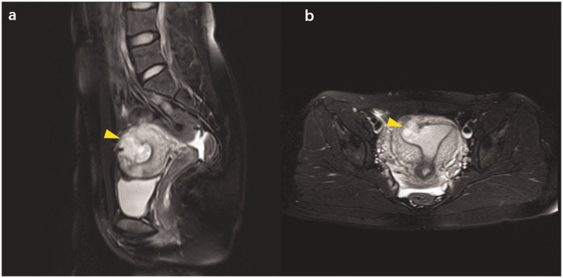
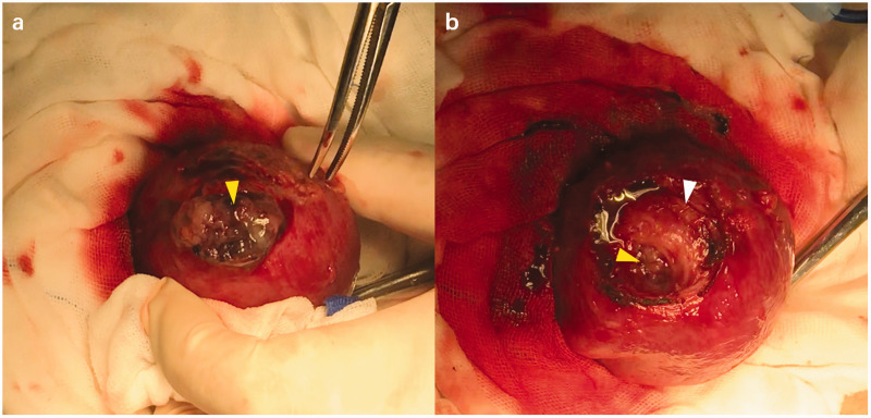
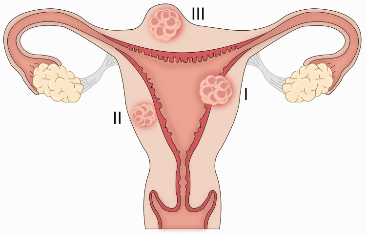
Myomectomy scar pregnancy (MSP) is a rare disease, which is defined as a gestational sac located within a previous myomectomy scar. MSP is an uncommon late complication of uterine fibroids after myomectomy. We report a case where the implantation site matched the site of the previous myomectomy, and review the existing literature. A 28-year-old pregnant woman presented with vaginal bleeding. She was diagnosed with MSP by ultrasound and magnetic resonance imaging, and then underwent laparotomic enucleation. The patient's postoperative course was uneventful. Taking into account the findings in our case and the seven other reported cases of MSP, we propose that MSP can be divided into three types and that surgical enucleation of the pregnancy mass is an effective treatment.
Keywords: Myomectomy scar pregnancy; beta-human chorionic gonadotrophin; ectopic pregnancy; gestational sac; surgical enucleation; uterine fibroids; vaginal bleeding.
Figures
Similar articles
-
Ultrasonographic Features of Uterine Scar after Laparoscopic and Laparoscopy-Assisted Minilaparotomy Myomectomy: A Comparative Study.J Minim Invasive Gynecol. 2020 Jan;27(1):148-154. doi: 10.1016/j.jmig.2019.03.026. Epub 2019 Jul 10. J Minim Invasive Gynecol. 2020. PMID: 31301467 Clinical Trial.
-
Conservative management of uterine artery pseudoaneurysm after laparoscopic-assisted myomectomy and subsequent pregnancy outcome: case series and review of the literature.Eur J Obstet Gynecol Reprod Biol. 2014 Nov;182:146-53. doi: 10.1016/j.ejogrb.2014.09.020. Epub 2014 Sep 19. Eur J Obstet Gynecol Reprod Biol. 2014. PMID: 25277771 Review.
-
Successful surgical treatment of postmyomectomy uterine diverticulum: a case report.BMC Womens Health. 2023 Aug 3;23(1):406. doi: 10.1186/s12905-023-02539-1. BMC Womens Health. 2023. PMID: 37537601 Free PMC article.
-
Ectopic pregnancies in caesarean section scars: 5 years experience.Clin Imaging. 2020 Oct;66:26-34. doi: 10.1016/j.clinimag.2020.04.037. Epub 2020 May 3. Clin Imaging. 2020. PMID: 32442857 Review.
-
The management of uterine leiomyomas.J Obstet Gynaecol Can. 2015 Feb;37(2):157-178. doi: 10.1016/S1701-2163(15)30338-8. J Obstet Gynaecol Can. 2015. PMID: 25767949
Cited by
-
Long-term obstetric, perinatal, and surgical complications in singleton pregnancies following previous cesarean myomectomy: a retrospective multicentric study.Front Surg. 2024 Aug 1;11:1430439. doi: 10.3389/fsurg.2024.1430439. eCollection 2024. Front Surg. 2024. PMID: 39149134 Free PMC article.
-
Case report of a giant cervical leiomyoma.Front Med (Lausanne). 2025 Apr 2;12:1521984. doi: 10.3389/fmed.2025.1521984. eCollection 2025. Front Med (Lausanne). 2025. PMID: 40241893 Free PMC article.
-
Undiagnosed placenta percreta complicated by bowel injury and puerperal infection: a case report.BMC Pregnancy Childbirth. 2025 Jul 19;25(1):771. doi: 10.1186/s12884-025-07897-2. BMC Pregnancy Childbirth. 2025. PMID: 40684104 Free PMC article.
-
Management of Myomectomy Scar Pregnancy: A Scoping Review.Medicina (Kaunas). 2025 Apr 29;61(5):817. doi: 10.3390/medicina61050817. Medicina (Kaunas). 2025. PMID: 40428776 Free PMC article.
References
-
- Stewart EA, Laughlin-Tommaso SK, Catherino WH, et al. Uterine fibroids. Nat Rev Dis Primers 2016; 2: 16043. - PubMed
-
- Paul PG, Mannur S, Shintre H, et al. Myomectomy scar pregnancy: a rare complication of myomectomy. J Gynecol Surg 2018; 34: 53–57.
-
- Park WI, Jeon YM, Lee JY, et al. Subserosal pregnancy in a previous myomectomy site: a variant of intramural pregnancy. J Minim Invasive Gynecol 2006; 13: 242–244. - PubMed
-
- Wong KS, Tan J, Ang C, et al. Myomectomy scar ectopic pregnancy. Aust N Z J Obstet Gynaecol 2010; 50: 93–94. - PubMed
-
- Bannon K, Fernandez C, Rojas D, et al. Diagnosis and management of intramural ectopic pregnancy. J Minim Invasive Gynecol 2013; 20: 697–700. - PubMed
Publication types
MeSH terms
LinkOut - more resources
Full Text Sources
Medical
Research Materials
