. 2019 Feb;2(1):e1133.
doi: 10.1002/cnr2.1133. Epub 2018 Sep 24.

Pluronic micelles encapsulated curcumin manifests apoptotic cell death and inhibits pro-inflammatory cytokines in human breast adenocarcinoma cells

Affiliations

Pluronic micelles encapsulated curcumin manifests apoptotic cell death and inhibits pro-inflammatory cytokines in human breast adenocarcinoma cells

Foram U Vaidya et al. Cancer Rep (Hoboken). 2019 Feb.

Abstract

Background: Curcumin is a natural derivative, which exhibits broad spectrum biological activities including anti-oxidant, anti-inflammatory, and anti-cancer. Since ancient times, it has been used for the treatment of various diseases. Many reports highlighted its potential as a chemopreventive and chemotherapeutic agent. Despite its imperative properties, the pharmacological application had been limited due to low solubility in the aqueous medium, limited tissue absorption, and rapid degradation at physiological pH.

Aims: Cytotoxicity of drugs and their undesirable side effects are major obstacles in the regimens of cancer therapy. Therefore, natural plant derivatives-based anti-cancer drug delivery systems are getting more attention as they are less toxic, safer, and effective. In the present study, Pluronic block copolymer encapsulated curcumin was developed as an improved curcumin delivery system with the aim to improve its efficacy and biological response against cancer cells.

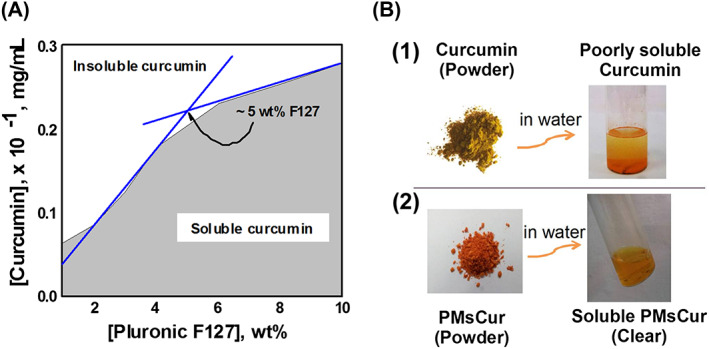
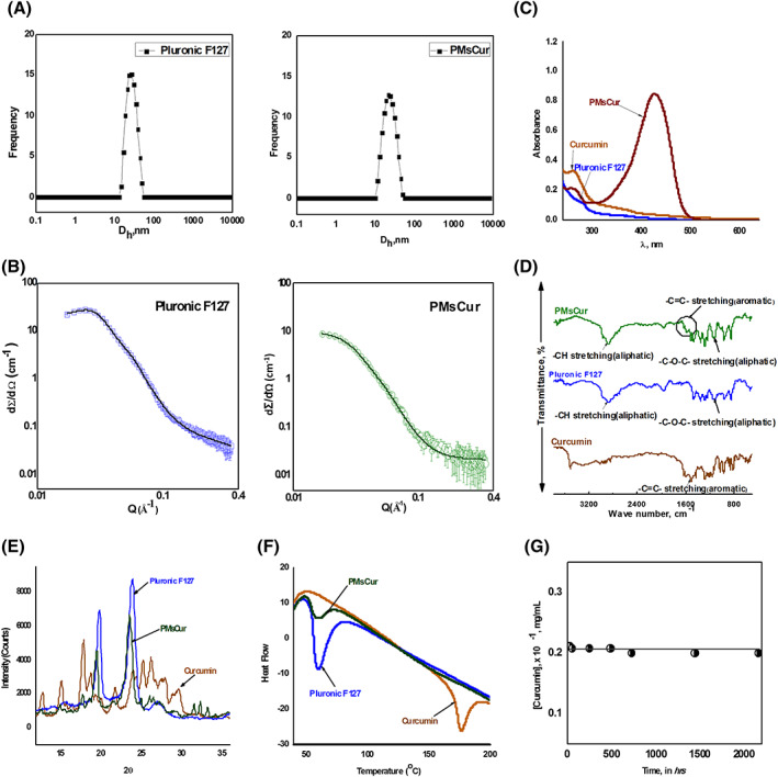
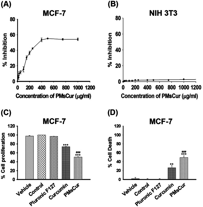
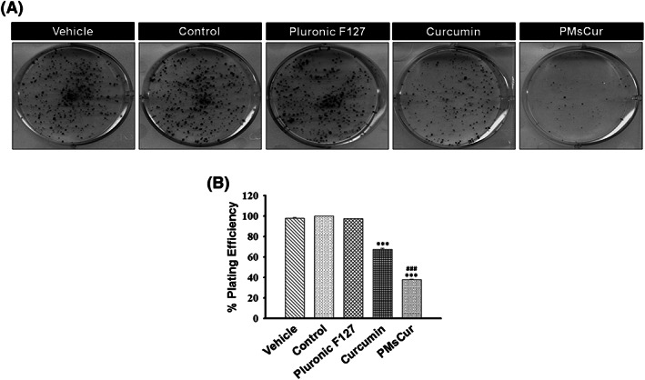
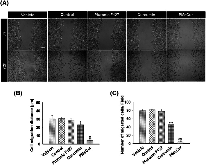
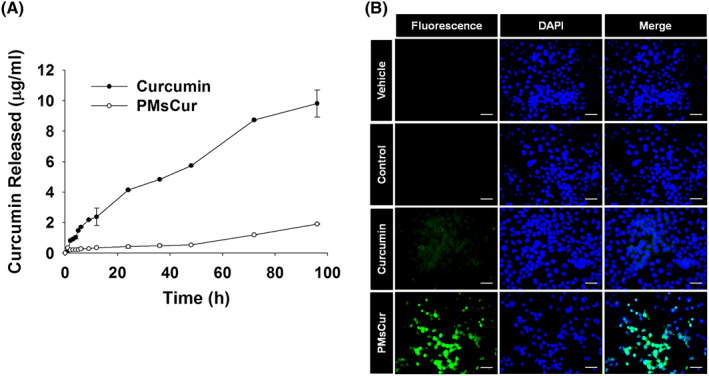
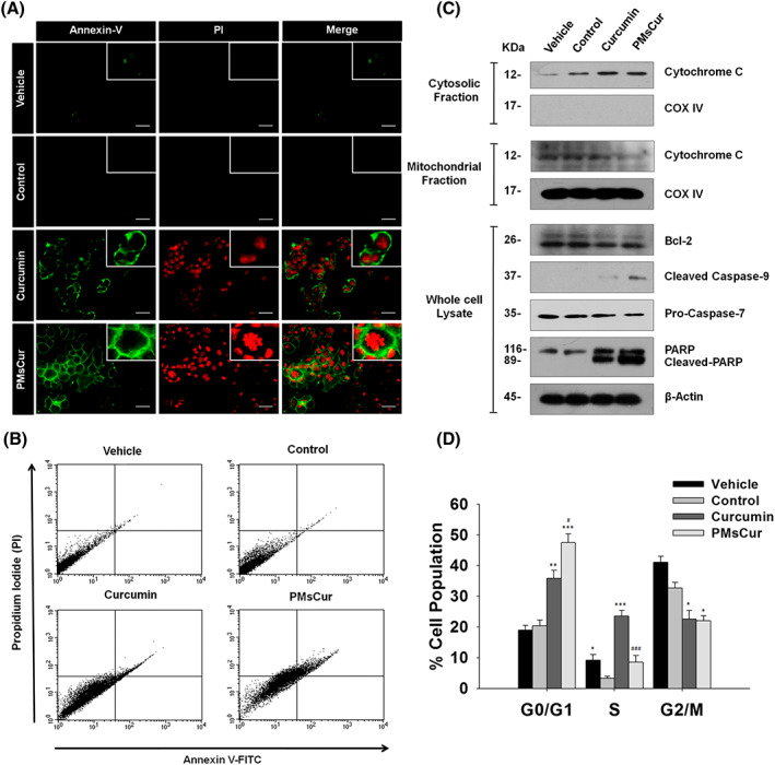
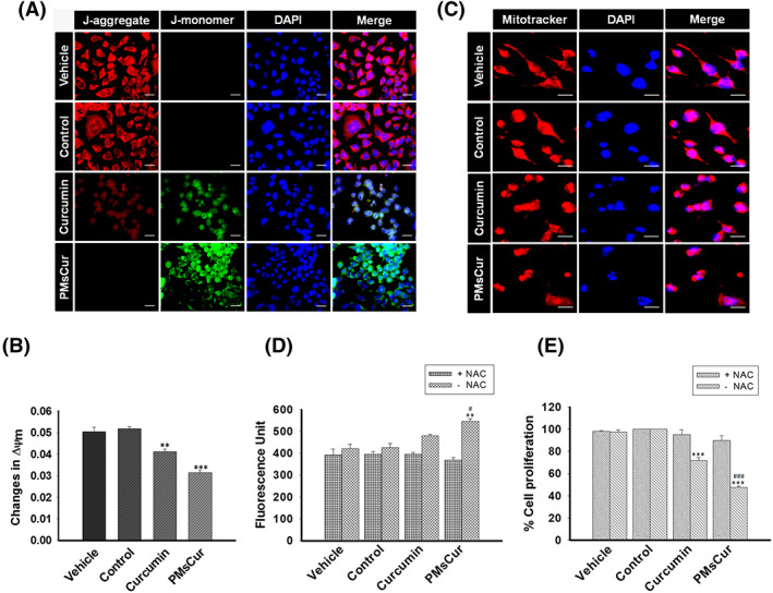
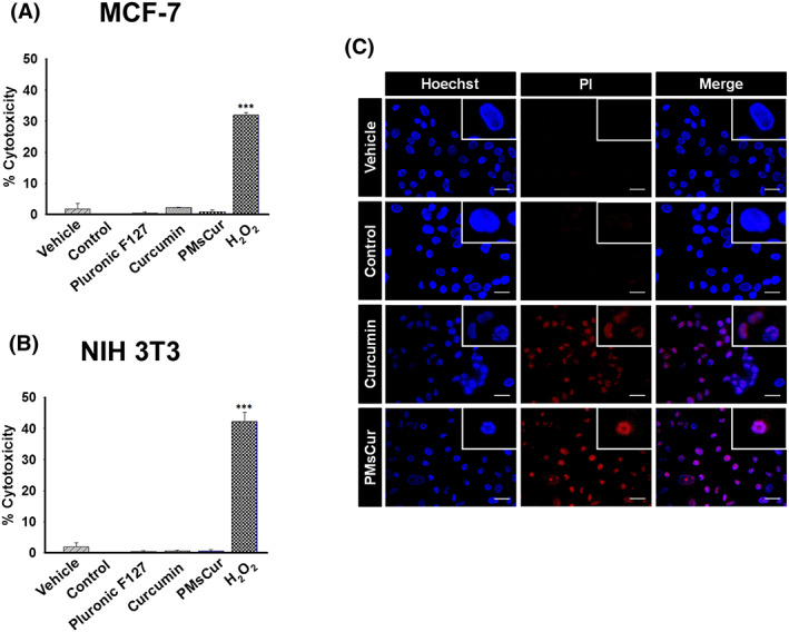
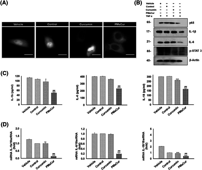
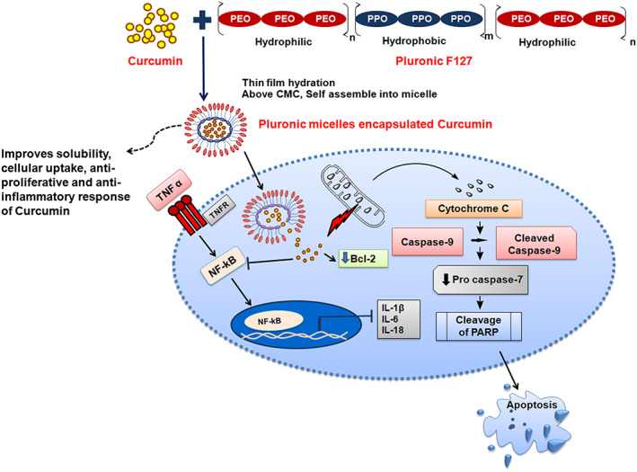
Methods and results: Pluronic micelles encapsulated curcumin was synthesized, and its characterization was done by particle size analysis, Fourier transform infrared spectroscopy, small-angle neutron scattering analysis, PXRD, and differential scanning calorimetry. Further, its biological activities were corroborated in cancer cells. Results indicate that Pluronic micelles encapsulated curcumin exemplify solubility and stability of curcumin in the aqueous medium. Biophysical characterization indicated that Pluronic F127 forms nanoparticle, and its micellar core radius was increased after incorporation of curcumin. Furthermore, biological studies show that Pluronic micelles encapsulated curcumin inhibits cell proliferation, improves cellular uptake of curcumin, arrests the cell cycle in G0/G1 phase, and inhibits the activation of NF-kB and release of pro-inflammatory cytokines to manifest apoptotic cell death rather than necrotic. This formulation was non-toxic to normal cells.

Conclusion: This study suggests that Pluronic micelles encapsulated curcumin is stable that can effectively inhibit cell proliferation and release of pro-inflammatory cytokines in cancer cells as compared with the free curcumin. This approach could be applied to improve the therapeutic index of anti-cancer agents.

Keywords: Pluronic F127; apoptosis; aqueous soluble Pluronic micelles encapsulated curcumin; cancer; curcumin.


Conflict of interest statement

None declared.

Figures

Figure 1
Figure 1
Solubility of curcumin. A, Solubility of curcumin in Pluronic F127 solution at 37°C. B, (1) Insoluble slurry of free curcumin added in water; (2) Synthesized PMsCur dispersed (soluble curcumin) in water
Figure 2
Figure 2
Characterization of PMsCur. A, Intensity vs hydrodynamic size of 5.0 wt% aqueous solution of Pluronic F127 and 1.0 wt% PMsCur at 37°C. B, SANS intensities of Pluronic F127 and PMsCur in D2O at 37°C. The concentration of F127 in both samples was 5 wt%. C, UV‐visible spectra of curcumin, Pluronic F127, and PMsCur in water at 37°C. D, FTIR spectra of curcumin, Pluronic F127, and PMsCur. E, PXRD patterns of curcumin, Pluronic F127, and PMsCur. F, DSC analysis of curcumin, Pluronic F127, and PMsCur. G, Solubility vs time plot of 1 wt% PMsCur
Figure 3
Figure 3
Anti‐proliferative effect of Pluronic F127, curcumin, and PMsCur. A,B, To determine IC50 value, A, MCF‐7 and B, NIH 3T3 cells were treated with wide range of concentrations (10, 20, 25, 50, 100, 150, 200, 250, 400, 500, 750, and 1000 μg/mL) of PMsCur for 24 h. Inhibition of cell proliferation was evaluated by MTT assay. C,D, The antiproliferative efficiency of PMsCur on MCF‐7 cells was evaluated by MTT and trypan blue assay. Cells were treated with Pluronic F127 (364 μg/mL), curcumin (5.66 μg/mL), and PMsCur (364 μg/mL) for 24 h. Vehicle control contained 0.02% DMSO, and control represents untreated cells. C, Evaluation of cell proliferation by MTT assay. The bar graphs represent percentage of cell proliferation. D, Evaluation of cell death by trypan blue assay. The bar graphs represent the percentage of cell death. Error bars represent ± SEM of three independent experiments. Significance indicated as **P ≤ 0.01, ***P ≤ 0.001 between untreated cells and treated cells and ### P ≤ 0.001 between free curcumin and PMsCur‐treated cells by performing one‐way ANOVA followed by Student‐Newman‐Keuls multiple comparisons test. Auto‐fluorescence of curcumin was normalized to avoid interference
Figure 4
Figure 4
Effect of Pluronic F127, curcumin, and PMsCur on the clonogenic ability of MCF‐7 cells. Cells were treated with Pluronic F127 (364 μg/mL), curcumin (5.66 μg/mL), and PMsCur (364 μg/mL) for 24 h. Vehicle control contained 0.02% DMSO, and control represents untreated cells. A, Colony formation assay was performed by crystal violet staining. B, The bar graph represents % of plating efficiency of survived cells. Error bars represent ±SEM of three independent experiments. Significance indicated as ***P ≤ 0.001 between untreated cells and treated cells and ### P ≤ 0.001 between free curcumin and PMsCur‐treated cells by performing one‐way ANOVA followed by Student‐Newman‐Keuls multiple comparisons test
Figure 5
Figure 5
Effect of Pluronic F127, curcumin, and PMsCur on migration ability of MCF‐7 cells. Cells were treated with Pluronic F127 (364 μg/mL), curcumin (5.66 μg/mL), and PMsCur (364 μg/mL) for 24 h. Vehicle control contained 0.02% DMSO, and control represents untreated cells. A, Microscopic analysis of wound healing after 0 and 72 h of treatment. Scale bar represents 50 μm. B, The bar graphs represent the distance migrated by the cells. C, The bar graph represents the number of migrated cells/field. Error bars represent ±SEM of three independent experiments. Significance indicated as**P ≤ 0.01,***P ≤ 0.001 between untreated cells and treated cells and ## P ≤ 0.01,### P ≤ 0.001 between free curcumin and PMsCur‐treated cells by performing one‐way ANOVA followed by Student‐Newman‐Keuls multiple comparisons test
Figure 6
Figure 6
In vitro release study and cellular uptake of curcumin from free curcumin and PMsCur. A, In vitro drug release study: The line graph represents the amount of curcumin released at respective time points. B, Cellular uptake study. MCF‐7 cells were treated with curcumin (5.66 μg/mL) and PMsCur (364 μg/mL) for 24 h and counterstained with DAPI (1 μg/mL). Vehicle control contained 0.02% DMSO, and control represents untreated cells. Cellular uptake of curcumin was analyzed qualitatively under fluorescence microscope. Scale bar represents 20 μm
Figure 7
Figure 7
Effect of PMsCur on apoptosis and cell cycle arrest. The MCF‐7 cells were treated with curcumin (5.66 μg/mL) and PMsCur (364 μg/mL) for 24 h. Vehicle control contained 0.02% DMSO, and control represents untreated cells. A, Images represent AnnexinV‐FITC/PI stained cells. Scale bar represents 20 μm. B, Flow cytometric analysis of AnnexinV‐FITC/PI stained cells under BD FACS caliber using BD CellQuest Pro software. C, Western blotting analysis of Bcl‐2, cytochrome c (from cytosolic and mitochondrial fractions), caspase‐9, procaspase‐7, and PARP. COX IV and β‐actin were used as a loading control. D, Cell cycle analyses by flow cytometry. Error bars represent ±SEM of three independent experiments. Significance indicated as *P ≤ 0.05,**P ≤ 0.01,***P ≤ 0.001 between untreated cells and treated cells and # P ≤ 0.05, ### P ≤ 0.001 between free curcumin and PMsCur‐treated cells by performing one‐way ANOVA followed by Student‐Newman‐Keuls multiple comparisons test
Figure 8
Figure 8
Effect of PMsCur on MMP and ROS mediated cell death. MCF‐7 cells were treated with curcumin (5.66 μg/mL) and PMsCur (364 μg/mL) for 24 h. Vehicle control contained 0.02% DMSO, and control represents untreated cells. A, Representative images of JC‐1 stained cells, counterstained with DAPI. Scale bar represents 20 μm. B, Analysis of the change in MMP (∆Ψm) by quantification of fluorescence intensity of JC‐1. The bar graph represents the ratio of red to green fluorescence. C, Change in MPT was analyzed using Mitotracker dye (100 nM), counter stained with DAPI, and cells were observed under high magnification of fluorescence microscope. More than 100 cells from three random fields were analyzed by Image J software (NIH, USA). Scale bar represents 10 μm. D, The bar graph represents the level of ROS with or without pretreatment of NAC. The dichlorofluorescein (DCF) fluorescence was recorded under multi‐mode plate reader. The graph represents fluorescence unit of the DCF dye, used to detect intracellular ROS level. E, The bar graph represents the percent inhibition of cell proliferation determined by MTT assay, with or without pretreatment of NAC. Error bars represent ±SEM of three independent experiments. Significance indicated as **P ≤ 0.01,***P ≤ 0.001 between untreated cells and treated cells and # P ≤ 0.05,### P ≤ 0.001 between free curcumin and PMsCur‐treated cells by performing one‐way ANOVA followed by Student‐Newman‐Keuls multiple comparisons test. Auto‐fluorescence of curcumin was normalized to avoid interference
Figure 9
Figure 9
Cytotoxicity analysis. A,B, The cytotoxicity of Pluronic F127 (364 μg/mL), curcumin (5.66 μg/mL), and PMsCur (364 μg/mL) was monitored using the LDH assay kit. A, MCF‐7 and B, NIH 3T3 cells were treated with Pluronic F127, curcumin, and PMsCur for 24 h. Vehicle control contained 0.02% DMSO, and control represents untreated cells. 2 mM H2O2 for 6 h was used as a positive control. After treatment, the cells were subjected for the release of LDH in a culture medium according to manufacturer's instructions. Cytotoxicity was represented in terms of % cytotoxicity, related to LDH release. Error bars represent ±SEM of three independent experiments. Significance indicated as ***P ≤ 0.001 between untreated cells and treated cells by performing one‐way ANOVA followed by Student‐Newman‐Keuls multiple comparisons test. C, For detection of apoptotic cell death, MCF‐7 cells were treated with curcumin (5.66 μg/mL) and PMsCur (364 μg/mL) for 24 h and stained with Hoechst‐PI. More than 150 cells from three random fields were analyzed. Scale bar represents 5 μm. Auto‐fluorescence of curcumin was normalized to avoid interference
Figure 10
Figure 10
PMsCur enhances the anti‐inflammatory efficacy of curcumin. A, Representative image of GFP‐p65 localization in MCF‐7 cells. MCF‐7 cells were transfected with GFP‐p65 (NF‐κB) for 24 h and then treated with curcumin (5.66 μg/mL) and PMsCur (364 μg/mL) for 24 h. Vehicle control contained 0.02% DMSO, and control represents untreated cells. Subsequently, after completion of incubation, the cells were washed and exposed to TNF‐α (10 ng/mL) for 1 h and analyzed under a fluorescence microscope. Scale bar represents 20 μm. (B‐D) The MCF‐7 cells were treated with curcumin (5.66 μg/mL) and PMsCur (364 μg/mL) for 24 h followed by exposure of TNF‐α (10 ng/mL) for 1 h. Vehicle control contained 0.02% DMSO, and control represents untreated cells. B, Western blotting of p65, IL‐1β, IL‐6, and pSTAT‐3. C, ELISA of pro‐inflammatory cytokines IL‐1β, IL‐6, and IL‐18. D, mRNA expression of pro‐inflammatory cytokines IL‐1β, IL‐6, and IL‐18 quantified by real‐time PCR
Figure 11
Figure 11
Illustration demonstrates that aqueous soluble PMsCur manifests apoptotic cell death, exerts an anti‐inflammatory effect by inhibiting NF‐κB activation and expression of pro‐inflammatory cytokines

References

    1. Nagai H, Kim YH. Cancer prevention from the perspective of global cancer burden patterns. J Thorac Dis. 2017;9(3):448‐451. - PMC - PubMed
    1. Batrakova EV, Kabanov AV. Pluronic block copolymers: evolution of drug delivery concept from inert nanocarriers to biological response modifiers. J Control Release. 2008;130(2):98‐106. - PMC - PubMed
    1. Juárez P. Plant‐derived anticancer agents: a promising treatment for bone metastasis. BoneKEy Reports. 2014;3:599. - PMC - PubMed
    1. Seca AM, Pinto DC. Plant secondary metabolites as anticancer agents: successes in clinical trials and therapeutic application. Int J Mol Sci. 2018;19(1):263. - PMC - PubMed
    1. Greenwell M, Rahman P. Medicinal plants: their use in anticancer treatment. Int J Pharm Sci Res. 2015;6(10):4103‐4112. - PMC - PubMed

Publication types