. 2020 Sep 3;107(3):499-513.
doi: 10.1016/j.ajhg.2020.06.018. Epub 2020 Jul 27.

Enhanced MAPK1 Function Causes a Neurodevelopmental Disorder within the RASopathy Clinical Spectrum

Affiliations

Enhanced MAPK1 Function Causes a Neurodevelopmental Disorder within the RASopathy Clinical Spectrum

Marialetizia Motta et al. Am J Hum Genet. .

Abstract

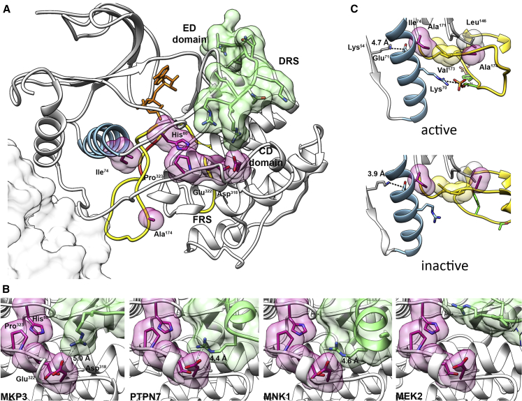
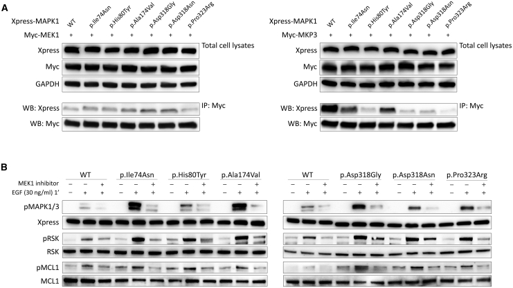
Signal transduction through the RAF-MEK-ERK pathway, the first described mitogen-associated protein kinase (MAPK) cascade, mediates multiple cellular processes and participates in early and late developmental programs. Aberrant signaling through this cascade contributes to oncogenesis and underlies the RASopathies, a family of cancer-prone disorders. Here, we report that de novo missense variants in MAPK1, encoding the mitogen-activated protein kinase 1 (i.e., extracellular signal-regulated protein kinase 2, ERK2), cause a neurodevelopmental disease within the RASopathy phenotypic spectrum, reminiscent of Noonan syndrome in some subjects. Pathogenic variants promote increased phosphorylation of the kinase, which enhances translocation to the nucleus and boosts MAPK signaling in vitro and in vivo. Two variant classes are identified, one of which directly disrupts binding to MKP3, a dual-specificity protein phosphatase negatively regulating ERK function. Importantly, signal dysregulation driven by pathogenic MAPK1 variants is stimulus reliant and retains dependence on MEK activity. Our data support a model in which the identified pathogenic variants operate with counteracting effects on MAPK1 function by differentially impacting the ability of the kinase to interact with regulators and substrates, which likely explains the minor role of these variants as driver events contributing to oncogenesis. After nearly 20 years from the discovery of the first gene implicated in Noonan syndrome, PTPN11, the last tier of the MAPK cascade joins the group of genes mutated in RASopathies.

Keywords: C. elegans; ERK2; MAPK cascade; MKP3; Noonan syndrome; RAS signaling; RASopathies; RSK; exome sequencing; intracellular signaling.


Conflict of interest statement

I.M.W. and M.M.M. are employees of GeneDx. S.Á. and M.M.-G. are employees of NimGenetics. All the other authors declare no competing interests.

Figures

Figure 1
Figure 1
Clinical Features of Individuals with De Novo MAPK1 Variants Facial features of subjects 1 to 7. Note the occurrence of hypertelorism, downslanting palpebral fissures, ptosis, low-set/posteriorly rotated ears with evident antitragus and earlobes with central depression, long philtrum with evident columns, marked upper lip vermilion and everted lower lip, and short/webbed neck. Craniofacial features resemble NS or a related RASopathy in subjects 1, 2, 3, 4, and 6. The evolving phenotype of subject 2 with age is reported in Figure S3.
Figure 2
Figure 2
Ectopic Expression of Disease-Causing MAPK1 Mutant Alleles Promotes a Variably Boosted Stimulus-Dependent Phosphorylation and Nuclear Translocation of MAPK1 (A) MAPK phosphorylation assays. Representative blots (below) and graphs reporting mean ± SD densitometry values (above) of three independent experiments are shown. Affected residues mapping at the DRS are shown in blue, while those mapping at the activation segment are colored in pink. HEK293T cells were transiently transfected with the indicated Xpress-tagged MAPK1 construct, serum starved (16 h), and stimulated with EGF (30 ng/mL) in time-course experiments or left untreated. Equal amounts of cell lysates were resolved on 10% polyacrylamide gels. Asterisks indicate statistically significant differences in the phosphorylation levels compared to cells overexpressing wild-type MAPK1 at the corresponding experimental points (p < 0.05, ∗∗p < 0.01; Student’s t test). (B) MAPK1 subcellular localization showed by confocal laser scanning microscopy (CLSM) observations. Panels represent central sections. Assays were performed in Lenti-X 293T cells transiently expressing Xpress-tagged wild-type and three representative MAPK1 mutants, basally (left) and after 5 min of EGF stimulation (right). Similarly to the wild type protein, all mutants localized in the cytoplasm basally, whereas they showed a variably more efficient nuclear translocation after EGF stimulation. Fixed cells were stained with an anti-Xpress mouse monoclonal antibody followed by goat anti-mouse Alexa Fluor-594 (red) and an anti-phalloidin antibody conjugated to green-fluorescent Alexa Fluor 488 dye (green). Nuclei are visualized by DAPI staining (blue). Scale bar is 10 μm. Images referred to the complete set of mutants are reported in Figure S5.
Figure 3
Figure 3
Disease-Causing MAPK1 Mutations Promote a Variably Enhanced Stimulus-Dependent Activation of the MAPK Signaling Cascade (A) Overexpression of MAPK1 mutants promote enhanced phosphorylation of RSKs and MCL1, two cytoplasmic substrates of the kinase, as assessed by time-course experiments. Representative blots (below) and graphs reporting mean ± SD densitometry values (above) of three independent experiments are shown. Affected residues mapping at the DRS are shown in blue, while those mapping at the activation segment are colored in pink. HEK293T cells were transiently transfected with the indicated Xpress-tagged MAPK1 construct, serum starved (16 h), and treated with EGF (30 ng/mL) in time-course experiments or left unstimulated. Equal amounts of cell lysates were resolved on 10% polyacrylamide gels. Asterisks indicate statistically significant differences in the phosphorylation levels compared to cells overexpressing wild-type MAPK1 at the corresponding experimental points (p < 0.05, ∗∗p < 0.01; Student’s t test). (B) Luciferase assay in Lenti-X 293T cells. Variably enhanced induction of the reporter after EGF stimulation was observed in cells expressing MAPK1 mutants, whereas no difference in the transactivation activity was documented in the unstimulated state. Elk1-induced expression of luciferase was estimated by measuring luciferase activity in lysates prepared from Lenti-X 293T cells cotransfected with pFR-Luc, pFA2-Elk1, pRL-TK-Renilla, and vectors expressing the wild-type or each of the six MAPK1 mutants. Each value represents luciferase activity in relative light units, which was normalized for Renilla luciferase activity. Three independent experiments were performed, each including duplicate samples. Bars indicate means ± SD. One-way ANOVA (p = 0.0001, R square = 0.8913) followed by Bonferroni’s multiple comparison test (∗∗p < 0.005, ∗∗∗p < 0.001).
Figure 4
Figure 4
Consequences of MAPK1 Disease-Causing Variants on C. elegans Vulval Development (A) Amino acid sequences of wild-type and p.Asp318Gly MAPK1, wild-type MPK-1 (ortholog of MAPK1), and mpk-1(pan14[D321G]) mutant engineered by CRISPR-Cas9. The modified residues are highlighted in bold. (B) mpk-1(pan14[D321G]) (p.Asp321Gly) knock-in animals displayed a low penetrant multivulva (Muv) phenotype. A variable prevalence of Muv was also observed in worms overexpressing a subset of disease-causing MAPK1 alleles under the control of plin-31, which drives expression principally in vulval precursor cells (VPCs). Asterisks specify significant differences from animals expressing MAPK1WT (p < 0.02, ∗∗p < 0.01, ∗∗∗p < 0.0005; two-tailed Fisher’s exact test). (C) Expression of the mutant alleles ameliorates the vulvaless (Vul) phenotype of animals carrying the hypomorphic let-23 (EGFR) sy1 allele. Asterisks specify significant differences from let-23(sy1) animals (p < 0.05, ∗∗∗∗p < 0.00002) or from let-23(sy1) animals expressing MAPK1WT (∗∗p < 0.02, ∗∗∗p < 0.002, ∗∗∗∗p < 0.00002). (D) Nomarski images show that a normal vulva develops in control adult hermaphrodites (above), whereas a number of multiple ectopic pseudovulvae are observed in mpk-1(pan14[D321G]) animals (below). Asterisks indicate the ectopic pseudovulvae. (E) Nomarski images of vulval precursor cells (VPCs) at late L3/early L4 larval stages. In control animals, only P6.p detaches from the cuticle generating a single, symmetric invagination (above). This process is altered in a minority of L3/L4 larvae carrying the mpk-1(pan14[D321G]) allele, which show multiple, asymmetric invaginations (below). This phenotype represents the prodromal sign of Muv. Black and white arrowheads point to normal and extra invaginations, respectively. Anterior is to the left and dorsal is up, in all images. Scale bars, 50 μm. EV, empty vector. Number of animals scored are reported in Table S4.
Figure 5
Figure 5
Location and Functional Impact of Pathogenic Variants (A) Structure of MAPK1 complexed with a peptide from the D-motif of MKP3 (PDB: 2FYS). Different colors and representations highlight functional important regions. Activation segment, yellow; phosphorylatable residues Thr185 and Tyr187, red; helix C, light blue; mutated residues, semi-transparent violet surfaces; MKP3 D-motif, semi-transparent green surface; putative position of the second monomer in the MAPK1 dimer (according to PDB: 2ERK), gray surface. CD and ED domains, part of the DRS, are indicated. The ATP analog phosphoaminophosphonic acid-adenylate ester, not present in the structure, has been added in the corresponding position from the MAPK1/MNK1 complex (PDB: 2Y9Q) (orange sticks). (B) Structures of MAPK1 complexes with peptides interacting with the DRS region, corresponding to D-motifs of phosphatases MKP3 (PDB: 2FYS) and PTPN7 (PDB: 2GPH), MAPK1 substrate MNK1 (PDB: 2Y9Q), and kinase MEK2 (PDB: 4H3Q). Representations and colors are used as above. The residues of the interacting peptides forming salt bridges with Asp318 are reported as sticks. In the case of MEK2, no residues form an ion pair with Asp318: the MEK2 residue closest to Asp318 is Arg4, whose side chain was not solved in the crystal structure, indicating that a stable salt bridge between MAPK1-Asp318 and MEK2-Arg4 residues is not present. A putative conformation for Arg4 has been modeled in the structure (thin sticks). (C) Location and interactions of residues Ile74 and Ala174 in the active and inactive structures of MAPK1 (PDB: 2ERK and 1ERK, respectively). Representations and colors are used as above. Ile74, located on helix C, participates in a hydrophobic cluster comprising activation segment residues Ala171 and Val173, in both protein conformations. This region is critical in the activation mechanism. In the active state, Lys70 forms an ion pair with the phosphorylated Thr185, causing a remodeling of helix C, and the formation of a stronger salt bridge between conserved residues Glu71 and β3 Lys54, which is a prerequisite for activation. Ala174 is in the activation segment and interacts with Leu146, which is in the catalytic region. In all cases, residue numbers correspond to the human sequence.
Figure 6
Figure 6
Disease-Causing Mutations Impair Binding of MAPK1 with MKP3 but Not Binding to MEK1, and Retain Dependence on MEK Activity in Their Upregulation of MAPK Signaling (A) Co-immunoprecipitation assays. Lysates from HEK293T cells transiently transfected to express wild-type and mutant Xpress-tagged MAPK1 protein with Myc-MEK1 or Myc-MKP3 were immunoprecipitated with an anti-Myc antibody and assayed by western blotting using the indicated antibodies. (B) MAPK1 mutation-promoted MAPK signal upregulation retains dependence on MEK activity. MAPK, RSK, and MCL1 phosphorylation assays were performed in transiently transfected HEK293T cells starved for 16 h and stimulated with EGF (30 ng/mL, 1 min) after treatment with the MEK inhibitor trametinib (1.5 ng/mL for 2 h). Blots show a decrease in pMAPK, pRSK and pMCL1 in cells overexpressing wild-type or mutant MAPK1 proteins in presence of trametinib, indicating that the mutation-driven signal upregulation of the MAPK cascade retains dependence on MEK activity.

References

    1. Yoon S., Seger R. The extracellular signal-regulated kinase: multiple substrates regulate diverse cellular functions. Growth Factors. 2006;24:21–44. - PubMed
    1. Kolch W. Coordinating ERK/MAPK signalling through scaffolds and inhibitors. Nat. Rev. Mol. Cell Biol. 2005;6:827–837. - PubMed
    1. Samatar A.A., Poulikakos P.I. Targeting RAS-ERK signalling in cancer: promises and challenges. Nat. Rev. Drug Discov. 2014;13:928–942. - PubMed
    1. Krens S.F., Spaink H.P., Snaar-Jagalska B.E. Functions of the MAPK family in vertebrate-development. FEBS Lett. 2006;580:4984–4990. - PubMed
    1. Hayashi S., Ogura Y. ERK signaling dynamics in the morphogenesis and homeostasis of Drosophila. Curr. Opin. Genet. Dev. 2020;63:9–15. - PubMed

Publication types

Substances