Review
. 2020 Jul 24;10(8):1441.
doi: 10.3390/nano10081441.

Surface Modification of Iron Oxide-Based Magnetic Nanoparticles for Cerebral Theranostics: Application and Prospection

Affiliations
Review

Surface Modification of Iron Oxide-Based Magnetic Nanoparticles for Cerebral Theranostics: Application and Prospection

Yanyue Wu et al. Nanomaterials (Basel). .

Abstract

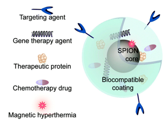
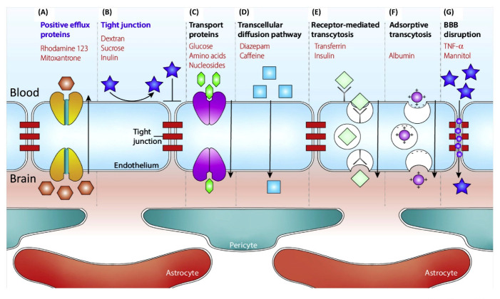
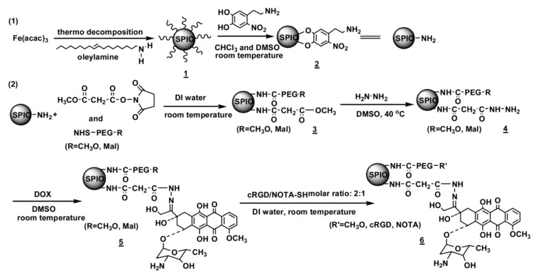
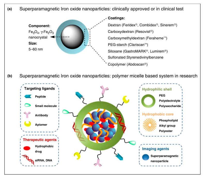
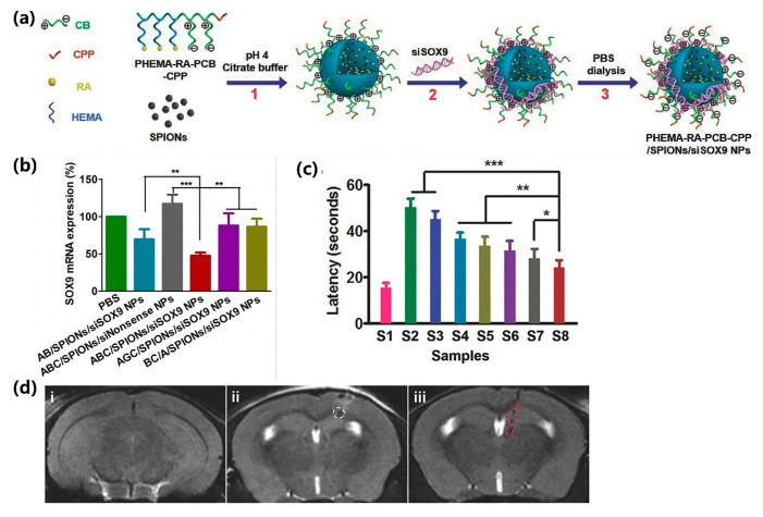
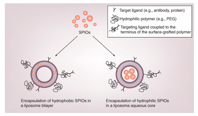
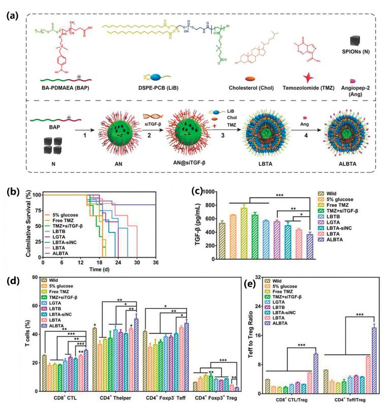
Combining diagnosis with therapy, magnetic iron oxide nanoparticles (INOPs) act as an important vehicle for drug delivery. However, poor biocompatibility of INOPs limits their application. To improve the shortcomings, various surface modifications have been developed, including small molecules coatings, polymers coatings, lipid coatings and lipopolymer coatings. These surface modifications facilitate iron nanoparticles to cross the blood-brain-barrier, which is essential for diagnosis and treatments of brain diseases. Here we focus on the characteristics of different coated INOPs and their application in brain disease, particularly gliomas, Alzheimer's disease (AD) and Parkinson's disease (PD). Moreover, we summarize the current progress and expect to provide help for future researches.

Keywords: blood-brain barrier; iron oxide magnetic nanoparticles; surface coating; target therapy; theranostics.


Conflict of interest statement

The authors declare no conflict of interest.

Figures

Figure 1
Figure 1
Architecture of second-generation iron-based nano-theranostics. Reproduced from “Surface Engineering of Iron Oxide Nanoparticles for Targeted Cancer Therapy”, with permission from American Chemical Society, 2011.
Figure 2
Figure 2
Nanoscale size effect of nanocrystals on magnetism and induced Magnetic Resonance (MR) signals. (a) Transmission Electron Microscope (TEM) images of Fe3O4 nanocrystals of 4 to 6, 9, and 12 nm. (b) Size-dependent T2-weighted MR images of nanocrystals in aqueous solution at 1.5 T. (c) Size-dependent changes from red to blue in color-coded MR images based on T2 values. (d) Graph of T2 value versus nanocrystal size. (e) Magnetization of nanocrystals measured by a superconducting quantum interference device (SQUID) magnetometer. Reproduced from “Nanoscale size effect of magnetic nanocrystals and their utilization for cancer diagnosis via magnetic resonance imaging”, with permission from American Chemical Society, 2005.
Figure 3
Figure 3
BBB transport pathways. A schematic diagram of endothelial cells (ECs), pericytes, and astrocytes forming the BBB. (A) The activity of efflux transport proteins such as p-glycoprotein (P-gp). (B) Selective permeability of tight BBB. (C) Carrier mediated pathway. (D) Passive transportation. (E) Receptor mediated pathway. (F) Adsorptive mediated pathway. (G) Natural occurring biomolecules increase the permeability of the brain endothelium. Reproduced from “Advanced in Microfluidic Blood-Brain Barrier (BBB) Models”, with permission from Elsevier, 2019.
Scheme 1
Scheme 1
Procedures used to prepare the multifunctional nitrodopamine anchored iron oxide nanoparticles (INOPs) nanocarriers. Reproduced from “cRGD-functionalized, DOX-conjugated, and 64Cu-labeled superparamagnetic iron oxide nanoparticles for targeted anticancer drug delivery and PET/MR imaging”, with permission from Elsevier, 2011.
Figure 4
Figure 4
Schematic illustration of the polymer micelle composite system of surface-engineered superparamagnetic iron oxide nanoparticles (SPION) for imaging and therapy application. (a) Clinically approved or in clinical test SPION with biocompatible coatings and iron oxide cores; for each kind of coating material, commercial products are listed aside. (b) Amphiphilic polymers to form a micelle structure with hydrophilic shell and hydrophobic core. INOPs and hydrophobic drugs are packed into the core, targeting ligands can be coupled onto the hydrophilic surface. Reproduced from “Superparamagnetic iron oxide nanoparticles for MR imaging and therapy: design considerations and clinical applications”, with permission from Elsevier, 2014.
Figure 5
Figure 5
(a) Preparation of siRNA-RA NPs. (b) Knockdown the expression level of SOX-9 mRNA. (c) The latency in target quadrant in Morris water maze (MWM). (d) Representative in vivo T2 MRI images of brains with PHEMA-RA-PCB-CPP/SPIONs/siSOX-9 NPs. (S1) wild, (S2) PBS, (S3) NSCs alone, (S4) PHEMA-RA-PCB-CPP/SPIONs/si-nonsense NPs NSCs, (S5) PHEMA-PCB-C PP/RA/SPIONs/siSOX-9 NPs NSCs, (S6) PHEMA-RA-PEG-CPP/SPIONs/siSOX-9 NPs NSCs, (S7) PHEMA-RA-PCB/SPIONs/siSOX-9 NPs NSCs, and (S8) PHEMA-RA-PCB-CPP/SPIONs/siSOX-9 NPs NSCs. * p < 0.05, ** p < 0.01, and *** p < 0.005 (n = 5) versus control. Reproduced from “Traceable Nanoparticle Delivery of Small Interfering RNA and Retinoic Acid with Temporally Release Ability to Control Neural Stem Cell Differentiation for Alzheimer’s Disease Therapy”, with permission from John Wiley and Sons, 2016.
Figure 6
Figure 6
Diagram of superparamagnetic iron oxide nanoparticles (SPIONs) encapsulated by liposome. Reproduced from “Liposome-nanoparticles hybrids for multimodal diagnostic and therapeutic applications”, with permission from Future Medicine Ltd., 2007.
Figure 7
Figure 7
(a) Preparation of DSPE-PCB coated BAP/SPIONs@siTGF-β NPs. (b) T cell composition of different groups. (c) CD8+ cytotoxic lymphocyte cells (CTL): Treg ratios and CD4+ effector T cells: Treg ratios in spleen upon various treatments. (d) The survival time of different groups of intracranial glioblastoma-bearing mice after various treatments. (e) The regulation of TGF-β secretion after treatment with various formulations in vivo. *: differences between ALBTA with other groups, *: p < 0.05, **: p < 0.01, ***: p < 0.001. Reproduced from “Traceable Nanoparticles with Dual Targeting and ROS Response for RNAi-Based Immunochemotherapy of Intracranial Glioblastoma Treatment”, with permission from John Wiley and Sons, 2018.
Figure 8
Figure 8
(a) Preparation of Au coated Fe3O4. (b) Cell viability of C6 cells after various treatments. (c) The C6 tumor growth curves of mice after various treatments, where the tumor volumes were normalized to their initial tumor sizes (mean ± SD, n = 4). (d) Photographs of (c). p values were calculated by the student’s test: * p < 0.05. Reproduced from “Multifunctional pDNA-Conjugated Polycationic Au Nanorod-Coated Fe3O4 Hierarchical Nanocomposites for Trimodal Imaging and Combined Photothermal/Gene Therapy”, with permission from John Wiley and Sons, 2016.

References

    1. Lu A.H., Salabas E.L., Schuth F. Magnetic nanoparticles: Synthesis, protection, functionalization, and application. Angew. Chem. Int. Ed. Engl. 2007;46:1222–1244. doi: 10.1002/anie.200602866. - DOI - PubMed
    1. Gao J., Gu H., Xu B. Multifunctional magnetic nanoparticles: Design, synthesis, and biomedical applications. Acc. Chem. Res. 2009;42:1097–1107. doi: 10.1021/ar9000026. - DOI - PubMed
    1. Hao R., Xing R., Xu Z., Hou Y., Gao S., Sun S. Synthesis, functionalization, and biomedical applications of multifunctional magnetic nanoparticles. Adv. Mater. 2010;22:2729–2742. doi: 10.1002/adma.201000260. - DOI - PubMed
    1. Ulbrich K., Hola K., Subr V., Bakandritsos A., Tucek J., Zboril R. Targeted drug delivery with polymers and magnetic nanoparticles: Covalent and noncovalent approaches, release control, and clinical studies. Chem. Rev. 2016;116:5338–5431. doi: 10.1021/acs.chemrev.5b00589. - DOI - PubMed
    1. Ho D., Sun X., Sun S. Monodisperse magnetic nanoparticles for theranostic applications. Acc. Chem. Res. 2011;44:875–882. doi: 10.1021/ar200090c. - DOI - PMC - PubMed

LinkOut - more resources