. 2020 Jul 24;21(15):5249.
doi: 10.3390/ijms21155249.

Bovine Organospecific Microvascular Endothelial Cell Lines as New and Relevant In Vitro Models to Study Viral Infections

Affiliations

Bovine Organospecific Microvascular Endothelial Cell Lines as New and Relevant In Vitro Models to Study Viral Infections

Anne-Claire Lagrée et al. Int J Mol Sci. .

Abstract

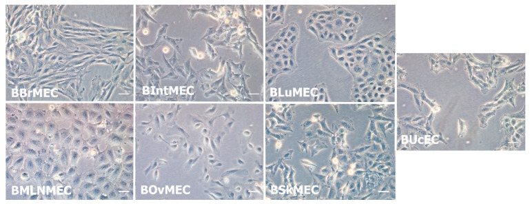
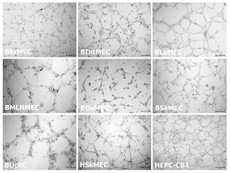
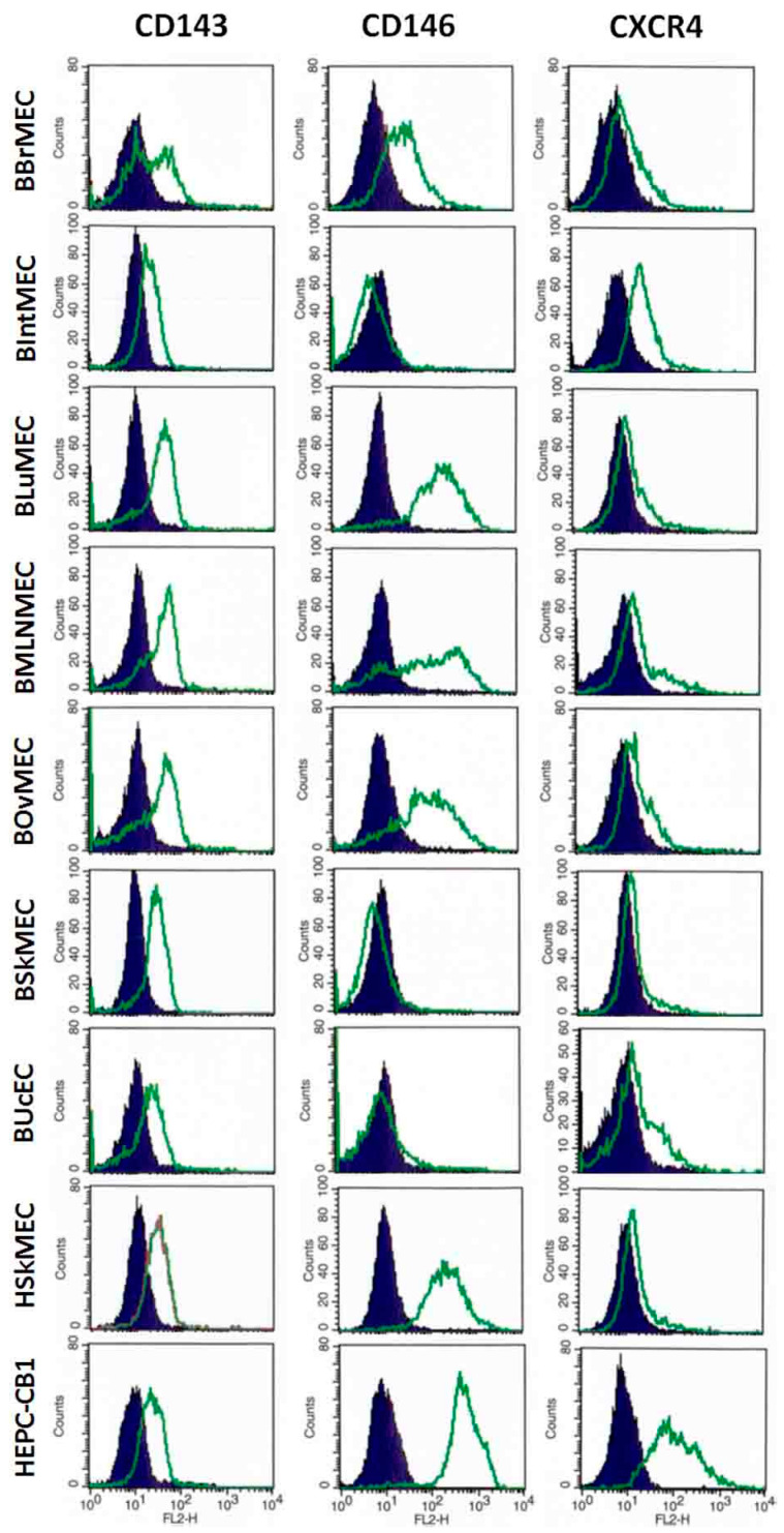
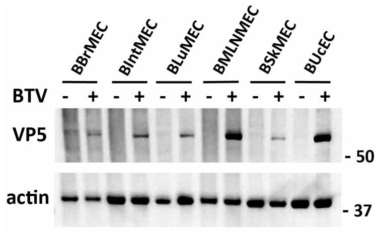
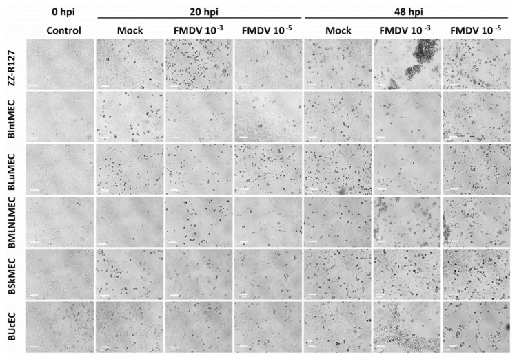
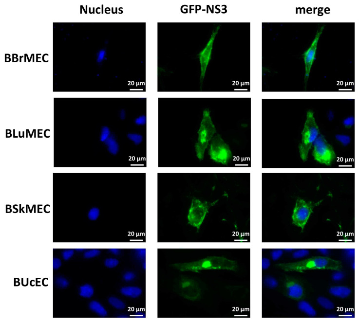
Microvascular endothelial cells constitute potential targets for exogenous microorganisms, in particular for vector-borne pathogens. Their phenotypic and functional variations according to the organs they are coming from provide an explanation of the organ selectivity expressed in vivo by pathogens. In order to make available relevant tools for in vitro studies of infection mechanisms, our aim was to immortalize bovine organospecific endothelial cells but also to assess their permissivity to viral infection. Using transfection with SV40 large T antigen, six bovine microvascular endothelial cell lines from various organs and one macrovascular cell line from an umbilical cord were established. They display their own panel of endothelial progenitor/mature markers, as assessed by flow cytometry and RT-qPCR, as well as the typical angiogenesis capacity. Using both Bluetongue and foot-and-mouth disease viruses, we demonstrate that some cell lines are preferentially infected. In addition, they can be transfected and are able to express viral proteins such as BTV8-NS3. Such microvascular endothelial cell lines bring innovative tools for in vitro studies of infection by viruses or bacteria, allowing for the study of host-pathogen interaction mechanisms with the actual in vivo target cells. They are also suitable for applications linked to microvascularization, such as anti-angiogenic and anti-tumor research, growing fields in veterinary medicine.

Keywords: cattle diseases; endothelium organospecificity; host-pathogen interactions; immortalization; microvasculature; viruses.


Conflict of interest statement

The authors declare no conflict of interest.

Figures

Figure 1
Figure 1
Morphological aspect of the seven established bovine endothelial cell lines. Representative optical microscopy photographs (×20) of endothelial cell lines in culture. Pictures of growing cells were taken using inverted Leica DMi1 microscope equipped with digital camera Leica MC120 HD. Scale bar of 50 μm.
Figure 2
Figure 2
Bovine endothelial cell line angiogenesis assessed by pseudovessel formation on Matrigel™. Cells were seeded on Matrigel™ and angiogenesis was monitored under a video microscope during 24 h. Presented pictures are at 1.5 h (for BBrMEC, BIntMEC, and BSkMEC), at 4.5 h (for BLuMEC, BOvMEC, BUcEC, HSkMEC, and HEPC-CB1) and at 5.5 h (for BMLNMEC) after seeding. Scale bar is 200 μm.
Figure 3
Figure 3
Main endothelial cell markers expressed in various bovine cell lines assessed by flow cytometry. Bovine endothelial cell lines and two human endothelial cell lines, as controls, were labeled with anti-CD143, anti-CD146, and anti-CXCR4 antibodies and then analyzed by flow cytometry. Results are shown as histograms showing the fluorescence intensity with the isotypic control in dark blue and the antibody labeling in green.
Figure 4
Figure 4
Viral infections of bovine endothelial cell lines by Bluetongue virus (BTV). BBrMEC, BIntMEC, BLuMEC, BMLNMEC, BSkMEC, and BUcEC were infected with BTV (multiplicity of infection (MOI) = 0.1). After 18 h, BTV infection was assessed by anti-VP5 immunoblotting and actin was used as a loading control in Western blot analysis. Originals of Western blot pictures are presented in Figure S1.
Figure 5
Figure 5
Viral infections of bovine endothelial cell lines by foot-and-mouth disease virus (FMDV). BIntMEC, BLuMEC, BMLNMEC, BSkMEC, and BUcEC were inoculated with FMDV strain of O serotype (2 viral doses: MOI 10−3 and 10−5) and monitored for cytopathic effect (CPE) during 48 h using an inverted LEICA Statif DM IL LED microscope (×10 magnification). ZZ-R127, fetal goat tongue epithelial cells, were used as positive controls. The pictures presented correspond to 20 and 48 h post-infection (hpi). Scale bars represent 100 μm.
Figure 6
Figure 6
NS3-encoding DNA transfection of bovine endothelial cell lines. BBrMEC, BLuMEC, BSkMEC, and BUcEC were transfected with the expression vector encoding GFP-tagged BTV8-NS3. Intracellular localization of Hoechst-stained nuclei and GFP-tagged NS3 fluorescence was visualized by fluorescence microscopy (×63 magnification) in blue and green, respectively. Scale bars represent 20 μm.

References

    1. Sahni A., Narra H.P., Patel J., Sahni S.K. MicroRNA-Regulated Rickettsial Invasion into Host Endothelium via Fibroblast Growth Factor 2 and Its Receptor FGFR1. Cells. 2018;7:240. doi: 10.3390/cells7120240. - DOI - PMC - PubMed
    1. Bazzoni G., Dejana E. Endothelial Cell-to-Cell Junctions: Molecular Organization and Role in Vascular Homeostasis. Physiol. Rev. 2004;84:869–901. doi: 10.1152/physrev.00035.2003. - DOI - PubMed
    1. Mehta D., Ravindran K., Kuebler W.M. Novel regulators of endothelial barrier function. Am. J. Physiol.-Lung Cell. Mol. Physiol. 2014;307:L924–L935. doi: 10.1152/ajplung.00318.2014. - DOI - PMC - PubMed
    1. Cai Z., Zhang Y., Zhang Y., Miao X., Li S., Yang H., Ling Q., Hoffmann P.R., Huang Z. Use of a Mouse Model and Human Umbilical Vein Endothelial Cells to Investigate the Effect of Arsenic Exposure on Vascular Endothelial Function and the Associated Role of Calpains. Environ. Health Perspect. 2019;127:077003. doi: 10.1289/EHP4538. - DOI - PMC - PubMed
    1. Talavera-Adame D., Ng T.T., Gupta A., Kurtovic S., Wu G.D., Dafoe D.C. Characterization of microvascular endothelial cells isolated from the dermis of adult mouse tails. Microvasc. Res. 2011;82:97–104. doi: 10.1016/j.mvr.2011.04.009. - DOI - PubMed