. 2020 Jul 25;8(8):246.
doi: 10.3390/biomedicines8080246.

Mass-Spectrometry Based Proteome Comparison of Extracellular Vesicle Isolation Methods: Comparison of ME-kit, Size-Exclusion Chromatography, and High-Speed Centrifugation

Affiliations

Mass-Spectrometry Based Proteome Comparison of Extracellular Vesicle Isolation Methods: Comparison of ME-kit, Size-Exclusion Chromatography, and High-Speed Centrifugation

Anders Askeland et al. Biomedicines. .

Abstract

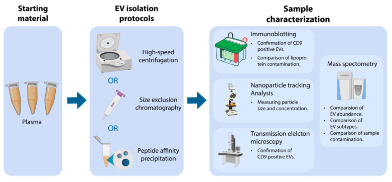
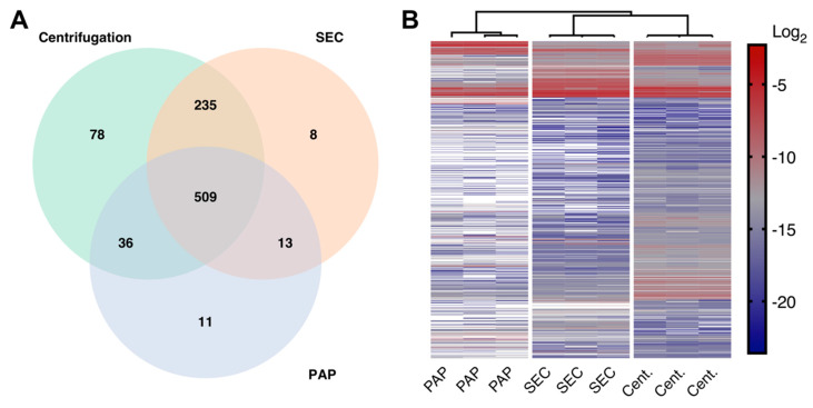
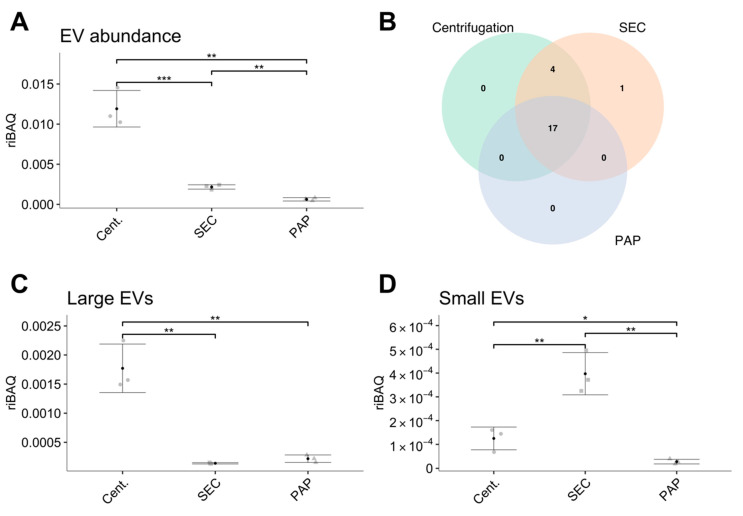
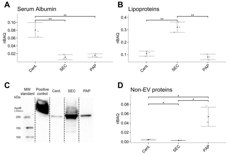
Extracellular vesicles (EVs) are small membrane-enclosed particles released by cells under various conditions specific to cells' biological states. Hence, mass-spectrometry (MS) based proteome analysis of EVs in plasma has gained much attention as a method to discover novel protein biomarkers. MS analysis of EVs in plasma is challenging and EV isolation is usually necessary. Therefore, we compared differences in abundance, subtypes, and contamination for EVs isolated by high-speed centrifugation, size exclusion chromatography (SEC), and peptide-affinity precipitation (PAP/ME kit) for subsequent MS-based proteome analysis. Successful EV isolation was evaluated by nanoparticle-tracking analysis, immunoblotting, and transmission electron microscopy, while EV abundance, EV subtypes, and contamination was evaluated by label-free tandem MS. High-speed centrifugation and SEC isolates showed high EV abundance at the expense of contamination by non-EV proteins and lipoproteins, respectively. These two methods also resulted in EVs of a similar type, however, with smaller EVs in SEC isolates. PAP isolates had a relatively low EV abundance and high contamination. We consider high-speed centrifugation and SEC suitable as EV isolation for MS biomarker studies, where the choice between the two should depend on the scientific questions and whether the focus is on larger or smaller EVs or a combination of both.

Keywords: EV isolation; Extracellular vesicles; ME kit; high-speed centrifugation; human plasma; mass spectrometry; peptide affinity; proteome; proteomics; size exclusion chromatography.


Conflict of interest statement

The authors declare no conflict of interest. The funders had no role in the design of the study; in the collection, analyses, or interpretation of data; in the writing of the manuscript, or in the decision to publish the results.

Figures

Figure 1
Figure 1
Overview of the applied workflow starting with plasma, isolation of extracellular vesicles (EV), followed by sample characterization. EVs were isolated using either high-speed centrifugation, size exclusion chromatography (SEC), or peptide affinity precipitation (PAP). EV isolates were characterized by nanoparticle tracking analysis (NTA), transmission electron microscopy (TEM), immunoblotting, and mass spectrometry (MS).
Figure 2
Figure 2
EV characterization by nanoparticle tracking analysis (NTA), immunoblotting, and transmission electron microscopy (TEM). (A) Differences in particle concentration measured by NTA. (B) Differences in particle mode size measured by NTA. (C) Immunoblot against CD9 (24 kDa) confirming the presence of the EV specific protein CD9 in isolates from all methods. (D) TEM images showing CD9 expression in vesicle-like particles in all three isolation methods. Error bars: Mean ± SD.
Figure 3
Figure 3
Differences in the EV proteomes in isolates produced by high-speed centrifugation (Cent.), size exclusion chromatography (SEC), and peptide affinity precipitation (PAP). (A) Venn diagram showing common and unique proteins identified in all three methods. (B) Hierarchical clustering of protein intensities from isolates obtained by high-speed centrifugation, SEC, and PAP showing that the three isolation methods resulted in three distinct EV proteomes. White = missing values.
Figure 4
Figure 4
Differences in the abundance and amount of extracellular vesicle (EV) specific proteins in isolates obtained by high-speed centrifugation (Cent.), size exclusion chromatography (SEC), and peptide affinity precipitation (PAP). (A) The difference in abundance of EV specific proteins. (B) Venn diagram showing the number of identified EV specific proteins per isolation method. (C,D) Comparison of relative abundances of EV subtypes divided into large EVs (C) and small EVs (D). Error bars: Mean ± SD. Significance levels: * (p-value < 0.05), ** (p-value < 0.01), and *** (p-value < 0.001).
Figure 5
Figure 5
Difference in the co-isolation of contaminants such as serum albumin, lipoproteins, and other non-EV proteins between isolates produced by high-speed centrifugation (Cent.), size-exclusion chromatography (SEC), and peptide affinity precipitation (PAP). (A) High-speed centrifugation produced EV isolates with the highest abundance of serum albumin. (B) SEC isolates contained the highest amount of lipoproteins. (C) Immunoblotting confirms elevated levels of apolipoprotein B in isolates produced by SEC. (D) PAP isolates contained the highest amount of non-EV proteins. Error bars: Mean ± SD. Significance levels: * (p-value < 0.05) and ** (p-value < 0.01).
Figure 6
Figure 6
Reproducibility of the extracellular vesicle (EV) isolation methods. Reproducibility was evaluated for each biological sample by technical replicates and was evaluated for protein identifications repeatability (A) and correlation matrix of all protein abundances (B+C). (A) Proteins identified in all technical replicates of EV samples isolated by using the same method. (B+C) Correlation matrix (coefficient of determination (R2)) of the reproducibility between technical replicates (Rep. 1 and Rep. 2) and biological controls (Con. 1, Con. 2, and Con. 3) of the evaluated EV isolation protocols.

References

    1. Rifai N., Gillette M.A., Carr S.A. Protein biomarker discovery and validation: The long and uncertain path to clinical utility. Nat. Biotechnol. 2006;24:971–983. doi: 10.1038/nbt1235. - DOI - PubMed
    1. Etzioni R., Urban N., Ramsey S., McIntosh M., Schwartz S., Reid B., Radich J., Anderson G., Hartwell L. Early detection: The case for early detection. Nat. Rev. Cancer. 2003;3:243–252. doi: 10.1038/nrc1041. - DOI - PubMed
    1. Anderson N.L., Anderson N.G. The human plasma proteome: History, character, and diagnostic prospects. Mol. Cell. Proteom. 2002;1:845–867. doi: 10.1074/mcp.R200007-MCP200. - DOI - PubMed
    1. Yáñez-Mó M., Siljander P.R.M., Andreu Z., Zavec A.B., Borràs F.E., Buzas E.I., Buzas K., Casal E., Cappello F., Carvalho J., et al. Biological properties of extracellular vesicles and their physiological functions. J. Extracell. Vesicles. 2015;4:1–60. doi: 10.3402/jev.v4.27066. - DOI - PMC - PubMed
    1. Kowal J., Arras G., Colombo M., Jouve M., Morath J.P., Primdal-Bengtson B., Dingli F., Loew D., Tkach M., Théry C. Proteomic comparison defines novel markers to characterize heterogeneous populations of extracellular vesicle subtypes. Proc. Natl. Acad. Sci. USA. 2016;113:E968–E977. doi: 10.1073/pnas.1521230113. - DOI - PMC - PubMed