. 2020 Jul 28;15(7):e0232565.
doi: 10.1371/journal.pone.0232565. eCollection 2020.

An image J plugin for the high throughput image analysis of in vitro scratch wound healing assays

Affiliations

An image J plugin for the high throughput image analysis of in vitro scratch wound healing assays

Alejandra Suarez-Arnedo et al. PLoS One. .

Abstract

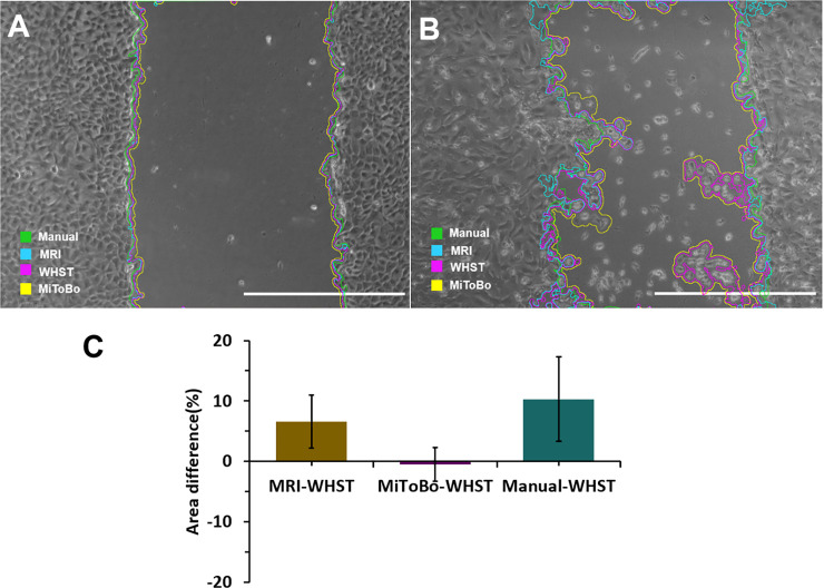
In vitro scratch wound healing assay, a simple and low-cost technique that works along with other image analysis tools, is one of the most widely used 2D methods to determine the cellular migration and proliferation in processes such as regeneration and disease. There are open-source programs such as imageJ to analyze images of in vitro scratch wound healing assays, but these tools require manual tuning of various parameters, which is time-consuming and limits image throughput. For that reason, we developed an optimized plugin for imageJ to automatically recognize the wound healing size, correct the average wound width by considering its inclination, and quantify other important parameters such as: area, wound area fraction, average wound width, and width deviation of the wound images obtained from a scratch/ wound healing assay. Our plugin is easy to install and can be used with different operating systems. It can be adapted to analyze both individual images and stacks. Additionally, it allows the analysis of images obtained from bright field, phase contrast, and fluorescence microscopes. In conclusion, this new imageJ plugin is a robust tool to automatically standardize and facilitate quantification of different in vitro wound parameters with high accuracy compared with other tools and manual identification.


Conflict of interest statement

The authors have declared that no competing interests exist.

Figures

Fig 1
Fig 1. Overall process of the image segmentation algorithm.
We show images from each of the main steps to illustrate how the wound area is separated from the cell monolayer area.
Fig 2
Fig 2. Wound healing size tool.
A. Interface window to adjust parameters B. Snapshot of the results in table format in pixels show area of the wound, wound coverage of total area, and average of the width and its standard deviation. C. Snapshot of the results in table format in μm show area of the wound, wound coverage of total area, and average of the width and its standard deviation.
Fig 3
Fig 3. Scratch validation using mold.
A Schematic of the use of mold for scratching wounds on 2D cultures. B. Percentage of coefficient of variation between scratch made with pipette tip of 200 μL with or without using the mold [n = 4 images per method]. C. Percentage of deviation from the straight line of each scratch made with pipette tip of 200 μL with or without using the mold [n = 4 images per method] p_value<0.05*.
Fig 4
Fig 4. Wound healing size tool test comparing HaCaT exposed to hAdMSCs conditioned medium and HaCaT cultured only with basic medium (DMEM +1%P/S) as control of the assay.
A. Time lapse (0, 12, 18 and 24 hours. Scale bar = 27550 μm) images of wound healing closure in HaCaT exposed to hAdMSCs conditioned medium. B. Scratch area in μm2.-C. Percentage of closure area. D. Scratch width in μm. E. Standard deviation of the scratch width in μm. F. Rate of cell migration in μm/hour. All the measurements and parameters were taken for 28 hours [n = 4 replicas per time for HaCaT exposed to hAdMSCs conditioned medium and n = 2 replicas per time for HaCaT exposed to control medium]. p-value<0.0001****, p-value<0.001***, p-value<0.01** Some error bars are shorter than the size of the symbols.
Fig 5
Fig 5. Differences in the area measured with the wound healing size tool (WHST) regarding the MRI wound healing tool (MRI), the ScratchAssayAnalyzer in MiToBo—a microscope image analysis toolbox (MiToBo) and manual drawing (Manual).
A. Scratch with low variation of calculated area between the different methods. Scale bar = 1000 pixels B. Scratch with high variation of calculated area between the different methods C. Quantification of the difference in measurements between methods with our plugin [n = 30 images].
Fig 6
Fig 6. Differences in the width measured with the wound healing size tool (WHST) regarding the manual drawing (Manual).
A Scratch identifies with our plugin. Wound with manual measurement of width. Manual data (average = 1149 pixels, standard deviation = 22.82 pixels) WHST data (average = 1165 pixels, standard deviation 102.35 pixels). Scale bar = 1000 pixels B. Quantification of the difference in measurements between wound healing size tool (WHST) and scratching manually [n = 30 images, 10 lines per area in the manual method] *p_value<0.01.

References

    1. Martin P. Wound healing—Aiming for perfect skin regeneration. Science (80-). 1997. April 4;276(5309):75–81. - PubMed
    1. Xu J, Song J, Yang X, Guo J, Wang T, Zhuo W. ProNGF siRNA inhibits cell proliferation and invasion of pancreatic cancer cells and promotes anoikis. Biomed Pharmacother. 2019. March 1;111:1066–73. 10.1016/j.biopha.2019.01.002 - DOI - PubMed
    1. Erdogan B, Ao M, White LM, Means AL, Brewer BM, Yang L, et al. Cancer-associated fibroblasts promote directional cancer cell migration by aligning fibronectin. J Cell Biol. 2017. November 1;216(11):3799–816. 10.1083/jcb.201704053 - DOI - PMC - PubMed
    1. Vang Mouritzen M, Jenssen H. Optimized scratch assay for in vitro testing of cell migration with an automated optical camera. J Vis Exp. 2018. August 8 (138). - PMC - PubMed
    1. van der Meer AD, Vermeul K, Poot AA, Feijen J, Vermes I. A microfluidic wound-healing assay for quantifying endothelial cell migration. Am J Physiol Circ Physiol. 2010. February;298(2):H719–25. - PubMed

Publication types

Substances