. 2020 Sep;53(9):e12795.
doi: 10.1111/cpr.12795. Epub 2020 Jul 28.

Exosome-transferred long non-coding RNA ASMTL-AS1 contributes to malignant phenotypes in residual hepatocellular carcinoma after insufficient radiofrequency ablation

Affiliations

Exosome-transferred long non-coding RNA ASMTL-AS1 contributes to malignant phenotypes in residual hepatocellular carcinoma after insufficient radiofrequency ablation

Dening Ma et al. Cell Prolif. 2020 Sep.

Abstract

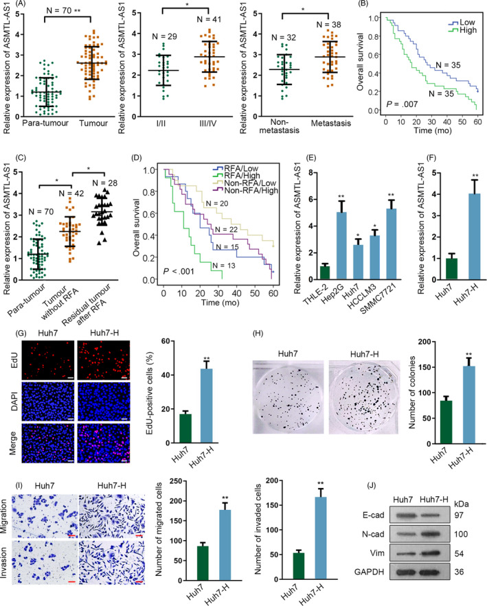
Objectives: Long non-coding RNAs (lncRNAs) are emerging RNA regulators in cancer progression, including in hepatocellular carcinoma (HCC). Recently, insufficient radiofrequency ablation (RFA) has been reported to lead to recurrence and metastasis of residual HCC tumours. Herein, we aimed to the role of ASMTL-AS1 in residual HCC after insufficient RFA.

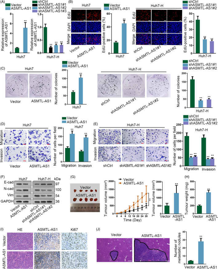
Materials and methods: In vitro insufficient RFA model was simulated in Huh7 cells and subsequently named Huh7-H cells. In vitro and in vivo assays were conducted to investigate ASMTL-AS1 function in HCC.

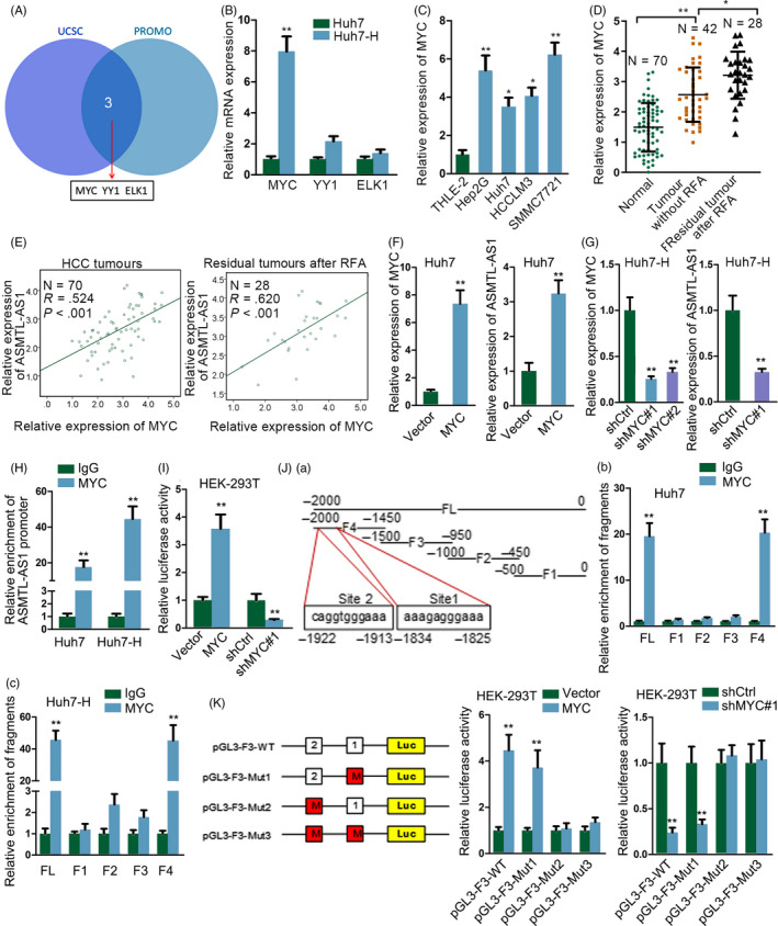
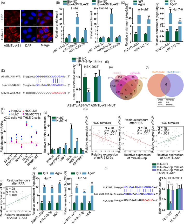
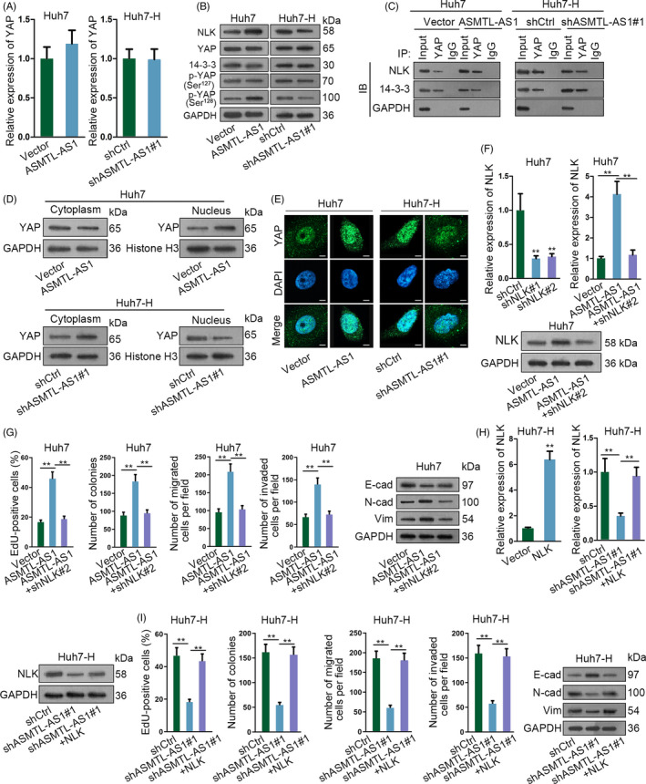
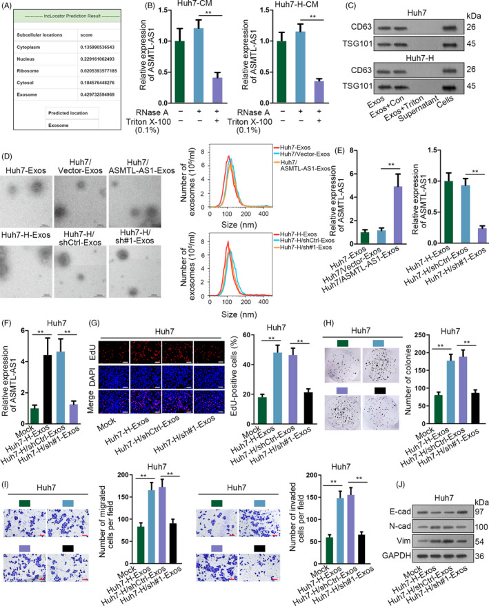
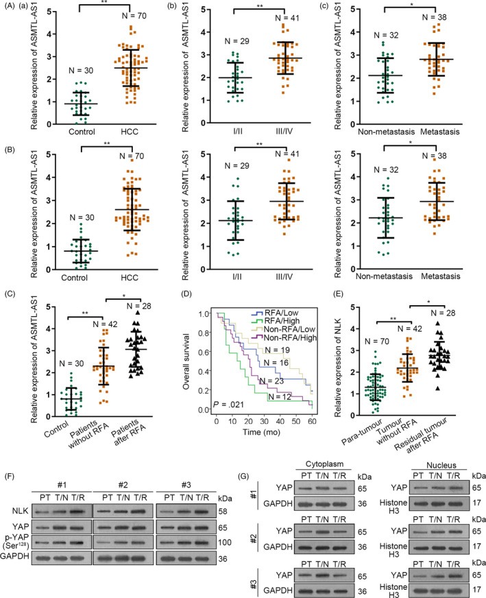
Results: LncRNA ASMTL-AS1 low expressed in normal human liver was found to be highly expressed in HCC tissues and further increased in tumours after insufficient RFA. ASMTL-AS1 expression was related to stage, metastasis and prognosis in HCC. Huh7-H possessed higher ASMTL-AS1 level and more aggressive than Huh7 cells. ASMTL-AS1 contributed to the malignancy of HCC cells both in vitro and in vivo. Mechanistically, ASMTL-AS1 was trans-activated by MYC and promoted NLK expression to activate YAP signalling via sequestering miR-342-3p in HCC. Interestingly, ASMTL-AS1 could be wrapped by exosomes and then convey malignancy through NLK/YAP axis between cells even in residual HCC after insufficient RFA.

Conclusions: Exosomal ASMTL-AS1 aggravates the malignancy in residual HCC after insufficient RFA via miR-342-3p/NLK/YAP signalling, opening a new road for the treatment of HCC and the prevention of recurrence or metastasis of residual HCC after insufficient RFA.

Keywords: ASMTL-AS1; HCC; NLK; YAP nuclear translocation; exosomes.


Conflict of interest statement

All authors ensure no conflicts of interest in our work.

Figures

Figure 1
Figure 1
ASMTL‐AS1 was highly expressed in residual HCC after insufficient RFA and Huh7 cells after heating. A, The expression pattern of ASMTL‐AS1 in indicated 70 of HCC tissues was evaluated by qRT‐PCR. B, Kaplan‐Meier curve determined the overall survival of HCC patients with high or low ASMTL‐AS1 level. C, qRT‐PCR result of ASMTL‐AS1 expression in 42 HCC tissues and 28 residual tumours after insufficient RFA, compared to 70 of all para‐tissues. D, Kaplan‐Meier curve assessed the overall survival of HCC patients suffered different treatment according to ASMTL‐AS1 expression. E, ASMTL‐AS1 level in HCC cell lines was analysed via qRT‐PCR. F, qRT‐PCR estimated the expression of ASMTL‐AS1 in Huh7 cells with or without heat treatment. G‐I, Biological behaviour of Huh7 and Huh7‐H cells was detected through conducting EdU assay (G; scale bar = 200 μm), colony formation assay (H) and transwell assay (I; scale bar = 200 μm). Western blot analysis of EMT‐related protein levels in Huh7 and Huh7‐H cells (J). *P < .05, **P < .01
Figure 2
Figure 2
ASMTL‐AS1 facilitated HCC cell growth and metastasis. A, Overexpression or knockdown efficiencies, respectively, in Huh7 or Huh7‐H cells were tested by qRT‐PCR analysis. B‐F, ASMTL‐AS1 function on HCC cell proliferation, motility and EMT was assessed by EdU (B; scale bar = 200 μm), colony formation (C) and transwell assays (D, E; scale bar = 200 μm) as well as Western blot (F), as needed. G, H, In vivo tumour growth experiments showed silencing ASMTL‐AS1 hindered tumour growth in vivo. I, The expression of ASMTL‐AS1 and Ki67 was, respectively, examined by ISH and IHC staining. Scale bar = 200 μm. J, HE staining and corresponding quantification of in vivo metastatic tumours in liver of mice in indicated groups. Scale bar = 400 μm. **P < .01
Figure 3
Figure 3
ASMTL‐AS1 was transcriptionally enhanced by MYC. A, Transcription factors for ASMTL‐AS1 were predicted by UCSC and PROMO. B, Relative expression of three potential transcription factors in Huh7 and Huh7‐H cells was tested through qRT‐PCR. C, D, qRT‐PCR results of MYC in HCC cell lines (C) or in indicated HCC tissues (D). E, Pearson's correlation analysis determined the association between MYC and ASMTL‐AS1 in clinical tissues from HCC patients with or without RFA treatment. F, G, qRT‐PCR analysis of the expression levels of MYC or ASMTL‐AS1 in MYC‐overexpressed Huh7 cells or in MYC‐inhibited Huh7‐H cells. H, I, The interaction of MYC with ASMTL‐AS1 promoter and its impact on ASMTL‐AS1 transcription were evaluated via ChIP and luciferase reporter assays, respectively. J, K, ChIP and luciferase reporter assays further confirmed the précised binding sequences for MYC to activate ASMTL‐AS1 transcription. *P < .05, **P < .01
Figure 4
Figure 4
ASMTL‐AS1 elevated the expression of NLK through competitively interacting with miR‐342‐3p. A, Subcellular localization of ASMTL‐AS1 in Huh7 and Huh7‐H cells was validated via FISH. Scale bar = 10 μm. B, RNA pull‐down assay confirmed miR‐342‐3p was sponged by ASMTL‐AS1 in HCC cells. C, RIP assay proved both ASMTL‐AS1 and miR‐342‐3p were enriched in RISC. D, Luciferase reporter assay validated miR‐342‐3p was the downstream of ASMTL‐AS1. E, Targets of miR‐342‐3p were predicted through bioinformatics analysis (a) and further screened out via loss‐ and gain‐of‐miR‐342‐3p analysis (b). F, qRT‐PCR showed the expression of indicated genes in HCC cell lines (a) and in Huh7 cells with or without heating (b). G, The relationship between NLK and either miR‐342‐3p or ASMTL‐AS1 was determined by Pearson's correlation analysis. H, I, RIP and luciferase reporter assays verified the competition between NLK and ASMTL‐AS1 in interacting with miR‐342‐3p‐guided RISCs. **P < .01
Figure 5
Figure 5
ASMTL‐AS1 regulated HCC malignancy via NLK‐activated YAP signalling. A, qRT‐PCR revealed ASMTL‐AS1 had no impact on the mRNA expression of YAP. B, The protein expression of NLK, YAP and p‐YAP at Ser127 or Ser128 in ASMTL‐AS1‐overexpressed Huh7 cells or ASMTL‐AS1‐depleted Huh7‐H cells was tested by Western blotting. C, Influence of ASMTL‐AS1 on the interaction of YAP with NLK or 14‐3‐3 was assayed via Co‐IP. D, E, Impact of ASMTL‐AS1 on YAP translocation was assessed by subcellular fractionation plus Western blot (D) and IF assay (E; scale bar = 20 μm). F, The mRNA or protein expression of NLK in indicated cells was analysed by qRT‐PCR or Western blotting, respectively. G, Rescue assays indicated that NLK knockdown recovered ASMTL‐AS1 overexpression‐accelerated proliferation and motility in Huh7 cells. H, I, Rescue assays proved overexpression of NLK countervailed the inhibitory effect of ASMTL‐AS1 depletion on the malignant behaviours of Huh7‐H cells. **P < .01
Figure 6
Figure 6
Exosome‐transferred ASMTL‐AS1 delivered malignancies between HCC cells. A, Online tool lncLocator predicted that ASMTL‐AS1 mainly located in exosomes. B, ASMTL‐AS1 level in Huh7 and Huh7‐H under diverse treatments was estimated by qRT‐PCR. C, Western blot identified exosomes through two markers including CD63 and TSG101. D, Representative images of exosomes from indicated cells were captured by electron microscopy, and the size and number of these exosomes were determined via NanoSight particle tracking analysis. Scale bar = 100 nm. E, The level of ASMTL‐AS1 in above exosomes was analysed through qRT‐PCR. F‐J, Impact of exosomes from control or ASMTL‐AS1‐silenced Huh7‐H cells on the cellular processes of Huh7 cells was estimated by EdU assay (scale bar = 200 μm), colony formation assay, transwell assay (scale bar = 200 μm) and Western blot. **P < .01
Figure 7
Figure 7
ASMTL‐AS1/NLK/YAP axis in clinical HCC patients. A‐C, qRT‐PCR analysis results of ASMTL‐AS1 expression in different groups of HCC patients: (A) The expression of ASMTL‐AS1 in the serum of indicated HCC patients (control referred to serums from 30 healthy individuals); (B) ASMTL‐AS1 expression in serum exosomes (SEs) of above HCC patients; and (C) ASMTL‐AS1 expression in SEs of HCC patients with or without RFA treatment. D, Overall survival rates of four indicated kinds of HCC patients were assessed through Kaplan‐Meier analysis. E, qRT‐PCR revealed the level of NLK in para‐tumours, primary HCC tumours without RFA treatment and residual HCC tumours after insufficient RFA. F, The levels of indicated proteins in above three kinds of tumours were analysed by Western blot. G, YAP localization in above tumours was also determined through Western blot. PT meant para‐carcinoma tissues. T/N referred to HCC tissues from patients with no RFA treatment, and T/R indicated HCC tissues from patients with insufficient RFA. *P < .05, **P < .01

References

    1. Siegel RL, Miller KD, Jemal A. Cancer statistics, 2016. CA Cancer J Clin. 2016;66(1):7‐30. - PubMed
    1. Chen W, Zheng R, Baade PD, et al. Cancer statistics in China, 2015. CA Cancer J Clin. 2016;66(2):115‐132. - PubMed
    1. Sapisochin G, Bruix J. Liver transplantation for hepatocellular carcinoma: outcomes and novel surgical approaches. Nature Rev Gastroenterol Hepatol. 2017;14:203‐217. - PubMed
    1. Villanueva A, Hernandez‐Gea V, Llovet JM. Medical therapies for hepatocellular carcinoma: a critical view of the evidence. Nat Rev Gastroenterol Hepatol. 2013;10(1):34‐42. - PubMed
    1. Cucchetti A, Piscaglia F, Cescon M, et al. Cost‐effectiveness of hepatic resection versus percutaneous radiofrequency ablation for early hepatocellular carcinoma. J Hepatol. 2013;59(2):300‐307. - PubMed