. 2020 Jul 28;24(1):467.
doi: 10.1186/s13054-020-03169-x.

Airway pressure morphology and respiratory muscle activity during end-inspiratory occlusions in pressure support ventilation

Affiliations

Airway pressure morphology and respiratory muscle activity during end-inspiratory occlusions in pressure support ventilation

Stella Soundoulounaki et al. Crit Care. .

Abstract

Background: The driving pressure of the respiratory system is a valuable indicator of global lung stress during passive mechanical ventilation. Monitoring lung stress in assisted ventilation is indispensable, but achieving passive conditions in spontaneously breathing patients to measure driving pressure is challenging. The accuracy of the morphology of airway pressure (Paw) during end-inspiratory occlusion to assure passive conditions during pressure support ventilation has not been examined.

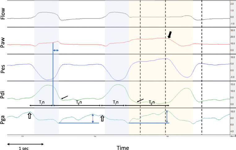
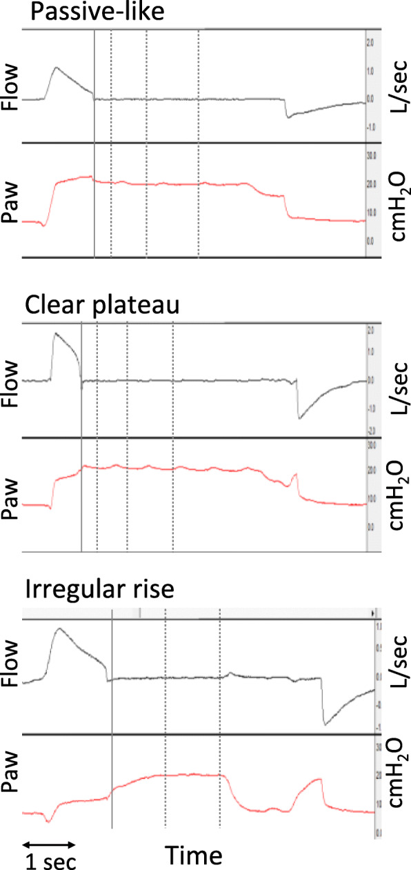
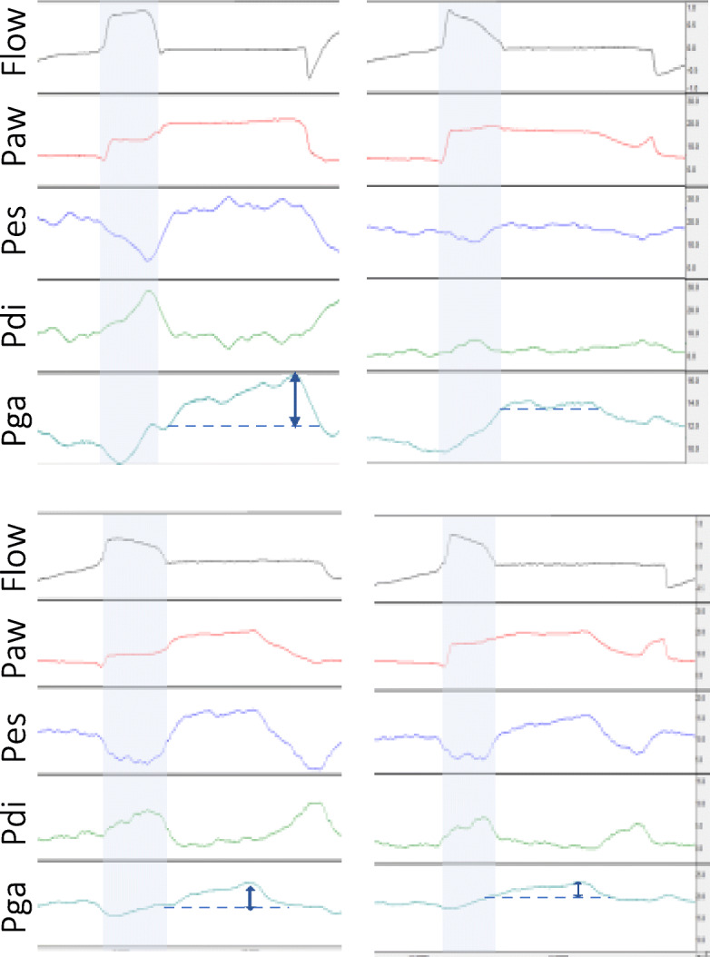
Methods: Retrospective analysis of end-inspiratory occlusions obtained from critically ill patients during pressure support ventilation. Flow, airway, esophageal, gastric, and transdiaphragmatic pressures were analyzed. The rise of gastric pressure during occlusion with a constant/decreasing transdiaphragmatic pressure was used to identify and quantify the expiratory muscle activity. The Paw during occlusion was classified in three patterns, based on the differences at three pre-defined points after occlusion (0.3, 1, and 2 s): a "passive-like" decrease followed by plateau, a pattern with "clear plateau," and an "irregular rise" pattern, which included all cases of late or continuous increase, with or without plateau.

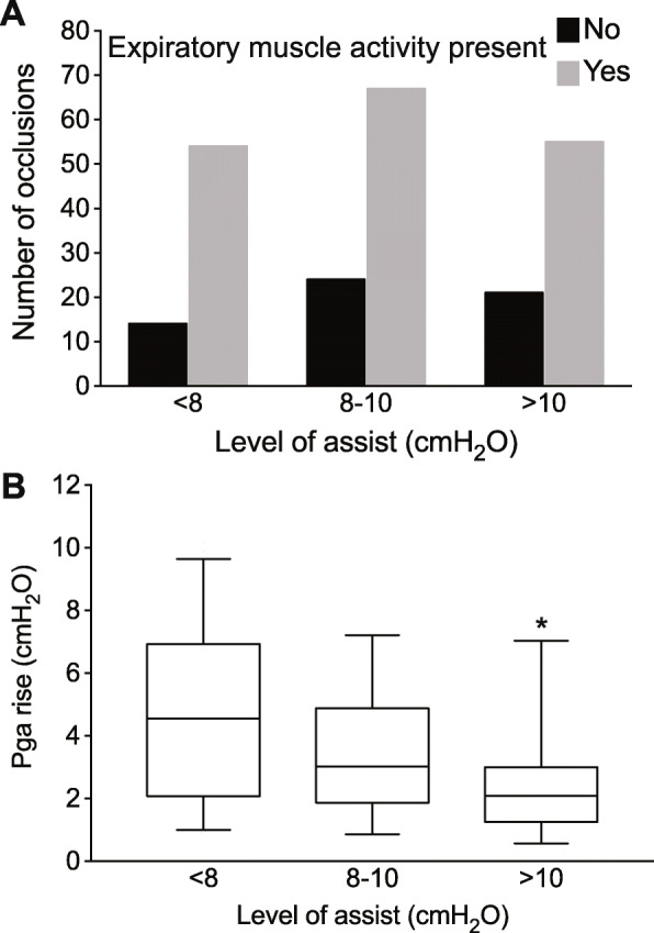
Results: Data from 40 patients and 227 occlusions were analyzed. Expiratory muscle activity during occlusion was identified in 79% of occlusions, and at all levels of assist. After classifying occlusions according to Paw pattern, expiratory muscle activity was identified in 52%, 67%, and 100% of cases of Paw of passive-like, clear plateau, or irregular rise pattern, respectively. The driving pressure was evaluated in the 133 occlusions having a passive-like or clear plateau pattern in Paw. An increase in gastric pressure was present in 46%, 62%, and 64% of cases at 0.3, 1, and 2 s, respectively, and it was greater than 2 cmH2O, in 10%, 20%, and 15% of cases at 0.3, 1, and 2 s, respectively.

Conclusions: The pattern of Paw during an end-inspiratory occlusion in pressure support cannot assure the absence of expiratory muscle activity and accurate measurement of driving pressure. Yet, because driving pressure can only be overestimated due to expiratory muscle contraction, in everyday practice, a low driving pressure indicates an absence of global lung over-stretch. A measurement of high driving pressure should prompt further diagnostic workup, such as a measurement of esophageal pressure.

Keywords: Driving pressure; Esophageal pressure; Gastric pressure; Protective ventilation.


Conflict of interest statement

S.S. has no competing interests to declare. E.A., E.K., K.V., and D.G. have received lecture fees from Medtronic.

Figures

Fig. 1
Fig. 1
Waveform analysis of end-inspiratory occlusion. Representative waveforms of flow (in l s), airway (Paw), esophageal (Pes), transdiaphragmatic (Pdi), and gastric (Pga) pressures (in cmH2O), from one patient, with an end-inspiratory occlusion on the second breath. The blue-shaded area indicates the mechanical inspiratory time, and the yellow-shaded area indicates the end-inspiratory occlusion. Horizontal black arrows indicate the start and end of neural inspiration (Tin) and expiration (TEn). The point of rapid decline of Pdi (end of neural inspiration) is indicated by the blue vertical line, and the horizontal blue double-headed arrow in Paw indicates the cycling off delay. The small black arrows in Pdi indicate the point of complete relaxation of the diaphragm. Black vertical dashed lines indicate the points at 0.3, 1, and 2 s post-occlusion. The thick black arrow in Paw shows the end of the plateau by the sudden decrease in Paw, due to diaphragmatic contraction during occlusion. Observe that the neural inspiratory and expiratory times are similar in the un-occluded and occluded breath, and the occlusion time is less than 2 s. Expiratory muscle activity is indicated by the rise of Pga, during expiration in the un-occluded breath, and during the end-inspiratory occlusion (blue double-headed arrows show the maximum change). Notice also the decrease of Pga at the onset of inspiration, suggesting the relaxation of expiratory muscles at this point (open arrows)
Fig. 2
Fig. 2
Classification of airway pressure waveform pattern during occlusion. Airway flow (in l/s) and airway pressure (Paw, in cmH2O) waveforms representative of the three patterns of Paw during occlusion, from three different patients. The solid vertical line indicates the point of occlusion and subsequent dotted lines the points at 0.3, 1, and 2 s post-occlusion. Each pattern was characterized by the relationships among Paw at different time points relative to the occlusion: occlusion, 0 s (Pawocc), 0.3 s (Paw0.3s), 1 s (Paw1s), and 2 s (Paw2s). Upper panel: a “passive-like” pattern with a rapid decrease in Paw (Pawocc > Paw0.3s), followed by plateau (Paw1s − Paw0.3s < 1 and Paw2s − Paw1s < 1 cmH2O). Middle panel: a “clear plateau” pattern with an early increase in Paw (Pawocc < Paw0.3s), followed by plateau (Paw1s − Paw0.3s < 1 and Paw2s − Paw1s < 1 cmH2O). Lower panel: an “irregular rise” pattern with a slow increase in Paw (Paw1s − Paw0.3s > 1 cmH2O) with plateau (Paw2s − Paw1s < 1 cmH2O)
Fig. 3
Fig. 3
Expiratory muscle activity at different levels of assist. a Number of occlusions with expiratory muscle activity present or not, at three different levels of assist. b Magnitude of expiratory muscle activity, as indicated by the rise of gastric pressure (Pga) after occlusion, at three levels of assist (only in cases with expiratory muscle activity). Box: interquartile range, whiskers: 5–95 range, line at median, *p < 0.05 for assist level > 10 vs 8–10 cmH2O, p < 0.0001 for assist level > 10 vs < 8 cmH2O
Fig. 4
Fig. 4
Changes in respiratory muscle activity to increase in pressure support. Representative waveforms of flow (in l/s), airway (Paw), esophageal (Pes), transdiaphragmatic (Pdi), and gastric (Pga) pressures (in cmH2O), during an end-inspiratory occlusion, from two patients (upper and lower panel), ventilated with low (left) and higher levels of PS (right panel). The blue arrows indicate the estimated magnitude of expiratory muscle activity

References

    1. Amato MBP, Meade MO, Slutsky AS, Brochard L, Costa ELV, Schoenfeld DA, et al. Driving pressure and survival in the acute respiratory distress syndrome. N Engl J Med. 2015;372:747–755. doi: 10.1056/NEJMsa1410639. - DOI - PubMed
    1. Chiumello D, Brioni M. Severe hypoxemia: which strategy to choose. Crit Care Lond Engl. 2016;20:132. doi: 10.1186/s13054-016-1304-7. - DOI - PMC - PubMed
    1. Russotto V, Bellani G, Foti G. Respiratory mechanics in patients with acute respiratory distress syndrome. Ann Transl Med. 2018;6:382. doi: 10.21037/atm.2018.08.32. - DOI - PMC - PubMed
    1. Henderson WR, Chen L, Amato MBP, Brochard LJ. Fifty years of research in ARDS. Respiratory mechanics in acute respiratory distress syndrome. Am J Respir Crit Care Med. 2017;196:822–833. doi: 10.1164/rccm.201612-2495CI. - DOI - PubMed
    1. Neto AS, Hemmes SNT, Barbas CSV, Beiderlinden M, Fernandez-Bustamante A, Futier E, et al. Association between driving pressure and development of postoperative pulmonary complications in patients undergoing mechanical ventilation for general anaesthesia: a meta-analysis of individual patient data. Lancet Respir Med. 2016;4:272–280. doi: 10.1016/S2213-2600(16)00057-6. - DOI - PubMed

MeSH terms

LinkOut - more resources