Case Reports
. 2020 Oct;103(4):1656-1659.
doi: 10.4269/ajtmh.20-0421.

Case Report: Multiorgan Involvement with Congenital Zika Syndrome

Affiliations
Case Reports

Case Report: Multiorgan Involvement with Congenital Zika Syndrome

Rodrigo Cachay et al. Am J Trop Med Hyg. 2020 Oct.

Abstract

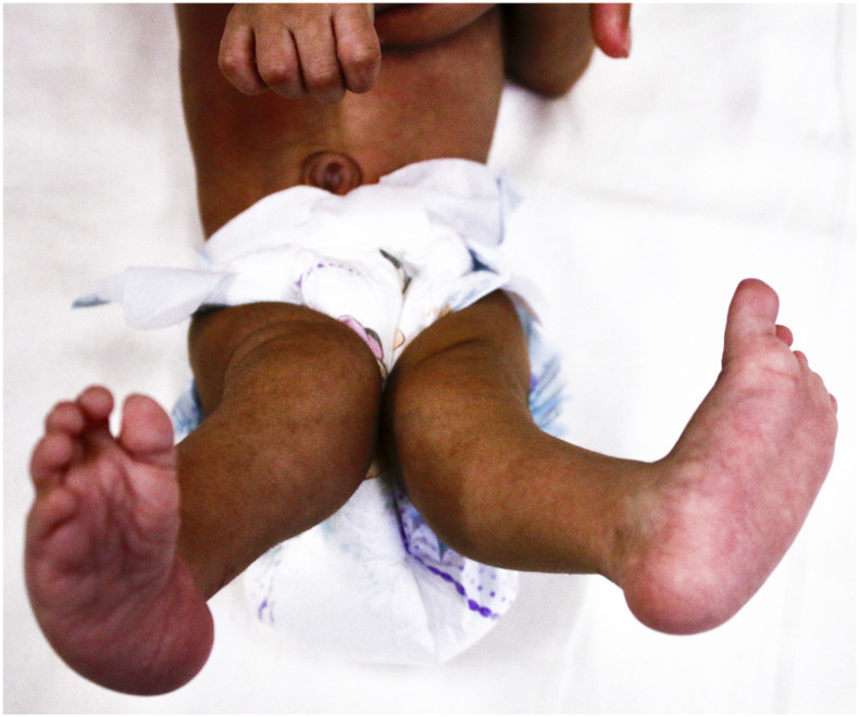
We report the case of an infant born with congenital Zika syndrome (CZS). During the largest Zika virus (ZIKV) outbreak in Peru, the mother presented with fever and rash that were confirmed to be due to ZIKV by real-time PCR. The infant was born with severe microcephaly. Imaging revealed corpus callosum dysgenesis, lissencephaly, ventriculomegaly, and calcifications. Mild hypertrophic cardiomyopathy with diastolic dysfunction was reported in the echocardiogram. Valgus deviation of the lower extremities and a left clubfoot were diagnosed at birth. The hip ultrasound showed incipient signs of Graf type II dysplasia. The findings confirm that CZS is a multiorgan phenotype in which microcephaly is merely the tip of the iceberg. A multidisciplinary approach is needed for the evaluation of these children.


Conflict of interest statement

Disclosure: The case was evaluated as part of the ZIKAlliance cohort (European Union's Horizon 2020 Research and Innovation Programme under grant agreement N. 734548) focused on the impact of ZIKV during pregnancy and the natural history of ZIKV in humans and their environment.

Figures

Figure 1.
Figure 1.
Prominent microcephaly and retrognathia. This figure appears in color at www.ajtmh.org.
Figure 2.
Figure 2.
Valgus deviation of lower extremities and left talipes equinovarus. This figure appears in color at www.ajtmh.org.
Figure 3.
Figure 3.
Computed tomography of the head. (A) Axial view, calcifications in the left thalamus. (B) Axial view, right occipital and bilateral temporal calcifications.
Figure 4.
Figure 4.
Magnetic resonance imaging of the head. (A) Sagittal view, absence of the corpus callosum and reduced brain volume at the expense of the frontal lobe. (B) Sagittal view, lissencephaly with hypoplasia of the temporal cortex. (C) Sagittal view, eyeball and optic nerve pathway. (D) Coronal view, smooth cortex with few grooves; absence of the corpus callosum without differentiation of the basal ganglia and the thalamus. (E) Coronal view, smooth cortex without cerebral grooves; evident dysmyelination. (F) Axial view, Probst bundles are seen by the occipital horns. (G) Axial view of temporal lobes with evident lissencephaly.

References

    1. Moore CA, et al. 2017. Characterizing the pattern of anomalies in congenital Zika syndrome for pediatric clinicians. JAMA Pediatr 171: 288–295. - PMC - PubMed
    1. Orofino DHG, et al. 2018. Cardiac findings in infants with in utero exposure to Zika virus- a cross sectional study. PLoS Negl Trop Dis 12: e0006362. - PMC - PubMed
    1. Mumtaz N, Koedam M, van den Doel PB, van Leeuwen JPTM, Koopmans MPG, van der Eerden BCJ, Rockx B, 2018. Zika virus infection perturbs osteoblast function. Sci Rep 8: 16975. - PMC - PubMed
    1. Brady OJ, et al. 2019. The association between Zika virus infection and microcephaly in Brazil 2015–2017: an observational analysis of over 4 million births. PLoS Med 16: e1002755. - PMC - PubMed
    1. DeSilva M, et al. Brighton Collaboration Congenital Microcephaly Working Group , 2017. Congenital microcephaly: case definition & guidelines for data collection, analysis, and presentation of safety data after maternal immunisation. Vaccine 35: 6472–6482. - PMC - PubMed

Publication types

MeSH terms