. 2020 Sep 18;295(38):13213-13223.
doi: 10.1074/jbc.RA120.014307. Epub 2020 Jul 28.

CHOP and c-JUN up-regulate the mutant Z α1-antitrypsin, exacerbating its aggregation and liver proteotoxicity

Affiliations

CHOP and c-JUN up-regulate the mutant Z α1-antitrypsin, exacerbating its aggregation and liver proteotoxicity

Sergio Attanasio et al. J Biol Chem. .

Abstract

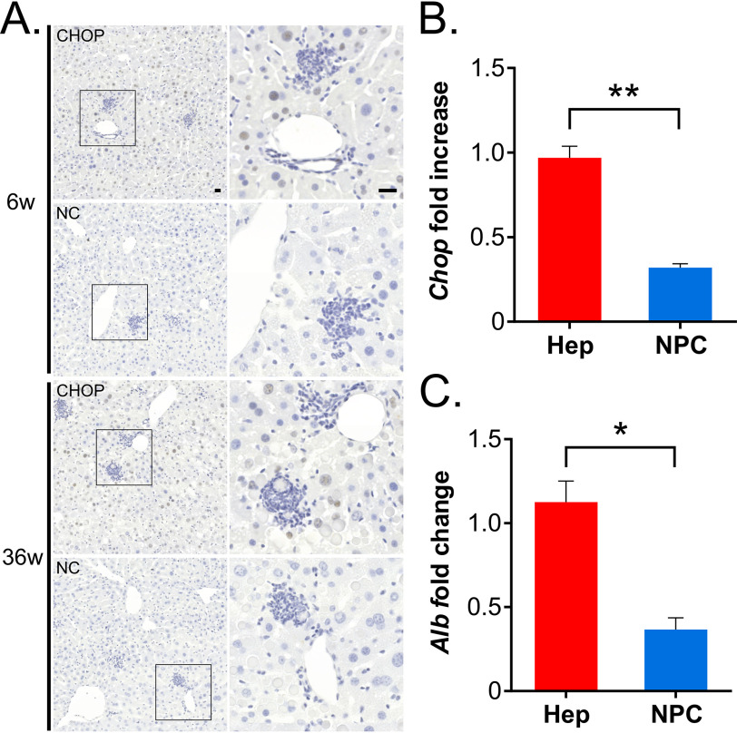
α1-Antitrypsin (AAT) encoded by the SERPINA1 gene is an acute-phase protein synthesized in the liver and secreted into the circulation. Its primary role is to protect lung tissue by inhibiting neutrophil elastase. The Z allele of SERPINA1 encodes a mutant AAT, named ATZ, that changes the protein structure and leads to its misfolding and polymerization, which cause endoplasmic reticulum (ER) stress and liver disease through a gain-of-function toxic mechanism. Hepatic retention of ATZ results in deficiency of one of the most important circulating proteinase inhibitors and predisposes to early-onset emphysema through a loss-of-function mechanism. The pathogenetic mechanisms underlying the liver disease are not completely understood. C/EBP-homologous protein (CHOP), a transcription factor induced by ER stress, was found among the most up-regulated genes in livers of PiZ mice that express ATZ and in human livers of patients homozygous for the Z allele. Compared with controls, juvenile PiZ/Chop-/- mice showed reduced hepatic ATZ and a transcriptional response indicative of decreased ER stress by RNA-Seq analysis. Livers of PiZ/Chop-/- mice also showed reduced SERPINA1 mRNA levels. By chromatin immunoprecipitations and luciferase reporter-based transfection assays, CHOP was found to up-regulate SERPINA1 cooperating with c-JUN, which was previously shown to up-regulate SERPINA1, thus aggravating hepatic accumulation of ATZ. Increased CHOP levels were detected in diseased livers of children homozygous for the Z allele. In summary, CHOP and c-JUN up-regulate SERPINA1 transcription and play an important role in hepatic disease by increasing the burden of proteotoxic ATZ, particularly in the pediatric population.

Keywords: CHOP; SERPINA1; alpha1-antitrypsin; alpha1-antitrypsin deficiency; c-JUN; c-Jun transcription factor; liver; liver injury; serpin; transcription regulation.


Conflict of interest statement

Conflict of interest—The authors declare that they have no conflicts of interest with the contents of this article.

Figures

Figure 1.
Figure 1.
CHOP is activated in PiZ mouse livers. A, Chop expression in livers of WT and PiZ mice of 6 and 36 weeks of age. B, Western blotting for CHOP in livers of WT and PiZ mice of 6 and 36 weeks of age (each lane corresponds to an independent mouse). A liver of a WT mouse injected intraperitoneally with 4 µg/g tunicamycin was used as a positive control. C, Western blotting for CHOP on nuclear extracts of 6-week-old WT and PiZ mouse livers. Lamin A was used as a nuclear marker and GAPDH as a cytosolic marker. D, representative immunohistochemistry with anti-CHOP antibody in 6- and 36-week-old WT and PiZ mouse livers (×40 magnification; scale bar, 100 μm; n = 3/group). E, heatmap from GSEA using a set of genes up-regulated by CHOP and differentially expressed genes between 6-week-old PiZ and WT mouse livers. The set of CHOP target genes was defined by combining ChIP-Seq and RNA-Seq data reported previously (44) from tunicamycin-treated WT mouse embryonic fibroblasts. Averages ± S. E. (error bars) are shown. Two-way ANOVA and Tukey's post-hoc tests were used: **, p < 0.001. 6w, 6 weeks old; 36w, 36 weeks old; N, nuclear extracts; C, cytosolic extracts; TUNI, tunicamycin.
Figure 2.
Figure 2.
CHOP expression detected in nuclei of hepatocytes but not of inflammatory infiltrating cells. A, representative images from immunohistochemistry with anti-CHOP antibody and negative control (NC) in livers of PiZ mice of 6 and 36 weeks of age. Images on the right correspond to the insets of the left panels. Scale bar, 20 μm; n = 3/group). 6w, 6 weeks old; 36w, 36 weeks old. B, Chop expression is greater in the parenchymal fraction enriched for hepatocytes (Hep) compared with the nonparenchymal cell (NPC) fraction. C Albumin, Albumin (Alb) gene expression was evaluated to confirm enrichment for hepatocytes. Averages ± S. E. (error bars) are shown; t test: *, p < 0.05; **, p < 0.01.
Figure 3.
Figure 3.
Genetic ablation of Chop reduces hepatic ATZ accumulation in juvenile PiZ mice. A, ELISA for serum ATZ in PiZ and PiZ/Chop−/− mice of 6 and 36 weeks of age (n ≥ 4/group). Serum was collected from the same mice at different ages. Averages ± S. D. (error bars) are shown. Two-way ANOVA and Tukey's post hoc tests were used: *, p < 0.05. B, representative PAS-D staining (×20 magnification; scale bar, 100 μm; n = 19 for 6-week-old PiZ/Chop−/− mice and n = 10 for 36-week-old PiZ/Chop−/− mice) and immunofluorescence (IF) for polymeric ATZ (×63 magnification; scale bar, 50 μm; n = 3/group) of livers of PiZ and PiZ/Chop−/− mice of 6 and 36 weeks of age. C, SERPINA1 expression in PiZ and PiZ/Chop−/− mice at 6 and 36 weeks of age (n = 3/group). β2-Microglobulin was used for normalization. Averages ± S. E. (error bars) are shown. Two-way ANOVA and Tukey's post hoc test were used: *, p < 0.05. D, representative Sirius Red staining on livers of 69-week-old PiZ and PiZ/Chop−/− mice (×10 magnification; scale bar, 200 μm; n = 10 PiZ and n = 5 PiZ/Chop−/− mice). E, Western blotting on soluble total ATZ and insoluble ATZ polymer of 6- and 36-week-old PiZ and PiZ/Chop−/− mouse livers. GAPDH and H3 were used for normalization. ns, not statistically significant; 6w, 6 weeks old; 36W, 36 weeks old; 69W, 69 weeks old.
Figure 4.
Figure 4.
A, Venn diagram comparing the gene data sets of PiZ versus WT, PiZ/Chop−/− versus WT, and PiZ/Chop−/− versus PiZ at 6 weeks of age. B and C, heatmaps showing the most relevant up- (B) and down-regulated (C) pathways in 6-week-old PiZ mouse livers and normalized to age-matched WT levels in PiZ/Chop−/− mice. DEG, differentially expressed genes; DW, down-regulated; UP, up-regulated.
Figure 5.
Figure 5.
CHOP and c-JUN up-regulate human SERPINA1 expression. A, schematic representation of AP-1–binding sites (1 and 2; shown in green) and CHOP-binding sites (1 and 2; shown in red) in the 5′-UTR of the human SERPINA1 gene (drawn to scale) retained in PiZ mouse genome. Nucleotide sequence is shown for the binding sites confirmed by luciferase assays. Nucleotide numbering is calculated as distance from the hepatic TSS. Arrows indicate the position of the primers for the qPCR on the liver chromatin immunoprecipitates shown in D and in Figs. S13 and S14. The dashed line indicates the promoter region cloned upstream of the luciferase reporter gene in the pAAT-Luc-AAT.3′UTR plasmid. Underlined nucleotides have been mutagenized. B, luciferase expression in HeLa cells co-transfected with the pAAT-Luc-AAT.3′UTR construct and the plasmids expressing human CHOP, c-JUN, c-FOS, or their combinations. An empty CMV-3XFLAG plasmid was used as control. Averages ± S. E. (error bars) are shown. One-way ANOVA and Tukey's post hoc test were used: a, p < 0.001 versus CMV-3XFLAG; b, p < 0.0001 versus c-JUN; c, p < 0.0001 versus CHOP; ns, not statistically significant versus CMV-3XFLAG. C, luciferase expression in HeLa cells co-transfected with the plasmids expressing human CHOP and c-JUN and the pAAT-Luc-AAT.3′UTR construct with mutagenized sites 1 and 2 of AP-1 sites. An empty CMV-3XFLAG plasmid was used as control. One-way ANOVA and Tukey's post hoc test were used: a, p < 0.001 versus CMV-3XFLAG; b, p < 0.0001 versus c-JUN + CHOP and versus AP-1 site 1wt-2M c-JUN + CHOP. D, ChIP on PiZ mouse livers using anti-CHOP antibody followed by qPCR on CHOP putative binding sites and AP-1 sites. p62 and Gapdh promoter regions were amplified as positive and negative control for CHOP enrichment, respectively. t test was used: *, p < 0.01. E, luciferase expression in HeLa cells co-transfected with the plasmids expressing human CHOP and c-JUN and the pAAT-Luc-AAT.3′UTR construct with mutagenized CHOP-binding sites 1 and 2. An empty CMV-3XFLAG plasmid was used as control. One-way ANOVA and Tukey's post hoc test were used: a, p < 0.001 versus CMV-3XFLAG; b, p < 0.0001 versus c-JUN + CHOP; c, p < 0.0001 versus AP-1 site 1wt-2M c-JUN + CHOP; d, p < 0.05 versus CMV-3XFLAG. F, luciferase expression in HeLa cells co-transfected with the plasmids expressing human CHOP, and the pAAT-Luc-AAT.3'UTR construct with mutagenized AP-1 site 2 and CHOP-binding site 2. An empty CMV-3XFLAG plasmid was used as control. One-way ANOVA and Tukey's post hoc test were used: a, p < 0.001 versus CMV-3XFLAG; b, p < 0.0001 versus CHOP; c, p < 0.0001 versus AP-1 site 1wt-2M CHOP. IP, immunoprecipitates; ns, not statistically significant.
Figure 6.
Figure 6.
Increased expression of CHOP in human livers expressing ATZ. Representative immunohistochemistry (IHC) with anti-CHOP antibody (top panels) and corresponding PAS-D staining (bottom panels) in the liver of a PiZZ patient with end-stage liver disease who underwent liver transplantation (PiZZ #3) and an age-matched PiMM control (PiMM #4) who also underwent liver transplantation as control (7) (×20 magnification; scale bar, 100 μm; insets, ×40 magnifications). Immunohistochemistry with anti-CHOP antibody and PAS-D staining for additional cases are shown in Fig. S15.
Figure 7.
Figure 7.
Age-dependent up-regulation of CHOP in human livers expressing ATZ. ddPCR for CHOP expression in human liver biopsies from healthy controls (adults: n = 6, age 50.7 ± 13.3 years; children: n = 12, age 6.8 ± 3.6 years), diseased controls (adults: n = 6, age 50.5 ± 13.0 years; children: n = 10, age 5.9 ± 4.3 years), and PiZZ patients (adults: n = 6, age 50.8 ± 13.0 years; children: n = 14, age 6.5 ± 3.7 years). Averages ± S. E. (error bars) are shown. Two-way ANOVA and Tukey's post hoc test were used: *, p < 0.05; **, p < 0.01.

References

    1. Carrell R. W., and Lomas D. A. (2002) α1-Antitrypsin deficiency—a model for conformational diseases. N. Engl. J. Med. 346, 45–53 10.1056/NEJMra010772 - DOI - PubMed
    1. Sveger T. (1988) The natural history of liver disease in α1-antitrypsin deficient children. Acta Paediatr. Scand. 77, 847–851 10.1111/j.1651-2227.1988.tb10767.x - DOI - PubMed
    1. Eriksson S., Carlson J., and Velez R. (1986) Risk of cirrhosis and primary liver cancer in alpha1-antitrypsin deficiency. N. Engl. J. Med. 314, 736–739 10.1056/NEJM198603203141202 - DOI - PubMed
    1. Sveger T. (1976) Liver disease in α1-antitrypsin deficiency detected by screening of 200,000 infants. N. Engl. J. Med. 294, 1316–1321 10.1056/NEJM197606102942404 - DOI - PubMed
    1. Perlmutter D. H. (2002) Liver injury in α1-antitrypsin deficiency: an aggregated protein induces mitochondrial injury. J. Clin. Invest. 110, 1579–1583 10.1172/JCI16787 - DOI - PMC - PubMed

Publication types

MeSH terms