Review
. 2020 Jul 28;11(1):3760.
doi: 10.1038/s41467-020-17575-w.

Origin and function of the yolk sac in primate embryogenesis

Affiliations
Review

Origin and function of the yolk sac in primate embryogenesis

Connor Ross et al. Nat Commun. .

Abstract

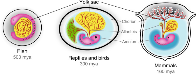
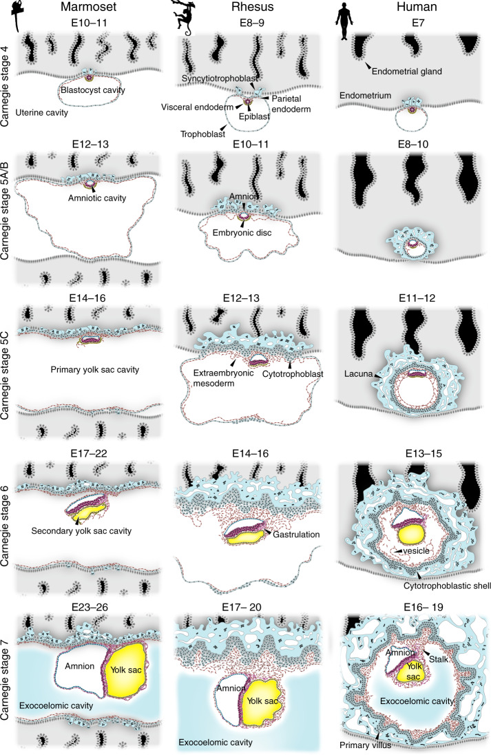
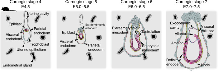
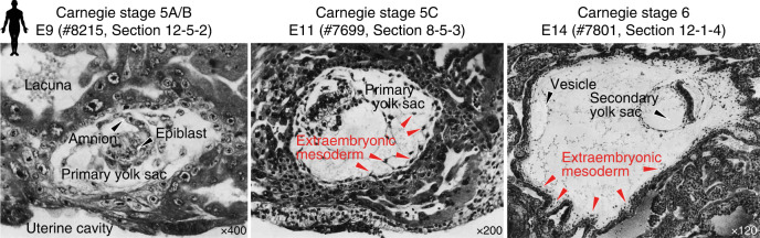
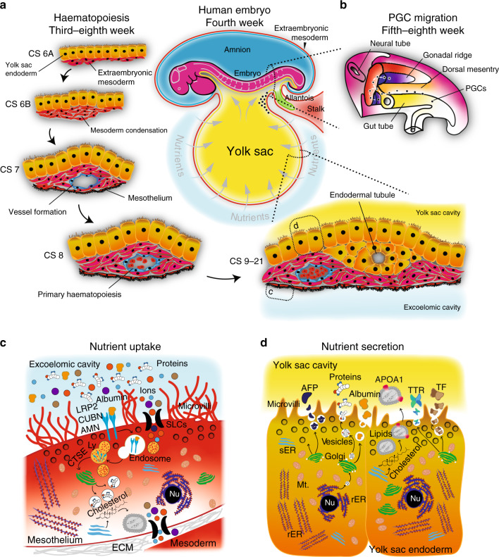
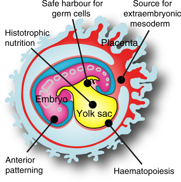
Human embryogenesis is hallmarked by two phases of yolk sac development. The primate hypoblast gives rise to a transient primary yolk sac, which is rapidly superseded by a secondary yolk sac during gastrulation. Moreover, primate embryos form extraembryonic mesoderm prior to gastrulation, in contrast to mouse. The function of the primary yolk sac and the origin of extraembryonic mesoderm remain unclear. Here, we hypothesise that the hypoblast-derived primary yolk sac serves as a source for early extraembryonic mesoderm, which is supplemented with mesoderm from the gastrulating embryo. We discuss the intricate relationship between the yolk sac and the primate embryo and highlight the pivotal role of the yolk sac as a multifunctional hub for haematopoiesis, germ cell development and nutritional supply.


Conflict of interest statement

The authors declare no competing interests.

Figures

Fig. 1
Fig. 1. Extraembryonic membranes in fish, birds and mammals.
The yolk sac dates back to our aquatic ancestors and thus represents the phylogenetically oldest extraembryonic tissue. Amnion, chorion and allantois are inventions of the amniotic egg, which have been subsequently adapted in mammals to support embryonic development inside the uterus. mya million years ago.
Fig. 2
Fig. 2. Yolk sac formation in primates.
Embryonic stages of common marmoset (Callithrix jacchus), rhesus monkey (Macaca mulatta) and human (Homo sapiens) are depicted in Carnegie stages and embryonic day (E) for each species. The definitive, secondary yolk sac is highlighted in yellow. Drawings are based on representative histological sections of common marmoset,, rhesus and human,,,, embryos.
Fig. 3
Fig. 3. Early postimplantation development in mouse.
Embryonic stages are depicted according to Carnegie stages and embryonic day (E),,,.
Fig. 4
Fig. 4. Histology of early implantation stages in human.
Embryonic stages are depicted according to Carnegie stage and embryonic day (E) with the Carnegie specimen and section ID indicated. Images are reproduced from ref. . Original annotations have been removed using the Adobe Photoshop Spot Healing Tool.
Fig. 5
Fig. 5. Functions of the primate secondary yolk sac.
a Vasculogenesis of the yolk sac forms an intricate vascular plexus that envelopes the yolk sac from Carnegie stage (CS) 6A. Haematopoiesis ensues in preparation for the onset of embryonic circulation, mediated by the developing heart. Reciprocal crosstalk signalling between the endoderm and mesoderm regionalises foci of condensed mesoderm, which form primitive blood islands. b PGCs are specified in the embryonic region and migrate via the yolk sac into the hindgut and up through the dorsal mesentery towards the genital ridges. Schematic drawn after ref. . The original images were published in Medical Physiology E-Book, Copyright Elsevier (Health Sciences, ISBN 1455733288, 9781455733286). c The mesothelium exhibits hallmarks of absorption, degradation and re-synthesis, evident by high concentration of LRP2–CUBN–AMN endovesicular complexes in the plasma membrane. Lysosomes contain hydrolytic enzymes, such as the Cathepsins, to mediate the degradation of maternal proteins and other complex molecules in the nutrient-rich exocoelomic cavity. Nutrients are either transported or re-synthesised and exocytosed either directly into surrounding blood vessels or transported through the extracellular environment towards the endodermal tubules and the yolk sac cavity. d The endoderm contains high concentrations of rER, sER, glycogen vesicles and exocytotic vesicles. Yolk sac endoderm cells synthesise and exocytose key carrier proteins such at AFP, TTR, ALB and TF into the yolk sac cavity, which are absorbed by primitive gut endoderm. Nu nucleus, rER rough endoplasmic reticulum, sER smooth endoplasmic reticulum, Mt Mitochondrion, SLC solute carrier family, Ly lysosome, CTSE Cathepsin family member E, LRP2 LDL-related receptor 2, CUBN Cubulin, ApoA apolipoprotein A, TTR transthyretin, TF transferrin, AFP alpha-fetoprotein, AMN amnionless.
Fig. 6
Fig. 6. Principal functions of the primate hypoblast and yolk sac.
Graphical illustration summarising the roles of the hypoblast—yolk sac lineage during early primate development.

References

    1. Ravi, V. & Venkatesh, B. The divergent genomes of teleosts. Annu. Rev. Anim. Biosci. 6, 47–68 (2018). - PubMed
    1. Jeffery, J. E., Bininda-Emonds, O. R. P., Coates, M. I. & Richardson, M. K. Analyzing evolutionary patterns in amniote embryonic development. Evol. Dev. 4, 292–302 (2002). - PubMed
    1. Sheng G, Foley AC. Diversification and conservation of the extraembryonic tissues in mediating nutrient uptake during amniote development. Ann. NY Acad. Sci. 2012;1271:97–103. - PMC - PubMed
    1. Bainbridge, D. R. J. The evolution of pregnancy. Early Hum. Dev. 90, 741–745 (2014). - PubMed
    1. Blackburn, D. G. in Encyclopedia of Reproduction, Vol. 4 (eds Knobil, E. & Neill, J. D.) 994–1003 (Academic, London, 1999).

Publication types