

Study of Kyasanur forest disease viremia, antibody kinetics, and virus infection in target organs of Macaca radiata
- PMID: 32724103
- PMCID: PMC7387489
- DOI: 10.1038/s41598-020-67599-x
Study of Kyasanur forest disease viremia, antibody kinetics, and virus infection in target organs of Macaca radiata
Abstract
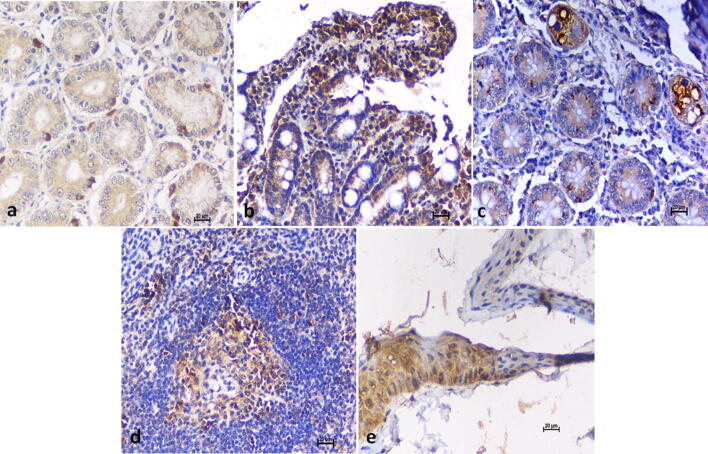
The present manuscript deals with experimental infections of bonnet macaques (Macaca radiata) to study disease progression for better insights into the Kyasanur Forest Disease (KFD) pathogenesis and transmission. Experimentally, 10 monkeys were inoculated with KFD virus (KFDV) (high or low dose) and were regularly monitored and sampled for various body fluids and tissues at preset time points. We found that only 2 out of the 10 animals showed marked clinical signs becoming moribund, both in the low dose group, even though viremia, virus shedding in the secretions and excretions were evident in all inoculated monkeys. Anti-KFDV immunoglobulin (Ig)M antibody response was observed around a week after inoculation and anti-KFDV IgG antibody response after two weeks. Anaemia, leucopenia, thrombocytopenia, monocytosis, increase in average clotting time, and reduction in the serum protein levels were evident. The virus could be re-isolated from the skin during the viremic period. The persistence of viral RNA in the gastrointestinal tract and lymph nodes was seen up to 53 and 81 days respectively. Neuro-invasion was observed only in moribund macaques. Re-challenge with the virus after 21 days of initial inoculation in a monkey did not result in virus shedding or immune response boosting.
Conflict of interest statement
The authors declare no competing interests.
Figures

References
-
- Work TH, Trapido H. Kyasanur forest disease a new virus disease in India: Summary of the preliminary report of investigations of the Virus Research Centre on an epidemic disease affecting forest villagers and wild monkeys of Shimoga district, Mysore. Indian J Med Sci. 1957;11:1–2. - PubMed
-
- Mourya DT, Yadav PD. The recent scenario of the emergence of Kyasanur forest disease in India and public health importance. Curr. Trop. Med. Rep. 2016;3:7–13.
-
- Jorge Boshell, M., Rajagopalan, P.K., Goverdhan, M.K. & Pavri, K.M. The isolation of Kyasanur forest disease virus from small mammals of the SagaSorab forests, Mysore state, India. Indian J. Med. Res.56(4), 569–72 (1968). - PubMed
-
- Rajagopalan PK, Paul SD, Sreenivasan MA. Involvement of Rattus blanfordi (rodentia: muridae) in the natural cycle of Kyasanur forest disease virus. Indian J Med. Res. 1969;57(6):999–1002. - PubMed
MeSH terms
Substances
LinkOut - more resources
Full Text Sources
