Hippocampal volume and hippocampal neuron density, number and size in schizophrenia: a systematic review and meta-analysis of postmortem studies
- PMID: 32724199
- PMCID: PMC7854798
- DOI: 10.1038/s41380-020-0853-y
Hippocampal volume and hippocampal neuron density, number and size in schizophrenia: a systematic review and meta-analysis of postmortem studies
Abstract
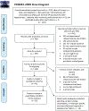
Reduced hippocampal volume is a consistent finding in neuroimaging studies of individuals with schizophrenia. While these studies have the advantage of large-sample sizes, they are unable to quantify the cellular basis of structural or functional changes. In contrast, postmortem studies are well suited to explore subfield and cellular alterations, but low sample sizes and subject heterogeneity impede establishment of statistically significant differences. Here we use a meta-analytic approach to synthesize the extant literature of hippocampal subfield volume and cellular composition in schizophrenia patients and healthy control subjects. Following pre-registration (PROSPERO CRD42019138280), PubMed, Web of Science, and PsycINFO were searched using the term: (schizophrenia OR schizoaffective) AND (post-mortem OR postmortem) AND hippocampus. Subjects were adult men and women with schizophrenia or schizoaffective disorder or non-psychiatric control subjects, and key outcomes, stratified by hippocampal hemisphere and subfield, were volume, neuron number, neuron density, and neuron size. A random effects meta-analysis was performed. Thirty-two studies were included (413 patients, 415 controls). In patients, volume and neuron number were significantly reduced in multiple hippocampal subfields in left, but not right hippocampus, whereas neuron density was not significantly different in any hippocampal subfield. Neuron size, averaged bilaterally, was also significantly reduced in all calculated subfields. Heterogeneity was minimal to moderate, with rare evidence of publication bias. Meta-regression of age and illness duration did not explain heterogeneity of total hippocampal volume effect sizes. These results extend neuroimaging findings of smaller hippocampal volume in schizophrenia patients and further our understanding of regional and cellular neuropathology in schizophrenia.
© 2020. The Author(s), under exclusive licence to Springer Nature Limited.
Conflict of interest statement
Conflict of interest
The authors declare no conflicts of interest.
Figures


References
-
- Adriano F, Caltagirone C, Spalletta G. Hippocampal volume reduction in first-episode and chronic schizophrenia: A review and meta-analysis. Neuroscientist. 2012;18(2):180–200. - PubMed
-
- Honea R, Crow TJ, Passingham D, Mackay CE. Regional deficits in brain volume in schizophrenia: A meta-analysis of voxel-based morphometry studies. Am J Psychiatry. 2005;162(12):2233–45. - PubMed
Publication types
MeSH terms
Grants and funding
LinkOut - more resources
Full Text Sources
Medical

