

Case Reports
doi: 10.12688/f1000research.23204.2.
eCollection 2020.
Appearance and management of COVID-19 laryngo-tracheitis: two case reports
Affiliations
- PMID: 32724561
- PMCID: PMC7364183
- DOI: 10.12688/f1000research.23204.2
Item in Clipboard
Case Reports
Appearance and management of COVID-19 laryngo-tracheitis: two case reports
F1000Res.
.
Abstract
We present two cases of coronavirus disease 2019 (COVID-19)-related laryngotracheitis in good-prognosis, ventilated patients who had failed extubation. As the pandemic continues to unfold across the globe and better management of those with respiratory failure develops, this may be an increasingly common scenario. Close ENT-intensivist liaison, meticulous team preparation, early consideration of rigid endoscopy and prospective data collection and case sharing are recommended.
Keywords: Airway management; COVID; Intensive care; SARS-CoV-19.
Copyright: © 2020 Oliver CM et al.
Conflict of interest statement
No competing interests were disclosed.
Figures
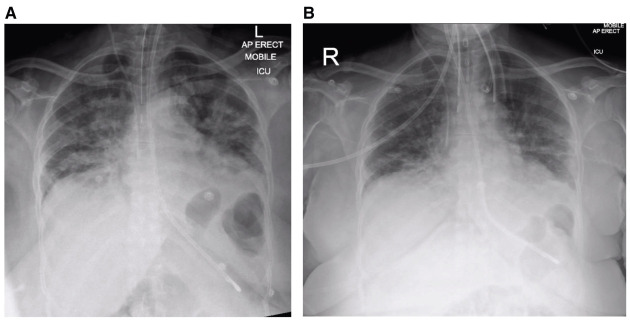
(
A) Post-intubation plain chest radiograph, on ICU on day 5 post-onset of symptoms. (
B) Plain chest radiograph on day 10 post-onset of symptoms.
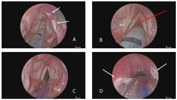
(
A) View of supraglottis showing ulcerated epiglottis. (
B) Glottis showing relative sparing of vocal cords and false cords, but profound subglottic oedema. (
C) Following change to size 6 endotracheal tube, there is some anterior glottic airway. (
D) However, the subglottis is also ulcerated and oedematous mucosa prevents rigid bronchoscopy (0
o Hopkins’ rod) beyond the third tracheal ring. White arrows indicate areas of ulceration and red arrow subglottic oedema.
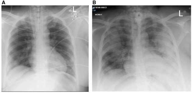
(
A) Post-intubation plain chest radiograph, on day 1 of hospital admission. (
B) Day 5 following re-intubation.
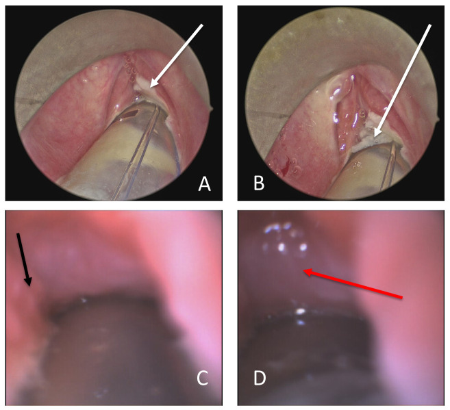
(
A) View of supraglottis showing ulcerated glottis. (
B) Glottis showing sparing of false cords, but profound glottic oedema and glottic and subglottic ulceration. (
C) Flexible bronchoscopy via the anterior commissure shows subglottic oedema and granulation tissue (black arrow). (
D) Oedematous mucosa prevents flexible bronchoscopy beyond the third tracheal ring. White arrows indicate areas of ulceration, black arrow granulation tissue and red arrow tracheal oedema.
References
-
- Davenport L: ICU Lessons on COVID-19 From Italian Front Line: Be Flexible.2020; (accessed 29.03.20 2020). Reference Source
-
- Royal College of Anaesthetists: Royal College of Anaesthetists COVID-19 clinical guidance.2020; (accessed 29.03.20 2020). Reference Source
Publication types
MeSH terms
LinkOut - more resources
Full Text Sources
Research Materials
Miscellaneous