Case Reports
. 2020 Jul 16:2020:8821332.
doi: 10.1155/2020/8821332. eCollection 2020.

An Unusual Case of Radicular Pain Caused by Bilateral Lumbar Synovial Cyst: A Case Report and Review of the Literature

Affiliations
Case Reports

An Unusual Case of Radicular Pain Caused by Bilateral Lumbar Synovial Cyst: A Case Report and Review of the Literature

David Ruiz-Picazo et al. Case Rep Orthop. .

Abstract

Introduction: Spinal synovial cysts (SSCs) constitute an uncommon degenerative lesion of the spine. They are usually asymptomatic but they may also cause symptoms of variable severity. SSCs are benign growths adjoining the facet joints that may induce low back pain, lumbar radiculopathy, and neurological deficit. There are different treatment options that range from conservative management to interventions like image-guided epidural steroid injection or direct cyst puncture and finally to open or endoscopic spinal canal decompression and spinal bone fusion with/without instrumentation. A discussion of current management options for this unusual disease is presented. Material and Methods. A 52-year-old female patient presented with low back pain and left leg pain. Plain radiography demonstrated instability at the L4-L5 level. Magnetic resonance images (MRIs) revealed a bilateral cystic lesion at the L4-L5 level with associated instability and degenerative disc disease at the level L5-S1. Initially, conservative treatment was performed by aspiration of the left cyst and infiltration with corticosteroids with improvement of the pain for 1 year. After this period, the radicular and the low back pain reoccurred.

Results: Following leg pain recurrence, a hybrid L4-S1 fusion was performed. After surgery, there was clinical improvement and six months later, the patient returned to daily activities. The radiological study after five-year follow-up shows adequate implant position, without signs of loosening, compatible with solid fusion.

Conclusion: After reviewing the literature, the optimal management for patients with symptomatic lumbar synovial cyst must be very individualized, which is essential to achieve a favorable outcome.


Conflict of interest statement

The authors declare that there is no conflict of interest regarding the publication of this paper.

Figures

Figure 1
Figure 1
(a) Standing lateral radiograph where L4-L5 segmentary instability (4 mm) can be seen. (b) Laminar inclination angle. In (a), we can appreciate vacuum phenomenon and facet hypertrophy, with 10° of facet tropism that suggests segmentary instability.
Figure 2
Figure 2
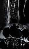
MR images on T1, T2, and STIR sequences. Hypointensity on T1 sequences and hyperintensity on T2 sequences. In sagittal cuts, we can see the cyst and how it compromises the spinal canal, as well as degenerative changes in L4-L5/L5-S1. In axial cuts, L4-L5 bilateral cysts can be seen (white arrows).
Figure 3
Figure 3
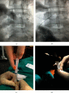
(a, b) Fluoroscopic images obtained during the aspiration and infiltration of the synovial cyst. The end of the needle was slightly bent to ease the access due to the morphology of the cyst. In (c) and (d), we can observe the cyst contents inside the syringe and the subsequent infiltration (2 cc of betamethasone + 2 cc of mepivacaine).
Figure 4
Figure 4
Standing radiographs in anterior-posterior and lateral projections. Postoperative study of the 5-year follow-up. We can observe the obtained fusion with and adequate position of the implants.

References

    1. Doyle A. J., Merrilees M. Synovial cysts of the lumbar facet joints in a symptomatic population: prevalence on magnetic resonance imaging. Spine. 2004;29(8):874–878. doi: 10.1097/00007632-200404150-00010. - DOI - PubMed
    1. Janssen S. J., Ogink P. T., Schwab J. H. The prevalence of incidental and symptomatic lumbar synovial facet cysts. Clinical Spine Surgery. 2018;31(5):E296–E301. doi: 10.1097/BSD.0000000000000648. - DOI - PubMed
    1. Chebib I., Chang C. Y., Schwab J. H., Kerr D. A., Deshpande V., Nielsen G. P. Histopathology of synovial cysts of the spine. Histopathology. 2018;72(6):923–929. doi: 10.1111/his.13465. - DOI - PubMed
    1. Epstein N., Baisden J. The diagnosis and management of synovial cysts: efficacy of surgery versus cyst aspiration. Surgical Neurology International. 2012;3(4):p. 157. doi: 10.4103/2152-7806.98576. - DOI - PMC - PubMed
    1. Ramhmdani S., Ishida W., Perdomo-Pantoja A., Witham T. F., Lo S.-F. L., Bydon A. Synovial cyst as a marker for lumbar instability: a systematic review and meta-analysis. World Neurosurgery. 2019;122:e1059–e1068. doi: 10.1016/j.wneu.2018.10.228. - DOI - PubMed

Publication types

LinkOut - more resources