Review
. 2020 Aug;68(Suppl 2):106-115.
doi: 10.1007/s00106-020-00877-2.

Implantation of a new active bone conduction hearing device with optimized geometry

Affiliations
Review

Implantation of a new active bone conduction hearing device with optimized geometry

S K Plontke et al. HNO. 2020 Aug.

Abstract

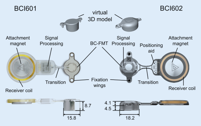
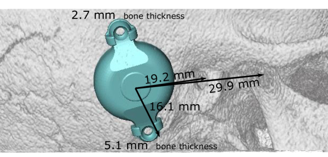
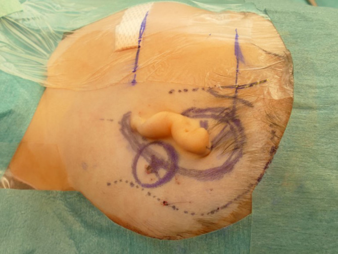
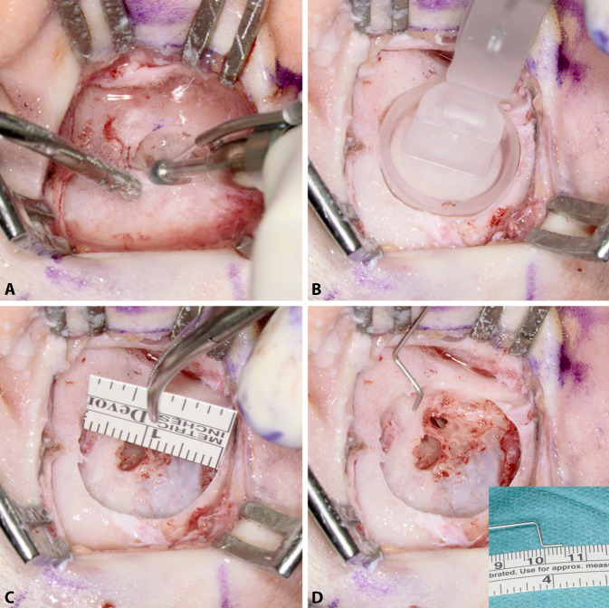
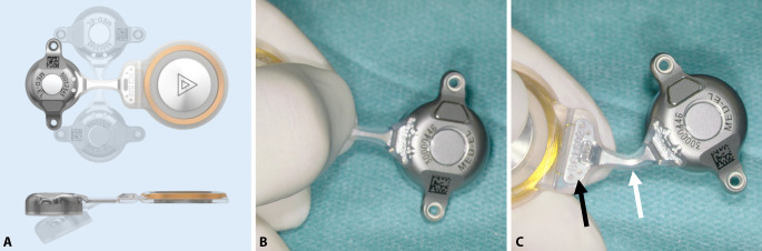
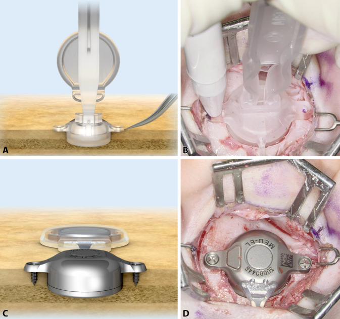
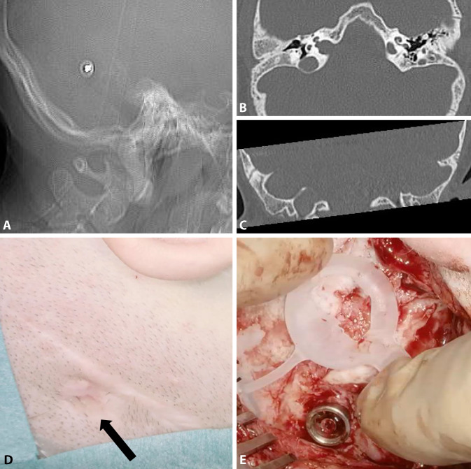
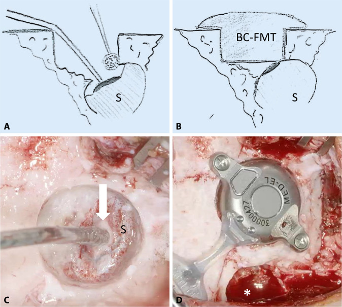
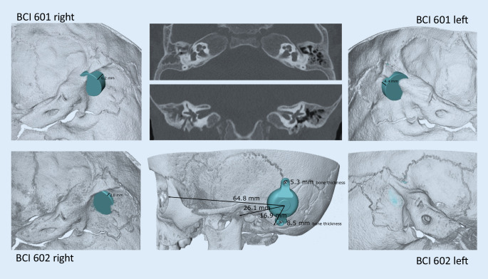
Here, we describe the surgical technique for implanting a new, active, transcutaneous bone conduction hearing aid. The implant technology is based on a system that has been in use reliably since 2012. The geometry of the new implant has been adapted based on experience with previously introduced implants. The surgery was feasible, standardized, and safe. Due to the optimized geometric design that improved the bone fit, it is not necessary to use specialized, detailed preoperative planning, except in challenging anatomical conditions; e.g., in young children, malformations, poor pneumatization, or after a canal wall down mastoidectomy.

Keywords: Hearing aids; Hearing loss, conductive; Hearing loss, mixed, conductive-sensorineural; Otologic surgical procedures; Patients.


Conflict of interest statement

S.K. Plontke: Audio Cure Pharma GmbH, Berlin (consultancy); Oticon Medical, Denmark; Cochlear Ltd., Australia; Federal Ministry of Research and Education (Research Projects); BV-HNOe.V.; MerckSerono; Infectopharm; Dr.Willmar Schwabe GmbH& Co. KG, Deutschland (honrary for lectures and travel support for lectures). T. Rahne: Oticon Medical,Denmark; Cochlear Ltd., Australia (Research Projects). S.K. Plontke, G. Götze, C. Wenzel, T. Rahne, and R.Mlynski: MED-EL, Austria, and MED-EL Germany (Research Projects); MED-EL Austria and MED-EL Germany (travel support for lectures).

Figures

Fig. 1
Fig. 1
Comparison of the bone conduction implants (BCI) 601 and 602. Images show the new active bone conduction hearing system, BCI 602 (right), and the previous model, BCI 601 (left), (© MED-EL, Innsbruck, Austria, with permission). Virtual three-dimensional (3D) models of the bone conduction-floating mass transducers (BC-FMT) are used for preoperative planning. All measurements are expressed in millimeters
Fig. 2
Fig. 2
Landmarks for transferring the preoperatively determined implant position into the surgical field. Suitable landmarks include the mastoid tip, the anterior wall of the external ear canal or its remnants, the lateral orbital rim (not shown here), or the temporomandibular joint [17]. During planning, bone thickness is measured in the area where the self-drilling cortical screws will be placed to fix the bone conduction-floating mass transducer [33]
Fig. 3
Fig. 3
Planning the incisions. The skin and pericranial incisions should not be superimposed. Exceptions include cases of atresia, with a possible aesthetic auricular reconstruction planned at a later stage. In these cases, the position of the ear is marked according to standard recommendations. The posterior incision is made through all layers at a distance of approximately 15 mm from the later helix [9]
Fig. 4
Fig. 4
Creating the implant bed. A Drilling of the implant bed is started in the center of the position planned for the bone conduction-floating mass transducer. The depth is 4.5 mm and the diameter is relatively small, approximately 5 mm. The implant bed is successively enlarged by moving the drill bit away from critical structures (sigmoid sinus, dura, ear canal, open mastoid cavity) to avoid damage. B The adequacy of the size and shape of the implant bed can be evaluated with a sterile “BCI 602 Sizer Kit.” Alternatively, the implant bed can be measured with a sterile ruler and a depth gauge (here, the authors used a 0.7-mm suction tip, off-label) (C, D)
Fig. 5
Fig. 5
Positioning the receiver coil and magnet. A The implant can be bent at the transition zone (white arrow in C) ±90° in the horizontal plane and −30° in the vertical plane. While bending, the implant should be held with the thumbs and index fingers of both hands; one hand should be placed at the positioning aid (B and black arrow in C), and the other hand should be placed at the bone conduction-floating mass transducer. (A: MED-EL, Innsbruck, Austria, with permission)
Fig. 6
Fig. 6
Fixing the bone conduction-floating mass transducer (BC-FMT) to the bone. A, B The fixation wings with anchor holes should lay flat on the bone. When pressing the BC-FMT down with a finger, it should not wiggle or wobble. C, D The BC-FMT is fixed with self-drilling cortical screws; this procedure is simpler in this model (BCI 602) than in the previous model (BCI 601). (AC: MED-EL, Innsbruck, Austria, with permission)
Fig. 7
Fig. 7
Preoperative plan for the implantation of an active, transcutaneous bone conduction hearing system in a 4.5-year-old patient with atresia and complete conductive loss (according to Plontke et al. 2014 [17]). The bone conduction-floating mass transducers required intracranial penetrations of: (A, B) 6–7 mm for the BCI 601 and (C, D) <2 mm for the BCI 602. Due to the expected skull growth [25], a computed tomography scan and planning were not repeated before surgery at the age of 5.5 years. Intraoperatively, the dura and sigmoid sinus were exposed, but not impressed. This patient is also shown in Fig. 4
Fig. 8
Fig. 8
Radiological planning and surgical field for the simultaneous explantation of a fixture from a percutaneous bone conduction implant and implantation of a BCI 602 (right ear). A The computed tomography (CT) scout view shows the fixture. Axial CT (B) and coronal (C) CT show mastoid hypoplasia, the lateralized sigmoid sinus, and minimal cortical bone thickness (<5 mm) at the bone conduction-floating mass transducer (BC-FMT) position. The abutment is completely overgrown with skin (black arrow) (D) and is removed before creating the implant bed (E). (A–C: CT images reproduced with permission from Prof. Dr. med. M.A. Weber, Institute for Diagnostic and Interventional Radiology, Pediatric and Neuro-Radiology, University Medicine Rostock)
Fig. 9
Fig. 9
Placing the bone conduction-floating mass transducer (BC-FMT) close to the sigmoid sinus. A, C Due to the close proximity of the sigmoid sinus (S), a bony island is created on the exposed sinus wall (white arrow). This can be used to protect the sinus from the drill bit by temporarily and gently pressing on the island with the suction tip. B, D Final position of the BC-FMT after removing the pre-existing fixture (asterisk)
Fig. 10
Fig. 10
Preoperative planning based on Plontke et al. 2014 [17] for the implantation of the active transcutaneous bone conduction hearing systems, BCI 601 and BCI 602, in a 3-year-old patient. The patient had bilateral atresia, poorly pneumatized mastoids, hypoplastic tympanic cavities, dysplastic ossicles, an atypical facial nerve course, and complete conductive loss. Without the BCI Lifts, the BCI 601 required intracranial penetrations of 6.2 mm (BCI 601 right) and 4.4 mm (BCI 601 left). The new implant with optimized geometry (BCI 602) required intracranial penetration, with an impression of the dura and/or sinus, of 2 mm (BCI 602right). However, intracranial penetration was completely avoided on the left side by using a 1-mm BCI Lift at the inferior fixation wing (bottom row center). After informed consent about the off-label use in this age group, and due to the explicit wishes of the parents, the BCI 602 was implanted in this patient at the age of 3 years and 4 months (this patient is also shown in Fig. 3). The axial computed tomography (CT) (center top) and coronal CT (center middle) are reproduced with permission from Prof. Dr. S. Kösling (Radiology, University Medicine Halle, Germany)

References

    1. Anonymous MED-EL Medical Electronics. Bonebridge (BCI 602) Surgical Guide V 2.0 (2019). In: MED-EL, Innsbruck, Austria
    1. Arnold H, Schulze M, Wolpert S, et al. Positioning a novel transcutaneous bone conduction hearing implant: a systematic anatomical and radiological study to standardize the retrosigmoid approach, correlating navigation-guided, and landmark-based surgery. Otol Neurotol. 2018;39:458–466. - PubMed
    1. Baumgartner WD, Hamzavi JS, Boheim K, et al. A new transcutaneous bone conduction hearing implant: short-term safety and efficacy in children. Otol Neurotol. 2016;37:713–720. doi: 10.1097/MAO.0000000000001038. - DOI - PubMed
    1. Beutner D, Delb W, Frenzel H, et al. Guideline “Implantable hearing aids”-short version : German S2k guideline of the Working Group of German-speaking Audiologists, Neurootologists and Otologists (ADANO), of the German Society of Oto-Rhino-Laryngology, Head and Neck Surgery (DGHNO) in collaboration with the German Society of Audiology (DGA), the German Society of Phoniatrics and Pediatric Audiology (DGPP), and patient representatives. HNO. 2018;66:654–659. doi: 10.1007/s00106-018-0531-4. - DOI - PubMed
    1. Brkic FF, Riss D, Scheuba K, et al. Medical, technical and audiological outcomes of hearing rehabilitation with the bonebridge transcutaneous bone-conduction implant: a single-center experience. J Clin Med. 2019;8:1614. doi: 10.3390/jcm8101614. - DOI - PMC - PubMed