. 2020 Oct;412(25):6777-6788.
doi: 10.1007/s00216-020-02820-4. Epub 2020 Jul 28.

Point-of-care-ready nanoscale ISFET arrays for sub-picomolar detection of cytokines in cell cultures

Affiliations

Point-of-care-ready nanoscale ISFET arrays for sub-picomolar detection of cytokines in cell cultures

Dipti Rani et al. Anal Bioanal Chem. 2020 Oct.

Abstract

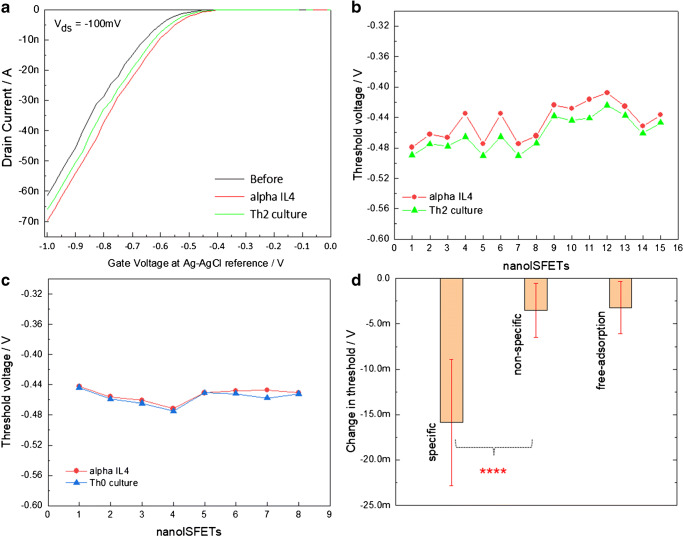
Rapid and frequent screening of cytokines as immunomodulation agents is necessary for precise interventions in severe pathophysiological conditions. In addition to high-sensitivity detection of such analytes in complex biological fluids such as blood, saliva, and cell culture medium samples, it is also crucial to work out miniaturized bioanalytical platforms with potential for high-density integration enabling screening of multiple analytes. In this work, we show a compact, point-of-care-ready bioanalytical platform for screening of cytokines such as interleukin-4 (IL-4) and interleukin-2 (IL-2) based on one-dimensional ion-sensitive field-effect transistors arrays (nanoISFETs) of silicon fabricated at wafer-scale via nanoimprint lithography. The nanoISFETs biofunctionalized with receptor proteins alpha IL-4 and alpha IL-2 were deployed for screening cytokine secretion in mouse T helper cell differentiation culture media, respectively. Our nanoISFETs showed robust sensor signals for specific molecular binding and can be readily deployed for real-time screening of cytokines. Quantitative analyses of the nanoISFET-based bioanalytical platform was carried out for IL-4 concentrations ranging from 25 fg/mL (1.92 fM) to 2.5 μg/mL (192 nM), showing a limit of detection down to 3-5 fM, which was found to be in agreement with ELISA results in determining IL-4 concentrations directly in complex cell culture media. Graphical abstract.

Keywords: Cytokines; Immunosensor; Ion-sensitive field-effect transistors; Label-free; Silicon nanowires.


Conflict of interest statement

The authors declare that they have no conflicts of interest.

Figures

None
Graphical abstract
Fig. 1
Fig. 1
Nanoscale silicon ISFETs for the assembly of a point-of-care electrical biosensor for monitoring of cytokines. (a) Scanning electron micrograph image of one set of 4 nanoISFETs with common source and individual drain electrodes; (b) photograph of a wire-bonded and encapsulated sensor chip ready for biosensor measurements. (c, d) Photographs showing a sensor chip connected to the miniaturized DC readout system
Fig. 2
Fig. 2
Biofunctional interface development for the label-free detection of interleukins using nanoISFETs. (a) IL-4 or IL-2-specific receptor molecules (anti-IL-4 or alpha IL-4 antibody and anti-IL-2 respectively) functionalized on the nanowire surface providing biospecific interaction for IL-4 and IL-2 from mouse T cell culture, (b) Th0 cultures with negligible concentrations of IL-4 and IL-2 were used for control experiments, (c) 3D illustrations of AFM measurements showing 4 nanowires before and after biofunctionalization, (d) graphs showing changes in the height profiles of nanoISFETs, and (e) histogram showing roughness analysis representing overall differences in heights for the scanned area shown, signifying the binding of receptor anti-IL-4
Fig. 3
Fig. 3
Electrical detection of IL-4 in complex mouse T cell culture medium. (a) Graph showing the transfer characteristics of an exemplary nanoISFET before and after biofunctionalization with anti-IL-4 (black and red curves), and after exposure to the culture medium with IL-4, (b) graph plots of Vth values associated with anti-IL-4-modified nanoISFETs (red) and after specific binding of IL-4 (green), (c) graph comparing threshold values for anti-IL-4-modified nanoISFETs (red) and mouse T cell culture media with negligible presence of IL-4 (blue), and (d) the graph showing average changes in the Vth values of anti-IL-4-modified nanoISFETs for specific binding (culture with IL-4) and non-specific binding (culture with other cytokines but negligible concentrations of IL-4) and free adsorption of IL-4s on non-modified nanoISFETs. An unpaired T test analysis of mean values for specific and non-specific binding showed the sensor signal as statistically extremely significant with P values of 0.0001
Fig. 4
Fig. 4
Electrical detection of IL-2 in complex mouse T cell culture medium (iTregs; differentiated using 2.5 ng/mL TGF-b and 5 ng/mL IL-2). (a) Graph showing typical transfer characteristics of an exemplary nanoISFET before and after biofunctionalization with anti-IL-4 (black and red curves), and after exposure to the culture medium with IL-2, (b) graph plots of Vths associated with alpha IL-2-modified nanoISFETs (red) and after specific binding of IL-2 (green), (c) graph comparing threshold values for anti-IL-2-modified nanoISFETs and mouse T cell culture with negligible presence of IL-2, and (d) the graph showing average changes in Vth of anti-IL-2-modified nanoISFETs for specific binding (culture with IL-2) and non-specific binding (culture with other cytokines, but negligible concentrations of IL-2). An unpaired T test analysis of mean values for specific and non-specific binding showed the sensor signal as statistically extremely significant with P values of 0.0001
Fig. 5
Fig. 5
Calibration of the nanoISFETs using known IL-4 concentrations spiked in PBS and real-time detection of IL-4 detection using nanoISFETs. (a) The graph shows a typical measurement, where drain output current from a nanoISFET is plotted before and after biofunctionalization steps and then after injections of different concentrations of IL-4 in PBS, (b) Average output current for the measurement shown in graph a, (c) average changes in equivalent threshold values which were calculated from output current values of 4 nanoISFETs for different IL-4 concentrations ranging from 1.92 fM up to 192 nM. The sigmoid fitting of this dose response curve was used for calibration of the sensors to compare the shift in Vth values from mouse T cell culture samples (with “unknown” IL-4 concentration), and (d) IL-4 concentrations in an “unknown” sample of mouse T cell culture measured by ELISA

References

    1. Stenken JA, Poschenrieder AJ. Bioanalytical chemistry of cytokines--a review. Anal Chim Acta. 2015;853:95–115. - PMC - PubMed
    1. Kelso A. Cytokines: principles and prospects. Immunol Cell Biol. 1998;76(4):300–317. - PubMed
    1. Banchereau J, Pascual V, O’Garra A. From IL-2 to IL-37: the expanding spectrum of anti-inflammatory cytokines. Nat Immunol. 2012;13(10):925–931. - PMC - PubMed
    1. Zamorano J, Rivas MD, Pérez-G M. Interleukin-4: a multifunctional cytokine. Inmunología. 2003;22(2):215–224.
    1. Arenas-Ramirez N, Woytschak J, Boyman O. Interleukin-2: biology, design and application. Trends Immunol. 2015;36(12):763–777. - PubMed

LinkOut - more resources