. 2020 Nov;25(6):979-991.
doi: 10.1007/s12192-020-01134-9. Epub 2020 Jul 28.

Interaction of HSPA5 (Grp78, BIP) with negatively charged phospholipid membranes via oligomerization involving the N-terminal end domain

Affiliations

Interaction of HSPA5 (Grp78, BIP) with negatively charged phospholipid membranes via oligomerization involving the N-terminal end domain

Paulo Roberto Dores-Silva et al. Cell Stress Chaperones. 2020 Nov.

Abstract

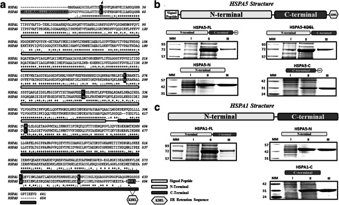
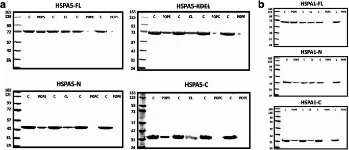
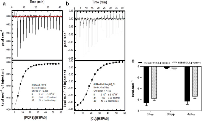
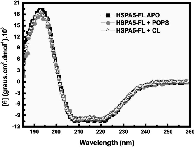
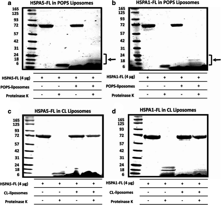
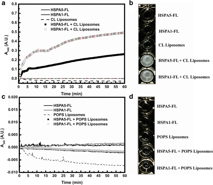
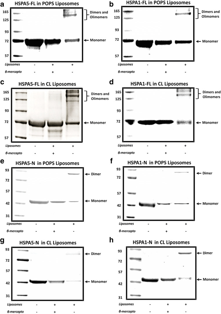
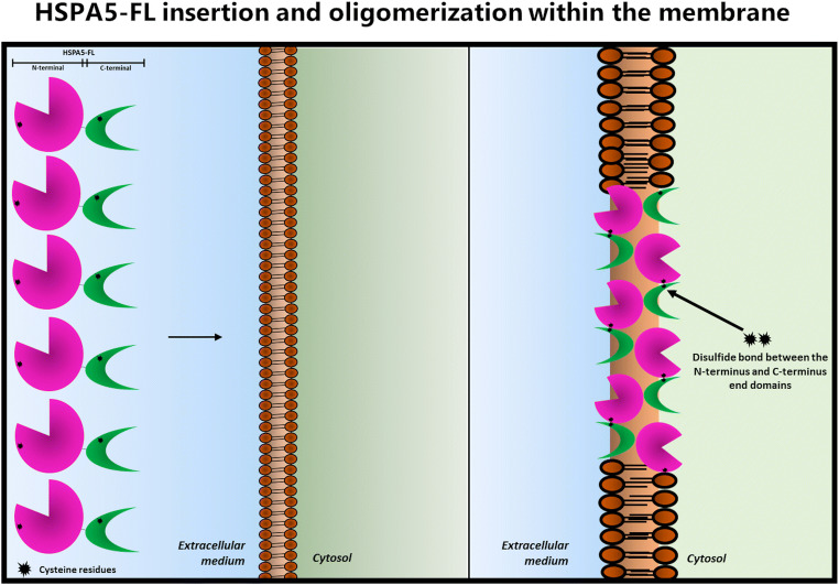
Heat shock proteins (HSPs) are ubiquitous polypeptides expressed in all living organisms that participate in several basic cellular processes, including protein folding, from which their denomination as molecular chaperones originated. There are several HSPs, including HSPA5, also known as 78-kDa glucose-regulated protein (GRP78) or binding immunoglobulin protein (BIP) that is an ER resident involved in the folding of polypeptides during their translocation into this compartment prior to the transition to the Golgi network. HSPA5 is detected on the surface of cells or secreted into the extracellular environment. Surface HSPA5 has been proposed to have various roles, such as receptor-mediated signal transduction, a co-receptor for soluble ligands, as well as a participant in tumor survival, proliferation, and resistance. Recently, surface HSPA5 has been reported to be a potential receptor of some viruses, including the novel SARS-CoV-2. In spite of these observations, the association of HSPA5 within the plasma membrane is still unclear. To gain information about this process, we studied the interaction of HSPA5 with liposomes made of different phospholipids. We found that HSPA5 has a high affinity for negatively charged phospholipids, such as palmitoyl-oleoyl phosphoserine (POPS) and cardiolipin (CL). The N-terminal and C-terminal domains of HSPA5 were independently capable of interacting with negatively charged phospholipids, but to a lesser extent than the full-length protein, suggesting that both domains are required for the maximum insertion into membranes. Interestingly, we found that the interaction of HSPA5 with negatively charged liposomes promotes an oligomerization process via intermolecular disulfide bonds in which the N-terminus end of the protein plays a critical role.

Keywords: Charged phospholipids; HSPA5; Hsp70; Liposomes; Membranes.


Figures

Fig. 1
Fig. 1
HSPA5 and HSPA1 share a high identity, and the proteins were isolated with high purity. a The global alignment between HSPA5 and HSPA1 indicates 64% of identity, which is considered high. The N-terminal of these proteins presents approximately 68% (flat gray rectangle), and the C-terminal presents 58% of identity (dark gray flat rectangle). Cysteine groups are indicated by black boxes, two for HSPA5 and five for HSPA1. The ER translocation signal peptide is displayed by the shadow area, and the ER retention signal (KDEL) is indicated by a hexagon. b HSPA5-FL, HSPA5-KDEL, HSPA5-N, and HSPA5-C. c HSPA1-FL, HSPA1-N, and HSPA1-C. Proteins were purified as described in Methods. All steps for the production and isolation process were visualized by LDS-PAGE and Coomassie blue staining. All proteins were obtained with more than 92% purity.
Fig. 2
Fig. 2
HSPA5 and HSPA1 and full-length and both N- and C-terminus end domains interact preferentially with negatively charged liposomes. a HSPA5-FL, HSPA5-KDEL, HSPA5-N, and HSPA5-C and b HSPA1-FL, HSPA1-N, and HSPA1-C (4 μg) were incubated with liposomes made of POPC, POPE, POPS, and CL (400 μg) in 50 mM Tris-HCl buffer (pH 7.5) for 30 min at 25 °C with continuous agitation. The mixture was centrifuged at 100,000×g for 1 h at 4 °C. The pellet was resuspended (300 μL) in 100 mM Na2CO3 buffer pH 11.5 and centrifuged at 100,000×g for 1 h at 4 °C. Proteoliposomes were solubilized in sample buffer containing 10 mM βM, and the proteins were resolved by LDS-PAGE and visualized by staining with Coomassie Brilliant Blue R-250. HSPA5-FL, HSPA5, and HSPA1-FL showed high affinity for the negatively charged liposomes (POPS and CL). The same behavior was observed for the N-terminus and C-terminus end domain of both proteins. Percentages of incorporation into liposomes are presented in Table 1.
Fig. 3
Fig. 3
HSPA5 interaction with POPS and CL liposomes is mainly driven by entropic changes. The thermodynamics parameters were obtained using an iTC200 microcalorimeter. Seventeen 2-μL aliquots of POPS or CL liposomes at 3 mM were injected into 203.8 μL of 10–15 μmol/L recombinant HSPA5-FL, at 25 °C. All solutions were prepared in 50 mM Tris-HCl (pH 7.4) buffer. The experimental isotherm curves were analyzed to obtain the KA and ΔHapp. a HSPA5-FL interaction with POPS liposomes. b HSPA5 interaction with CL liposomes. Protein insertion into liposomes was entropically and enthalpically driven, with a higher entropic contribution. c Thermodynamic signatures for HSPA5 interaction with POPS and CL shown that the interaction of HSPA5-FL with POPS or CL liposomes has a discrete enthalpy contribution, and it is pretty much entropically driven.
Fig. 4
Fig. 4
HSPA5 secondary structure did not change after insertion into liposomes. Circular dichroism performed for HSPA5-FL (5–10 μM) before and after incorporation in POPS or CL liposomes (3 mM) in 50 mM Tris-HCl buffer pH 7.5. A J-815 spectropolarimeter coupled to the Peltier system, at 25 °C, was used in these experiments. The secondary structures of HSPA5-FL APO and after incorporated in POPS or CL liposomes presented no differences, suggesting that incorporation of protein into liposomes had no effect in the secondary structure.
Fig. 5
Fig. 5
HSPA5 and HSPA1 interact differently with POPS or CL liposomes. HSPA5-FL or HSPA1-FL (4 μg) were incubated with POPS or CL liposomes (400 μg) in 50 mM Tris-HCl buffer (pH 7.5) for 30 min at 25 °C with continuous agitation. The mixture was centrifuged at 100,000×g for 1 h at 4 °C. The pellet was resuspended (300 μL) in 100 mM Na2CO3 buffer pH 11.5 and centrifuged again at 100,000×g for 1 h at 4 °C. The pellet was resuspended in 50 mM Tris-HCl buffer pH 7.4 and incubated with proteinase K (5 μg/mL) for 30 min at 25 °C and centrifuged for 1 h at 100,000×g. The pellet was solubilized in sample buffer containing 10 mM βM, and the proteins were resolved by LDS-PAGE and visualized by staining with Coomassie Brilliant Blue R-250. a HSPA5 in POPS liposomes. b HSPA1-FL in POPS liposomes. c HSPA5-FL in CL liposomes. d HSPA1-FL in CL liposomes. Brackets indicate low molecular weight peptides retained within the liposomes after protease digestion.
Fig. 6
Fig. 6
CL liposomes aggregated in the presence of Hsp70s. The aggregation of POPS or CL liposomes with or without incorporation of HSPA5-FL (a, b) or HSPA1-FL (c, d) in 50 mM Tris-HCl 50 buffer (pH 7.5) was monitored by the changes in absorbance at 340 nm every 10 s for 1 h at 25 °C. a and c aggregation pattern. b and d visualization of aggregates in 96-well plates.
Fig. 7
Fig. 7
Aggregation profile of HSPA5 and HSPA1 and the respective N-terminal end domain after incorporation into liposomes. HSPA5-FL and HSPA5-N or HSPA1-FL and HSPA1-N and HSPA1-N were incorporated into POPS (a, b, e, f) or CL (c ,d ,g ,h) liposomes in 50 mM Tris-HCl buffer (pH 7.5) for 30 min at 25 °C. The mixture was centrifuged at 100,000×g for 1 h at 4 °C. The pellet was resuspended (300 μL) in 100 mM Na2CO3 buffer (pH 11.5) and centrifuged at 100,000×g for 1 h at 4 °C. The pellet was solubilized in sample buffer containing or not 10 mM βM, and the proteins were resolved by LDS-PAGE and visualized by staining with Coomassie Brilliant Blue R-250. The presence of dimers and oligomers is indicated.
Fig. 8
Fig. 8
Model for the presence of antiparallel oligomers of HSPA5 or HSPA1 after incorporation into the lipid bilayer via formation of disulfide bonds

References

    1. Angelidis CE, Lazaridis I, Pagoulatos GN. Aggregation of hsp70 and hsc70 in vivo is distinct and temperature-dependent and their chaperone function is directly related to non-aggregated forms. Eur J Biochem. 1999;259:505–512. - PubMed
    1. Aprile FA, Dhulesia A, Stengel F, Roodveldt C, Benesch JL, Tortora P, Robinson CV, et al. Hsp70 oligomerization is mediated by an interaction between the interdomain linker and the substrate-binding domain. PLoS One. 2013;8:e67961. - PMC - PubMed
    1. Arispe N, De Maio A. ATP and ADP modulate a cation channel formed by Hsc70 I acidic phospholipid membranes. J Biol Chem. 2000;275:30839–30843. - PubMed
    1. Arispe N, Doh M, Simakova O, Kurganov B, De Maio A. Hsc70 and Hsp70 interact with phosphatidylserine on the surface of PC12 cells resulting in a decrease in viability. FASEB J. 2004;18:1636–1645. - PubMed
    1. Armijo G, Okerblom J, Cauvi DM, Lopez V, Schlamadinger DE, Kim J, Arispe N, de Maio A. Interaction of heat shock protein 70 with membranes depends on the lipid environment. Cell Stress Chaperones. 2014;19:877–886. - PMC - PubMed

Publication types

MeSH terms