. 2020 Nov;587(7835):657-662.
doi: 10.1038/s41586-020-2601-5. Epub 2020 Jul 29.

Papain-like protease regulates SARS-CoV-2 viral spread and innate immunity

Affiliations

Papain-like protease regulates SARS-CoV-2 viral spread and innate immunity

Donghyuk Shin et al. Nature. 2020 Nov.

Abstract

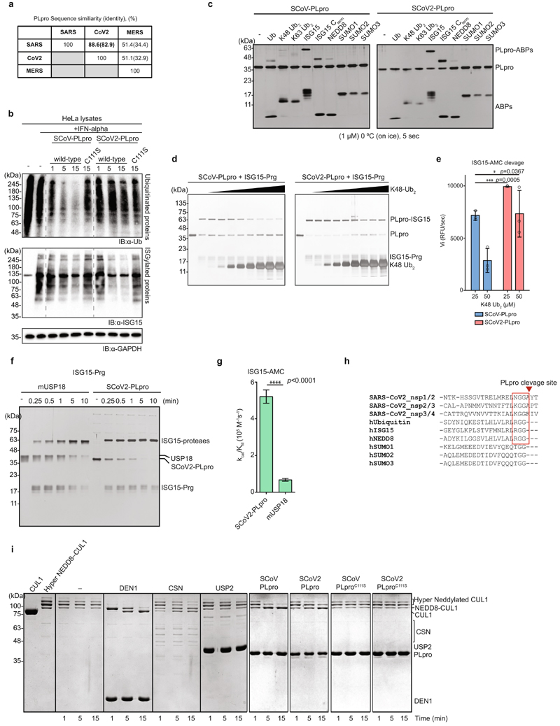
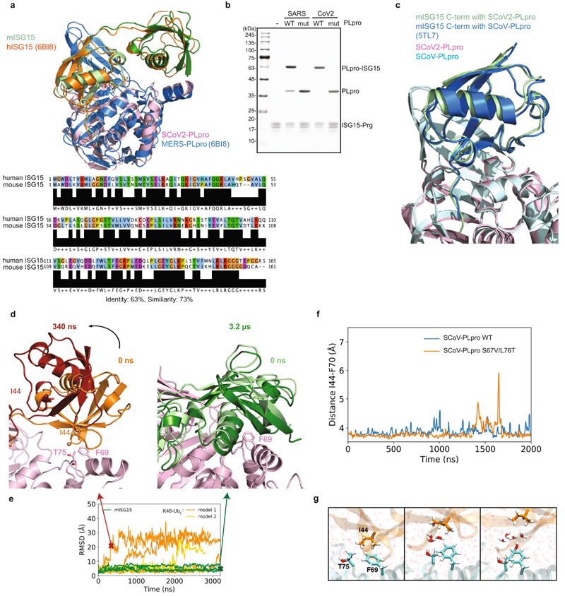
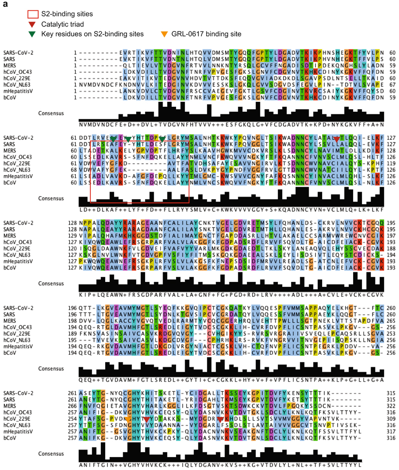
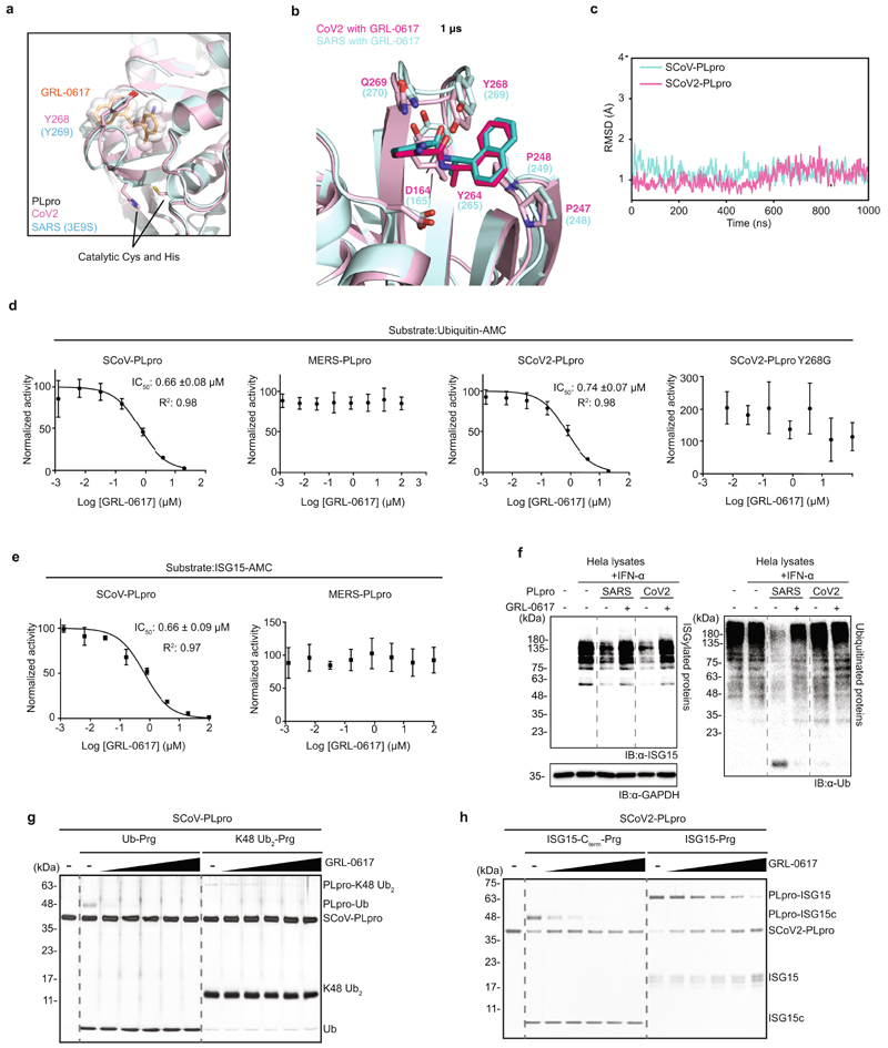
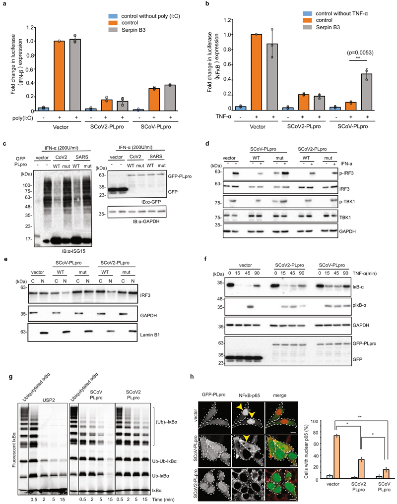
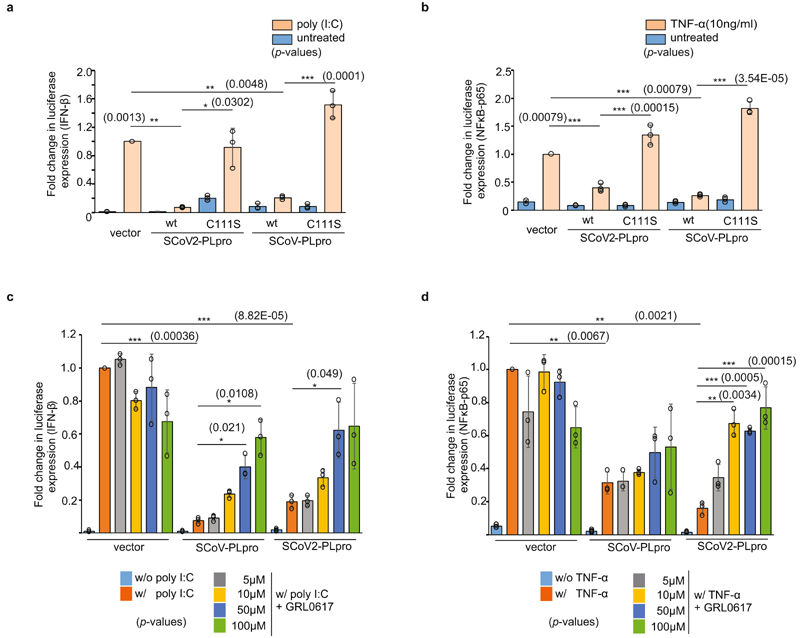
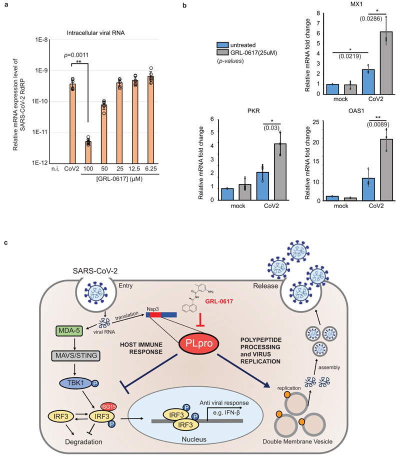
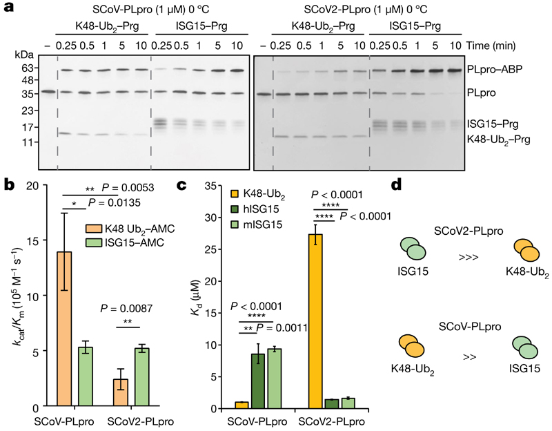
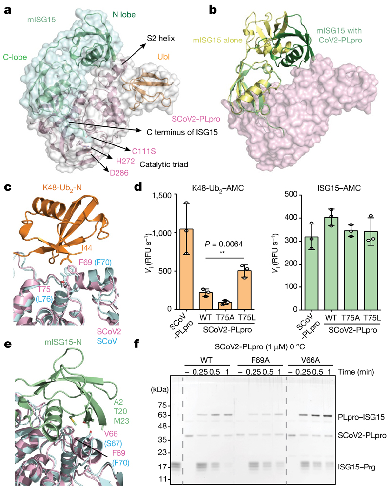
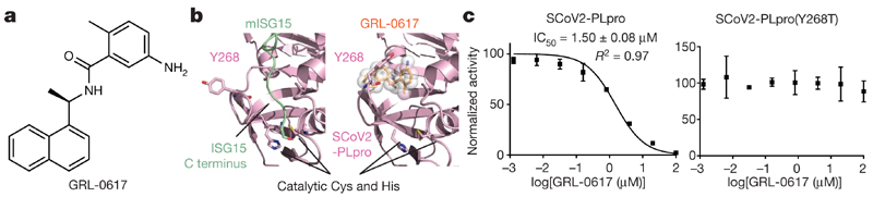
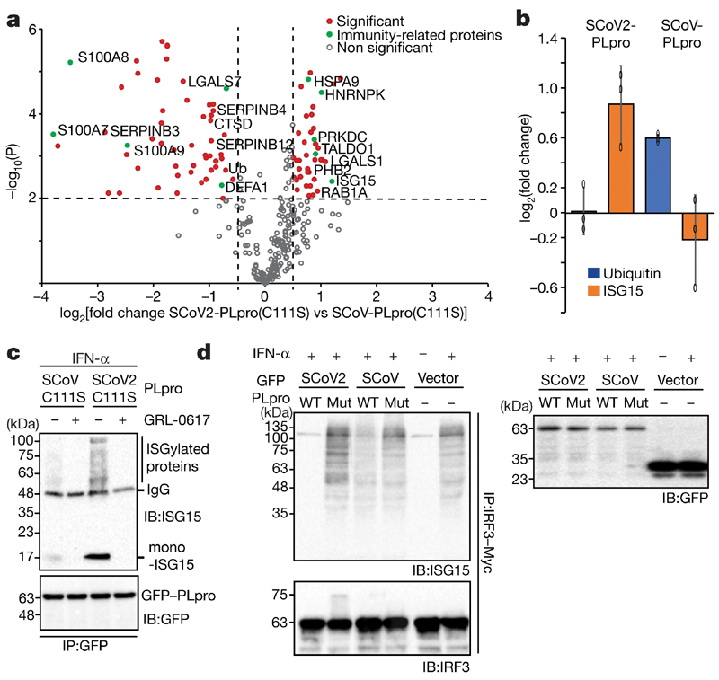
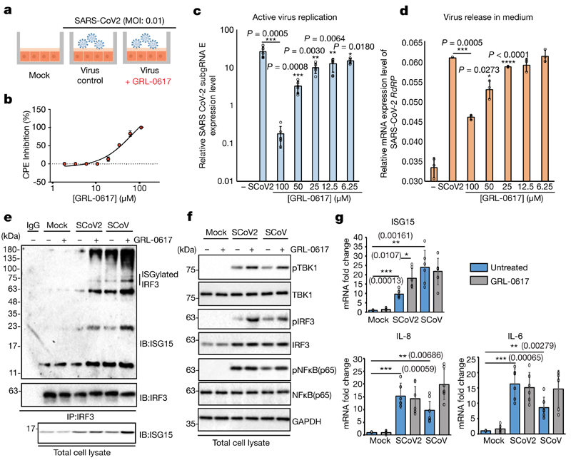
The papain-like protease PLpro is an essential coronavirus enzyme that is required for processing viral polyproteins to generate a functional replicase complex and enable viral spread1,2. PLpro is also implicated in cleaving proteinaceous post-translational modifications on host proteins as an evasion mechanism against host antiviral immune responses3-5. Here we perform biochemical, structural and functional characterization of the severe acute respiratory syndrome coronavirus 2 (SARS-CoV-2) PLpro (SCoV2-PLpro) and outline differences with SARS-CoV PLpro (SCoV-PLpro) in regulation of host interferon and NF-κB pathways. SCoV2-PLpro and SCoV-PLpro share 83% sequence identity but exhibit different host substrate preferences; SCoV2-PLpro preferentially cleaves the ubiquitin-like interferon-stimulated gene 15 protein (ISG15), whereas SCoV-PLpro predominantly targets ubiquitin chains. The crystal structure of SCoV2-PLpro in complex with ISG15 reveals distinctive interactions with the amino-terminal ubiquitin-like domain of ISG15, highlighting the high affinity and specificity of these interactions. Furthermore, upon infection, SCoV2-PLpro contributes to the cleavage of ISG15 from interferon responsive factor 3 (IRF3) and attenuates type I interferon responses. Notably, inhibition of SCoV2-PLpro with GRL-0617 impairs the virus-induced cytopathogenic effect, maintains the antiviral interferon pathway and reduces viral replication in infected cells. These results highlight a potential dual therapeutic strategy in which targeting of SCoV2-PLpro can suppress SARS-CoV-2 infection and promote antiviral immunity.


Conflict of interest statement

Competing interests The authors declare no competing interests.

Figures

Extended Data Fig. 1
Extended Data Fig. 1. Biochemical properties of SCoV2-PLpro.
a, Sequence similarity of PLpro from SARS, MERS and SARS-CoV-2. b, IFN-α-treated HeLa cell lysates were incubated with PLpro for indicated time points and analysed by immunoblot c, Propargylamide-activity based probes of ubiquitin like modifiers were reacted with (left) SCoV-PLpro (right) PLproCoV2. d, ISG15-Prg were incubated with SCoV-PLpro (left) or SCoV2-PLpro (right) with increasing amount of non-hydrolysable K48-Ub2. e, Initial AMC release rate from ISG15-AMC. Purified SCoV-PLpro and SCoV2-PLpro were incubated with ISG15-AMC and indicated amounts of K48-Ub2. The release of AMC was measured by increase of fluorescence at (Ex./Em. 360/487 nm). f, Purified mUSP18 (left) and SCoV2-PLpro (right) were incubated with ISG15-propargylamide activity-based probes for indicated time points. g, Catalytic efficiency (k cat/K m) of mUSP18 and SCoV2-PLpro on ISG15-AMC cleavage. h, Sequence alignment of PLpro cleavage site of Nsp1/2, Nsp2/3, Nsp3/4 from SARS-CoV2 and human ubiquitin like modifiers. i, Hyper-NEDDylated CUL1-RBX1 was incubated with purified PLpro proteins for indicated time points at 37 °C. Reactions were performed side-by-side by with well-characterized deneddylating enzymes (DEN1 with broad specificity or COP9 Signallosome CSN specific for NEDD8 linked directly to a cullin), or the broad specificity deubiquitinating enzyme USP2 as controls. Data in e, g are presented as mean ± s.d. (n = 3, independent experiments). **P < 0.01, ***P < 0.001, ****P < 0.0001; two-tailed paired t-tests. Experiments in b–d, f, i were repeated three times independently with similar results.
Extended Data Fig. 2
Extended Data Fig. 2. Complex structure of SCoV2-PLpro with mouseISG15.
a, Structural comparison of mouseISG15:SCoV2-PLpro with humanISG15: MERS-PLpro (PDB: 6BI8) and sequence alignment of human and mouse ISG15. b, Activity test of wild type or catalytically inactive mutant (C111S) of SCoV-PLpro and SCoV2-PLpro. ISG15 Propargyl-activity based probes were mixed with indicated PLpro proteins. Experiments were repeated three times independently with similar results. c, Structural comparison of C-terminal domain of ISG15 in complex with SCoV2-PLpro and SCoV-PLpro (PDB: 5TL7). d, Snapshots from molecular dynamics simulations of SCoV2-PLpro (light pink cartoon) with (left) K48-Ub2 at 340 ns and (right) mISG15 at 3.2 μs. Key residues in the interface are highlighted. e, Backbone r.m.s.d. of the N-terminal domain of mISG15 (green) and of the distal ubiquitin in K48-Ub2 in an apo-like model (orange, model 1, SCoV2-PLpro coordinates from substrate unbound form, PDB: 6W9C) and in an mISG15-like model (yellow, model 2, SCoV2-PLpro coordinates from substrate bound form, PDB: 6YVA) from their respective SCoV2-PLpro-bound starting structures as function of time. The r.m.s.d. was calculated after superimposing the helix backbone atoms of SCoV2-PLpro. Time points for structural snapshots in e) are marked with a cross. f, Minimum heavy atom distance between F70 (SARS) and I44(Ub) in wild type and double mutant (S67V/L76T) of SCoV-PLpro:K48-Ub2 as function of time. g, Water mediated dissociation pathway. Left, initial hydrophobic interactions between F69(CoV2), T75(CoV2) and I44(Ub). Middle, water wedges in between T75(CoV2) and I44(Ub). Right, water penetration between T75(CoV2)/F69 (CoV2) and I44(Ub) leads to dissociation.
Extended Data Fig. 3
Extended Data Fig. 3. Sequence alignment of papain like protease domain from corona viruses.
The amino acid sequences of papain-like protease domain from eight different coronaviruses (SARS-CoV-2, SARS, MERS, humanCoV-OC43, humanCoV-229E, humanCoV-NL63, murine HepatitisV, bovine CoV) were aligned with Clustal Omega. Accession numbers: SARS-CoV-2 (NC_045512), SARS (PDB: 3MJ5), MERS (PDB: 5W8U), hCoV-OC43 (AY585228), hCoV-229E (X69721), hCoV-NL63 (NC_005831), murine HepatitisV (NC_001846), bCoV (NC_003045).
Extended Data Fig. 4
Extended Data Fig. 4. Structural analysis of GRL-0167, SCoV2-PLpro complex.
a, Structural model of GRL-0617 bound SCoV2-PLpro. The conformation of Tyr268 on SCoV2-PLpro and the coordinates of GRL-0617 is obtained from the SCoV-PLpro:GRL-0617 structure (PDB: 3E9S) b, Snapshots of SCoV-PLpro (light cyan) and SCoV2-PLpro (light pink) with bound GRL-0617 (dark colours) after 1 μs of molecular dynamics simulation. The protein backbones are shown in cartoon representation, and the ligand with contacting residues as sticks. c, r.m.s.d. of the GRL-0617 bound to SCoV-PLpro (light blue) and SCoV2-PLpro (light pink) as a function of time. The r.m.s.d. was calculated for non-hydrogen atoms of GRL-0617 with respect to the starting structures in the MD simulations after superimposing the helix backbone atoms of PLpro. d, In vitro PLpro inhibition assay. Initial velocity of AMC release from ubiquitin-AMC in different concentration of GRL-0617 was measured and normalized to DMSO control. IC50 value of GRL-0617 to SCoV-PLpro and SCoV2-PLpro were presented. Data are presented as mean ± s.d. (n = 3, independent experiments). e, In vitro PLpro inhibition assay. Initial velocity of AMC release from ISG15-AMC in different concentration of GRL-0617 was measured and normalized to DMSO control. IC50 value of GRL-0617 to SCoV-PLpro were presented. Data are presented as mean ± s.d. (n = 3, independent experiments). f, Effects of GRL-0617 on (left) deISGylase or (right) deubiquitinase activity of PLpro of SARS and SARS-CoV-2. g, Effects of GRL-0617 on SCoV-PLpro activity to (left) ubiquitin or (right) K48-Ub2 propargyl activity-based probes. Inhibitory effect of GRL-0617 on ubiquitin species was tested with various concentration of GRL-0617 (0-400 μM). h, Effects of GRL-0617 on SCoV2-PLpro activity to (left) ISG15-Cterm or (right) ISG15 propargylamide activity-based probes. Inhibitory effect of GRL-0617 on ISG15 was tested with various concentration of GRL-0617 (0-400 μM). Experiments in fh were repeated three times independently with similar results.
Extended Data Fig. 5
Extended Data Fig. 5. Physiological roles of PLpro in cells.
a, b, Effect of SERPIN B3 on PLpro mediated IFN-β (a) or NF-κB p65 (b) expression level. A549 Cells were co-transfected with indicated GFP-PLpro and Myc-SERPINs and treated with either poly(I:C) or TNF-α to induce IFN-β and NF-κB p65 expression, respectively. Fold changes of luciferase level are presented. c, Effect of PLpro on IFN-induced cellular ISGylation. A549 cells were transfected with indicated PLpro plasmids and treated with IFN-α. Lysates were analysed by immune-blotting with indicated antibodies. d, e, Effect of PLpro on IFN-signalling pathway. d, A549 cells were transfected with indicated PLpro plasmids and treated with IFN-α. Lysates were analysed by immune-blotting with indicated antibodies. e, Effect of PLpro on cellular localization of IRF3. Cells from d were fractionated into cytosol and nucleus and the level of IRF3 was analysed. Lamin B1 was used for nuclear fraction control. f, Effect of PLpro on the NF-κB pathway. IκB-α phosphorylation and degradation were examined from A549 cells expressing indicated GFP-PLpro under treatment of TNF-α. g, in vitro IκBα deubiquitylation assay. Ubiquitinated IκBα were incubated with SCoV-PLpro or SCoV2-PLpro. USP2 were used as positive control. h, Effect of PLpro on NF-κB p65 cellular localization. Scale bar, 10 μm. Data in a, b, h are presented as mean ± s.d. (n = 3, independent experiments). *P < 0.05, **P < 0.01; two-tailed paired t-tests. Experiments in ch were repeated three times independently with similar results. e, Effect of PLpro on the NF-κB pathway. IκB-α phosphorylation and degradation were examined from A549 cells expressing indicated GFP-PLpro under treatment of TNF-α.
Extended Data Fig. 6
Extended Data Fig. 6. Effect of PLpro on IFN-β or NF-κB p65 expression level.
a, b, Effect of PLpro on IFN-β (a) or NF-κB p65 (b) expression level. A549 Cells were transfected with indicated GFP-PLpro and treated with either poly(I:C) or TNF-α to induce IFN-β and NF-κB p65 expression, respectively. c, d, Effect of GRL-0617 on PLpro mediated IFN-β (c) or NF-κB p65 (d) expression level. A549 Cells were transfected with indicated GFP-PLpro and treated with either poly (I:C) or TNF-α to induce IFN-β and NF-κB p65 expression, respectively. GRL-0617 is treated as indicated. All data are presented as mean ± s.d. (n = 3, independent experiments). *P < 0.05, **P < 0.01, ***P < 0.001; two-tailed paired t-tests.
Extended Data Fig. 7
Extended Data Fig. 7. Inhibitory effects of GRL-0617 on SARS-CoV2 infection.
a, Intracellular virus production was analysed by PCR targeting SARS-CoV-2 RdRP mRNA. Relative expression level of SARS-CoV2-2 genomic RNA was normalized to cellular GAPDH level. b, Intracellular RNA was isolated from cells without infection or cells infected with SARS-CoV-2 with or without treatment of GRL-0617. Relative mRNA-level fold change of indicated genes were analysed in a qRT–PCR analysis and normalized to ACTB levels. Data in a, b are presented as mean ± s.d. (n = 3, independent experiments). *P < 0.05, **P < 0.01; two-tailed paired t-tests. c, Schematic representation of the role of SARS-CoV-2 PLpro in the viral life cycle. The physiological role of SCoV2-PLpro in both host-immune response and polypeptide processing is shown. Inhibition of PLpro by GRL-0617 is also presented.
Fig. 1
Fig. 1. DeISGylating and deubiquitylating activities of SCoV-PLpro and SCoV2-PLpro.
a, SCoV-PLpro (left) or SCoV2-PLpro (right) were incubated with indicated Prg probes. Experiments were repeated three times independently with similar results. b, Catalytic efficiency (k cat/K m) of SCoV2-PLpro and SCoV-PLpro cleavage of K48-Ub2-AMC or ISG15-AMC. c, Dissociation constant (K d) of SCoV2-PLpro and SCoV-PLpro. Data in c, d, are mean ± s.d. or mean ± s.e.m. (n = 3 independent experiments). *P < 0.05, **P < 0.01, ****P < 0.0001; two-tailed paired t-test. d, Schematic representation of substrate specificity of SCoV2-PLpro (red) and SCoV-PLpro (blue). The preferred substrate is shown on the left.
Fig. 2
Fig. 2. Structural analysis of SARS-CoV-2 PLpro in complex with full length ISG15.
a, Crystal structure of SARS-CoV-2 PLpro(C111S) in complex with mouse ISG15. The C-terminal glycine of ISG15 and catalytic triad of SCoV2-PLpro are highlighted as stick model. The ubiquitin like domain (Ubl) is coloured orange. b, Comparison of unbound form of ISG15 with ISG15 in complex with SCoV2-PLpro. c, Comparison of N-terminal half of K48-linked di-ubiquitin (K48 Ub2-N)–SCoV-PLpro complex structure (PDB ID: 5E6J) with ISG15–SCoV2-PLpro. Residues forming hydrophobic interactions are highlighted as stick model. d, Initial velocity (V i) of AMC release from AMC probes (K48-Ub2–AMC and ISG15–AMC) with the indicated wild-type (WT) and mutant PLpro. Data are mean ± s.d. (n = 3 independent experiments). **P < 0.01; two-tailed paired t-test. e, Comparison of N-terminal half of mouse ISG15 (ISG15-N) and SCoV2-PLpro and SCoV-PLpro (PDB ID:5E6J). Residues forming hydrophobic interactions are highlighted as stick model. f, ISG15–Prg was incubated with wild type and mutant SCoV2-PLpro. Experiments were repeated three times independently with similar results.
Fig. 3
Fig. 3. Effect of GRL-0617 on SCoV2-PLpro.
a, Structure of GRL-0617. b, Comparison of ISG15-bound (left) and GRL-0617 bound (right) structure. Blocking loop 2 (BL2 loop) of SCoV2-PLpro is modelled on the basis of GRL-0617 bound SCoV-PLpro and SCoV2-PLpro structures (PDB ID: 3E9S and 6W9C). GRL-0617-interacting Tyr268 and catalytic Cys. His residues are highlighted as stick model. c, Cleavage of ISG15–AMC was measured and normalized to DMSO control. IC50 value of GRL-0617 in relation to SCoV2-PLpro activity is presented. Data are mean ± s.d. n = 3 independent experiments.
Fig. 4
Fig. 4. Effect on PLpros on IFN and NF-κB pathways.
a, Interactome analysis comparing SCoV2-PLpro(C111S) and SCoV-PLpro(C111S). Statistically significant and immunity-related proteins are highlighted. b, log2(fold change) of ubiquitin and ISG15 proteins enriched by SCoV2-PLpro or SCoV-PLpro immunoprecipitates versus empty vector. Data are mean ± s.d. (n = 3, independent experiments). c, ISGylated proteins were enriched from A549 cells treated with IFN-α (200 U ml-1) by immunoprcipitation of the indicated C111S mutant PLPro. d, ISGylation level of Myc–IRF3 in A549 cells expressing the indicated GFP–PLpro. Experiments in c, d, were repeated three times independently with similar results.
Fig. 5
Fig. 5. Inhibitory effects of GRL-0617 on SARS-CoV2.
a, Schematic representation of the SARS-CoV-2 (strain FFM1) growth inhibition test with GRL-0617. MOI, multiplicity of infection. b, CPE inhibition rate of GRL-0617 on CaCo-2 cells infected with SARS-CoV2. c, Intracellular active virus replication was analysed by measuring SARS-CoV-2 subgenomic RNA (subgRNA E) level and normalized to the cellular ACTB gene. d, Release of viral particles in culture medium was analysed by PCR targeting the open reading frame of the RNA-dependent RNA polymerase (RdRP) gene of SARS-CoV-2. e, f, The effect of GRL-0617 on the type I IFN pathway. CaCo-2 cells were infected with SARS-CoV-2 or SARS-CoV with or without GRL-0617 (50 μM). pTBK1, phosphorylated TBK1; pIRF3, phosphorylated IRF3; pNF-κB p65, phosphorylated pNF-κB p65. e, Endogenous IRF3 was immunoprecipitated and analysed by immunoblotting. f, Phosphorylation of TBK1 was analysed by immunoblotting. g, Relative mRNA levels of indicated genes from infected cells with or without GRL-0617 (25 μM) treatment were analysed and normalized to 18S RNA. P values in parentheses. Data in c, d, g, are mean ± s.d.; n = 3 independent experiments. *P < 0.05, **P < 0.01, ***P < 0.001, ****P < 0.0001; two-tailed paired t-test. Experiments in e, f, were repeated three times independently with similar results.

Comment in

  • SARS-CoV-2: the many pros of targeting PLpro.
    McClain CB, Vabret N. McClain CB, et al. Signal Transduct Target Ther. 2020 Oct 6;5(1):223. doi: 10.1038/s41392-020-00335-z. Signal Transduct Target Ther. 2020. PMID: 33024071 Free PMC article. No abstract available.

References

    1. Harcourt BH, et al. Identification of severe acute respiratory syndrome coronavirus replicase products and characterization of papain-like protease activity. J Virol. 2004;78:13600–13612. - PMC - PubMed
    1. Lim KP, Ng LFP, Liu DX. Identification of a novel cleavage activity of the first papain-like proteinase domain encoded by open reading frame 1a of the coronavirus Avian infectious bronchitis virus and characterization of the cleavage products. J Virol. 2000;74:1674–1685. - PMC - PubMed
    1. Frieman M, Ratia K, Johnston RE, Mesecar AD, Baric RS. Severe acute respiratory syndrome coronavirus papain-like protease ubiquitin-like domain and catalytic domain regulate antagonism of IRF3 and NF-κB signaling. J Virol. 2009;83:6689–6705. - PMC - PubMed
    1. Devaraj SG, et al. Regulation of IRF-3-dependent innate immunity by the papain-like protease domain of the severe acute respiratory syndrome coronavirus. J Biol Chem. 2007;282:32208–32221. - PMC - PubMed
    1. Bailey-Elkin BA, et al. Crystal structure of the Middle East respiratory syndrome coronavirus (MERS-CoV) papain-like protease bound to ubiquitin facilitates targeted disruption of deubiquitinating activity to demonstrate its role in innate immune suppression. J Biol Chem. 2014;289:34667–34682. - PMC - PubMed

MeSH terms