Review
. 2020 Jul 27;9(8):1785.
doi: 10.3390/cells9081785.

Role of Tumor-Associated Myeloid Cells in Breast Cancer

Affiliations
Review

Role of Tumor-Associated Myeloid Cells in Breast Cancer

Yoon Jin Cha et al. Cells. .

Abstract

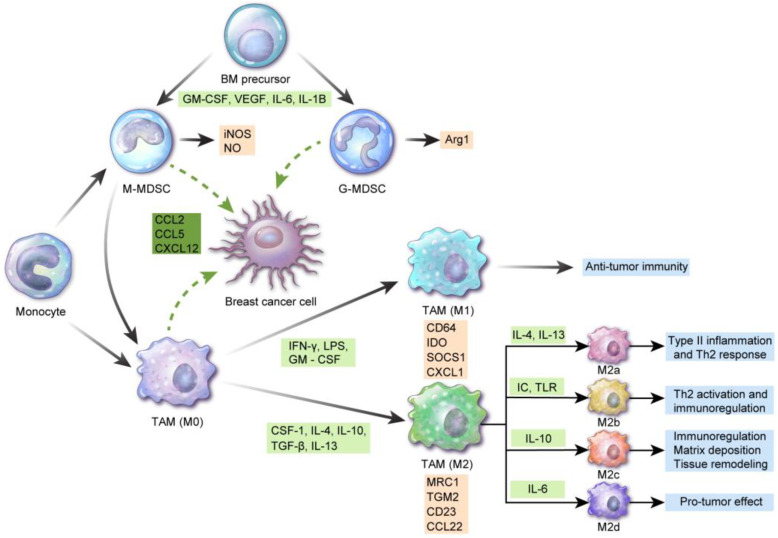
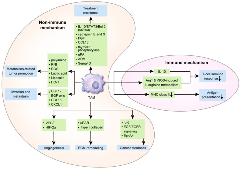
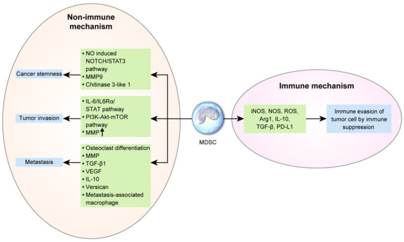
Stromal immune cells constitute the tumor microenvironment. These immune cell subsets include myeloid cells, the so-called tumor-associated myeloid cells (TAMCs), which are of two types: tumor-associated macrophages (TAMs) and myeloid-derived suppressor cells (MDSCs). Breast tumors, particularly those in human epidermal growth factor receptor 2 (HER-2)-positive breast cancer and triple-negative breast cancer, are solid tumors containing immune cell stroma. TAMCs drive breast cancer progression via immune mediated, nonimmune-mediated, and metabolic interactions, thus serving as a potential therapeutic target for breast cancer. TAMC-associated breast cancer treatment approaches potentially involve the inhibition of TAM recruitment, modulation of TAM polarization/differentiation, reduction of TAM products, elimination of MDSCs, and reduction of MDSC products. Furthermore, TAMCs can enhance or restore immune responses during cancer immunotherapy. This review describes the role of TAMs and MDSCs in breast cancer and elucidates the clinical implications of TAMs and MDSCs as potential targets for breast cancer treatment.

Keywords: breast cancer; myeloid-derived suppressor cells; tumor-associated macrophage; tumor-associated myeloid cells.


Conflict of interest statement

The authors declare no conflict of interest.

Figures

Figure 1
Figure 1
Differentiation and characteristics of tumor-associated myeloid cells: interferon (IFN)-γ, lipopolysaccharide, and granulocyte-macrophage colony-stimulating factor (GM-CSF) induce M1 tumor-associated macrophage (TAM) polarization from monocytes, which are involved in antitumor immunity. CSF-1, interleukin (IL)-4, IL-10, transforming growth factor (TGF)-β, and IL-13 contribute to M2 TAM polarization. M2 macrophages are further differentiated into M2a by IL-4 and IL-13 and are involved in type II inflammation and the Th2 response. Differentiation of M2 macrophages into M2b leads to Th2 activation and immunoregulation via immune complex and toll-like receptor ligand. M2c and M2d differentiation by IL-10 and IL-6 is involved in immunoregulation, matrix deposition and tissue remodeling, and induction and growth of tumor cell masses, respectively. Surface antigens of M1 macrophages include CD64, indoleamine 2,3-dioxygenase (IDO), suppressor of cytokine signaling 1 (SOCS1), and chemokine (C-X-C motif) ligand 1 (CXCL1). Mannose receptor C-type 1 (MRC1), transglutaminase 2 (TGM2), CD23, and C-C Chemokine Ligand 2 (CCL2) are considered M2 macrophage markers. Myeloid-derived suppressor cells (MDSCs) originate from bone marrow precursor cells in the presence of GM-CSF, vascular endothelial growth factor (VEGF), IL-6, and IL-1B and are divided into CD11b+CD14+HLA-DR−/low CD15 monocytic MDSCs and CD11b+CD14HLA-DRlow/− CD15+ granulocytic MDSCs, the former secreting inducible nitric oxide synthase (iNOS) and NO and the latter releasing reactive oxygen species (ROS) and Arg1. Among these, monocytic MDSCs can differentiate to TAMs. In breast cancer, CCL2, CCL5, and CXCL12 are involved in TAM and/or MDSC recruitment.
Figure 2
Figure 2
The role of tumor-associated macrophages (TAMs) in breast cancer: One of the immunogenic mechanisms underlying the secretion of IL-10, Arg1, and iNOS-related L-arginine by TAMs in breast cancer, which suppress the T-cell response and antigen presentation by decreasing major histocompatibility complex (MHC) class II levels. Non-immunogenic mechanisms include angiogenesis via the secretion of VEGF and hypoxia-inducible factor (HIF)-2α; extracellular matrix remodeling via releasing urokinase receptor (uPAR) and type I collagen; and evoking cancer stemness through IL-6, epidermal growth factor (EGF)/EGF receptor(EGFR) signaling, and EphA4. TAM contributes to invasion and metastasis via the CSF1-EGF axis, CCL18, and CXCL1. Polyamine, reactive nitrogen intermediates (RNI), ROS, lactic acid, lipocalin (LCN), and heme oxygenase-1 (HO-1), which are TAM metabolites, also promote breast cancer progression. Finally, treatment resistance mechanisms via TAMs are supported by the IL-10/STAT3/Bcl-2 pathway, cathepsin B and S, fibroblast growth factor, CCL18, thymidine phosphorylase, urokinase-type plasminogen activator (uPA), adrenomedullin (ADM), and Sema4D.
Figure 3
Figure 3
The role of myeloid-derived suppressor cells (MDSCs) in breast cancer: Common immunogenic pathways of MDSCs in breast cancer progression are the induction of immunosuppression by iNOS, NOS, ROS, Arg1, IL-10, TGF-β, and PD-L1, thus facilitating immune evasion of tumor cells. Non-immunogenic mechanisms include the enhancement of cancer stemness by the nitric oxide (NO)-induced Notch/ signal transducer and activator of transcription 3 (STAT3) pathway, matrix metallopeptidase (MMP) 9, and chitinase 3-like 1 and the promotion of tumor invasiveness by the IL-6/IL6Rα/STAT pathway, phosphoinositide 3-kinase (PI3K)-Akt-mammalian target of rapamycin (mTOR) pathway, and MMP upregulation. During metastasis, MDSCs differentiate into osteoclasts, which increases osteolytic bone metastasis and promotes MMP, TGF-β1, VEGF, IL-10, and versican secretion, and into metastasis-associated macrophages.

References

    1. Benson J.R., Jatoi I. The global breast cancer burden. Future Oncol. 2012;8:697–702. doi: 10.2217/fon.12.61. - DOI - PubMed
    1. Ismail-Khan R., Bui M.M. A review of triple-negative breast cancer. Cancer Control. 2010;17:173–176. doi: 10.1177/107327481001700305. - DOI - PubMed
    1. Burugu S., Asleh-Aburaya K., Nielsen T.O. Immune infiltrates in the breast cancer microenvironment: Detection, characterization and clinical implication. Breast Cancer. 2017;24:3–15. doi: 10.1007/s12282-016-0698-z. - DOI - PubMed
    1. Lewis C.E., Pollard J.W. Distinct role of macrophages in different tumor microenvironments. Cancer Res. 2006;66:605–612. doi: 10.1158/0008-5472.CAN-05-4005. - DOI - PubMed
    1. Hu M., Yao J., Carroll D.K., Weremowicz S., Chen H., Carrasco D., Richardson A., Violette S., Nikolskaya T., Nikolsky Y., et al. Regulation of in situ to invasive breast carcinoma transition. Cancer Cell. 2008;13:394–406. doi: 10.1016/j.ccr.2008.03.007. - DOI - PMC - PubMed