Review
. 2020 Jul 27;21(15):5329.
doi: 10.3390/ijms21155329.

RORγ Structural Plasticity and Druggability

Affiliations
Review

RORγ Structural Plasticity and Druggability

Mian Huang et al. Int J Mol Sci. .

Abstract

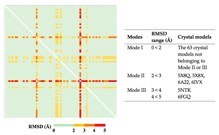
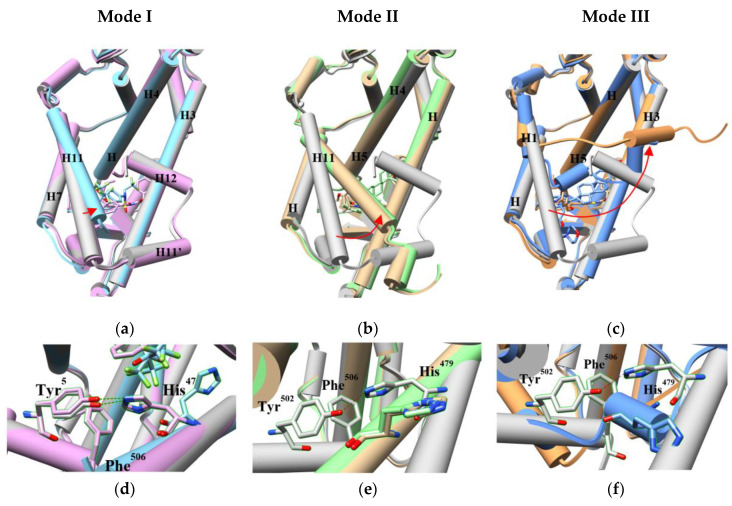
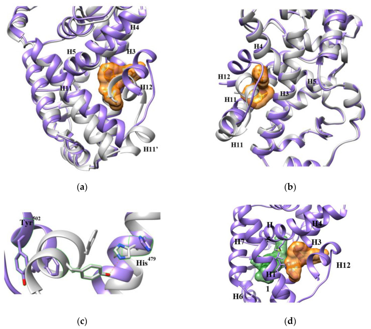
Retinoic acid receptor-related orphan receptor γ (RORγ) is a transcription factor regulating the expression of the pro-inflammatory cytokine IL-17 in human T helper 17 (Th17) cells. Activating RORγ can induce multiple IL-17-mediated autoimmune diseases but may also be useful for anticancer therapy. Its deep immunological functions make RORɣ an attractive drug target. Over 100 crystal structures have been published describing atomic interactions between RORɣ and agonists and inverse agonists. In this review, we focus on the role of dynamic properties and plasticity of the RORɣ orthosteric and allosteric binding sites by examining structural information from crystal structures and simulated models. We discuss the possible influences of allosteric ligands on the orthosteric binding site. We find that high structural plasticity favors the druggability of RORɣ, especially for allosteric ligands.

Keywords: RORγ; allosteric binding pocket; druggability; orthosteric binding pocket; plasticity.


Conflict of interest statement

The authors declare no conflict of interest.

Figures

Figure 1
Figure 1
The globular structure of the apo RORγ ligand-binding domain (LBD) (Protein Data Bank (PDB) 5X8U) with a coactivator (orange). H3, H5, H6, H7, H11 and H12 are presented in green color to indicate the orthosteric binding pocket, while other helices are presented in blue color.
Figure 2
Figure 2
The agonist lock stabilizes H12 through a hydrogen bond (green dashed line) in the agonist-bound RORγ LBD (PDB 4S14).
Figure 3
Figure 3
Physical features of the RORγ orthosteric modulators, including 18 agonists (Supplementary Table S1) and 69 inverse agonists (Supplementary Table S2). For every compound, the Van der Waals (VdW) volume of its binding pose in the orthosteric pocket was measured with YASARA [30].
Figure 4
Figure 4
The conformational change of RORγ LBD before and after agonist binding in the orthosteric pocket is minimal. The LBD (PDB 4S14, cyan) bound with the agonist (purple) is superimposed against the apo LBD (grey). The backbones of the LBD are presented in cylinder shape, while the agonist and the residues His479, Tyr502 and Phe506 are in stick shape. The agonist retains the architecture of the pocket and the hydrogen bond of the agonist lock (shown as black dashed lines).
Figure 5
Figure 5
Conformational comparison of RORγ orthosteric pockets induced by 69 inverse agonists. For each ligand-bound LBD, the H1–H11 (265–490 a.a.) portion was extracted for pairwise comparison. The root-mean-square deviation (RMSD) value between two models was measured in the Cα manner by YASARA and then labeled on the figure based on the color scale. According to their RMSD values, the conformations of the pockets were assigned into three modes, which are briefly listed in the right table. The detailed list can be found in Supplementary Table S3.
Figure 6
Figure 6
Three different conformation modes of the RORγ orthosteric pocket (ac) and the changes of the agonist lock (df) induced by orthosteric inverse agonists. The apo LBD model is shown in grey. The H11 movement is demonstrated by red arrows. (a,d) Two crystal models in mode I with bound ligands are presented in cyan (PDB 4NB6) and plum (PDB 4NIE). (b,e) Two crystal models in mode II with bound ligands are presented in light green (PDB 5X8Q) and tan (PDB 6A22). (c,f) Two crystal models in mode III with bound ligands are presented in blue (PDB 5NTK) and light brown (PDB 6FGQ).
Figure 7
Figure 7
Allosteric inverse agonists binding in the AF-2 site. The corresponding PDB codes for the co-crystal models of the compounds binding in the RORγ LBD are listed as follows: (1) 4YPQ, 5C4O [11]; (2) 5C4S [11]; (3) 5C4U [11]; (4) 5C4T [11]; (5) 6UCG [37]; (6) 6TLM [38]; (7) 5LWP [39]; (8) 6SAL [40]. No PDB code is available for compound (9) [41].
Figure 8
Figure 8
An allosteric inverse agonist 1 binds in the AF-2 binding pocket. The structure of RORγ LBD bound with 1 (orange surface) is presented in purple (PDB 4YPQ), while the apo LBD is in grey. (a,b) Two views of the pocket from different orientations display the movement of the helices. Especially, 1 partially occupies the space of H12 and forces it to shift away from other helices. (c) Affected by 1, Tyr502 is pushed to a position too far away from His479 to maintain the agonist lock. (d) The AF-2 site is adjacent to the orthosteric site (green) with a short tunnel connecting them, which is surrounded by hydrophobic residues shown in sticks. The orthosteric site is demonstrated with an orthosteric inverse agonist from PDB 5UFO.
Figure 9
Figure 9
An inverse agonist (10) potentially binds in an allosteric pocket of RORγ hinge domain (HD). The details of the full-length RORγ homology models and molecular docking experiment were described in Supplementary Protocol S1. (a) The top-ranked poses of 10 docked against the HD site in three different homology models are displayed in purple, tan and salmon colors, respectively. The binding energy of the poses in the homology models was analyzed (Supplementary Table S5). (b) In the purple model, 10 poses at a position closed to Lao’s. It hydrogen-bonds to Gln223 but stays far from Leu244. Ser113 and Leu114 are presented in orange color and Gln223 and Leu244 in light blue. (c) In the tan model, 10 shows no contact to Ser113, Leu114, Gln223 or Leu244. (c,d) In the salmon model, two poses of 10 were docked in the site. None of them is closed to Ser113, Leu114, Gln223 or Leu244. (f) 10 may bind in the orthosteric pocket of RORγ LBD (PDB 4NIE) through π-stacking (pink) and hydrophobic (grey) interactions. It may contact the agonist lock. More details are shown in Supplementary Figure S3.
Figure 10
Figure 10
The coactivator-binding pocket ((a), PDB 3KYT) and the extended AF-2 binding pocket ((b), PDB 4YPQ). The structure models are presented by hydrophobic surface with red color for high level of hydrophobicity, blue color for high level of hydrophilicity. The extended AF-2 pocket is circled by green line.
Figure 11
Figure 11
Schematic presentations of the conformational difference of H11, H11′ and H12 in RORγ LBD bound with an agonist (pink triangle) and an inverse agonist (blue diamond).

References

    1. Ivanov I.I., McKenzie B.S., Zhou L., Tadokoro C.E., Lepelley A., Lafaille J.J., Cua D.J., Littman D.R. The Orphan Nuclear Receptor RORγt Directs the Differentiation Program of Proinflammatory IL-17+ T Helper Cells. Cell. 2006;126:1121–1133. doi: 10.1016/j.cell.2006.07.035. - DOI - PubMed
    1. Meissburger B., Ukropec J., Roeder E., Beaton N., Geiger M., Teupser D., Civan B., Langhans W., Nawroth P.P., Gasperikova D., et al. Adipogenesis and insulin sensitivity in obesity are regulated by retinoid-related orphan receptor gamma. EMBO Mol. Med. 2011;3:637–651. doi: 10.1002/emmm.201100172. - DOI - PMC - PubMed
    1. Ruan Q., Kameswaran V., Zhang Y., Zheng S., Sun J., Wang J., DeVirgiliis J., Liou H.-C., Beg A.A., Chen Y.H. The Th17 immune response is controlled by the Rel–RORγ–RORγT transcriptional axis. J. Exp. Med. 2011;208:2321–2333. doi: 10.1084/jem.20110462. - DOI - PMC - PubMed
    1. Hu X., Liu X., Moisan J., Wang Y., Lesch C.A., Spooner C., Morgan R.W., Zawidzka E.M., Mertz D., Bousley D., et al. Synthetic RORγ agonists regulate multiple pathways to enhance antitumor immunity. OncoImmunology. 2016;5:e1254854. doi: 10.1080/2162402X.2016.1254854. - DOI - PMC - PubMed
    1. Hu X., Majchrzak K., Liu X., Wyatt M.M., Spooner C.J., Moisan J., Zou W., Carter L.L., Paulos C.M. In vitro priming of adoptively transferred T cells with a RORγ agonist confers durable memory and stemness In vivo. Cancer Res. 2018;78:3888–3898. doi: 10.1158/0008-5472.CAN-17-3973. - DOI - PMC - PubMed

MeSH terms

Substances

LinkOut - more resources