Review
. 2020 Jul;7(1):e000428.
doi: 10.1136/bmjgast-2020-000428.

Relief of biliary obstruction: choosing between endoscopic ultrasound and endoscopic retrograde cholangiopancreatography

Affiliations
Review

Relief of biliary obstruction: choosing between endoscopic ultrasound and endoscopic retrograde cholangiopancreatography

Andrew Canakis et al. BMJ Open Gastroenterol. 2020 Jul.

Abstract

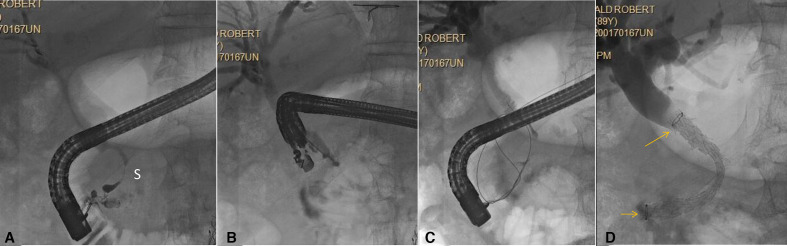
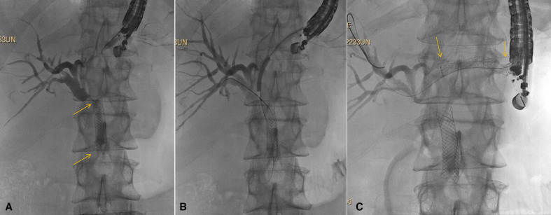
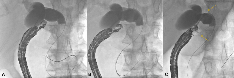
Endoscopic ultrasound (EUS) was originally devised as a novel diagnostic technique to enable endoscopists to stage malignancies and acquire tissue. However, it rapidly advanced toward therapeutic applications and has provided gastroenterologists with the ability to effectively treat and manage advanced diseases in a minimally invasive manner. EUS-guided biliary drainage (EUS-BD) has gained considerable attention as an approach to provide relief in malignant and benign biliary obstruction for patients when endoscopic retrograde cholangiopancreatography (ERCP) fails or is not feasible. Such instances occur in those with surgically altered anatomy, gastroduodenal obstruction, periampullary diverticulum or prior transampullary duodenal stenting. While ERCP remains the gold standard, a multitude of studies are showing that EUS-BD can be used as an alternative modality even in patients who could successfully undergo ERCP. This review will shed light on recent EUS-guided advancements and techniques in malignant and benign biliary obstruction.

Keywords: biliary obstruction; diagnostic and therapeutic endoscopy; endoscopic retrograde pancreatography; endoscopic ultrasonography; stents.


Conflict of interest statement

Competing interests: THB: consultant and speaker for Boston Scientific, WLG, Cook Endoscopy, Medtronic and Olympus America.

Figures

Figure 1
Figure 1
Biliary rendezvous. Patient with history of chronic pancreatitis, CBD stricture (S) and failed ERCP. (A) ERCP shows distal CBD stricture (S). Deep cannulation failed; (B) EUS-guided injection for rendezvous. Note the needle is pointing distally, which is optimal for this approach; (C) wire passed antegrade into the duodenum; (D) duodenoscope has been reinserted and covered self-expandable metal stent is placed transpapillary (stent ends seen at arrows). CBD, common bile duct; ERCP, endoscopic retrograde cholangiopancreatography; EUS, endoscopic ultrasound.
Figure 2
Figure 2
Hepaticogastrostomy (HGS). Patient with pancreatic cancer for more than 1 year and prior ERCP with metal stent placement (stent ends seen at arrows). Now with occluded stent and complete duodenal obstruction due to cancer progression. (A) Transgastric puncture and cholangiogram show indwelling self-expandable metal biliary stent (stent ends seen at arrows) with tumour overgrowth and tumour ingrowth; (B) Guidewire passage into the biliary tree with balloon dilation being performed. Note balloon dilation always begins well distal to the puncture site and progresses proximally; (C) Placement of covered self-expandable metal stent across the HGS (stent ends seen at arrows). A 7 Fr double pigtail was subsequently placed through the SEMS (not shown). Note, the patient underwent endoscopic gastrojejunostomy at the same session following HGS. ERCP, endoscopic retrograde cholangiopancreatography; SEMS, self-expanding metal stents.
Figure 3
Figure 3
Choledochoduodenostomy. Patient with malignant biliary obstruction and failed ERCP due to periampullary mass. (A) Transduodenal injection of contrast followed by wire placement. Note in this case the wire passes distally in the duct but preferably is passed proximally toward the bifurcation; (B) the delivery system of a cautery-enhanced LAMS is passed into the bile duct; (C) radiograph immediately after deployment of LAMS across choledochoduodenostomy (stent ends seen at arrows). A 7Fr double pigtail was subsequently placed through the LAMS (not shown). ERCP, endoscopic retrograde cholangiopancreatography; LAMS, lumen-apposing metal stents.

References

    1. Leung Ki E-L, Napoleon B. Endoscopic ultrasound-guided biliary drainage: a change in paradigm? World J Gastrointest Endosc 2019;11:345–53. 10.4253/wjge.v11.i5.345 - DOI - PMC - PubMed
    1. Shah SL, Perez-Miranda M, Kahaleh M, et al. . Updates in therapeutic endoscopic ultrasonography. J Clin Gastroenterol 2018;52:765–72. 10.1097/MCG.0000000000001104 - DOI - PubMed
    1. Tarantino I, Barresi L, Fabbri C. Endoscopic ultrasound guided biliary drainage. World J Gastrointest Endosc 2012;4:306–11. 10.4253/wjge.v4.i7.306 - DOI - PMC - PubMed
    1. Dumonceau J-M, Tringali A, Papanikolaou IS, et al. . Endoscopic biliary stenting: indications, choice of stents, and results: European Society of Gastrointestinal Endoscopy (ESGE) Clinical Guideline - Updated October 2017. Endoscopy 2018;50:910–30. 10.1055/a-0659-9864 - DOI - PubMed
    1. Baniya R, Upadhaya S, Madala S, et al. . Endoscopic ultrasound-guided biliary drainage versus percutaneous transhepatic biliary drainage after failed endoscopic retrograde cholangiopancreatography: a meta-analysis. Clin Exp Gastroenterol 2017;10:67–74. 10.2147/CEG.S132004 - DOI - PMC - PubMed

MeSH terms

LinkOut - more resources