. 2020 Sep 29;94(20):e00532-20.
doi: 10.1128/JVI.00532-20. Print 2020 Sep 29.

ISGylation of Hepatitis C Virus NS5A Protein Promotes Viral RNA Replication via Recruitment of Cyclophilin A

Affiliations

ISGylation of Hepatitis C Virus NS5A Protein Promotes Viral RNA Replication via Recruitment of Cyclophilin A

Takayuki Abe et al. J Virol. .

Abstract

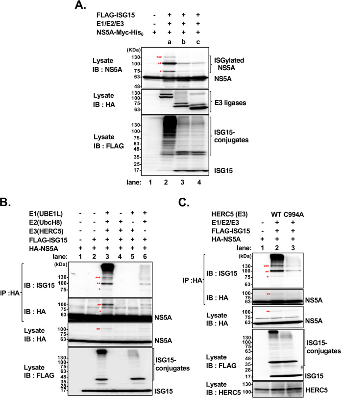
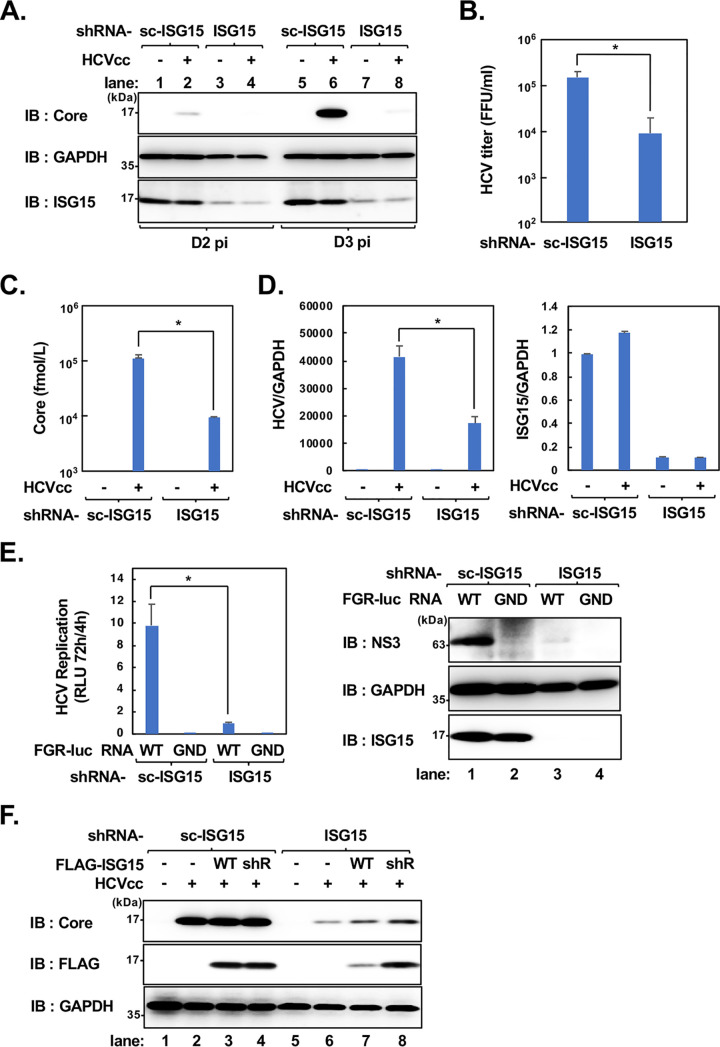
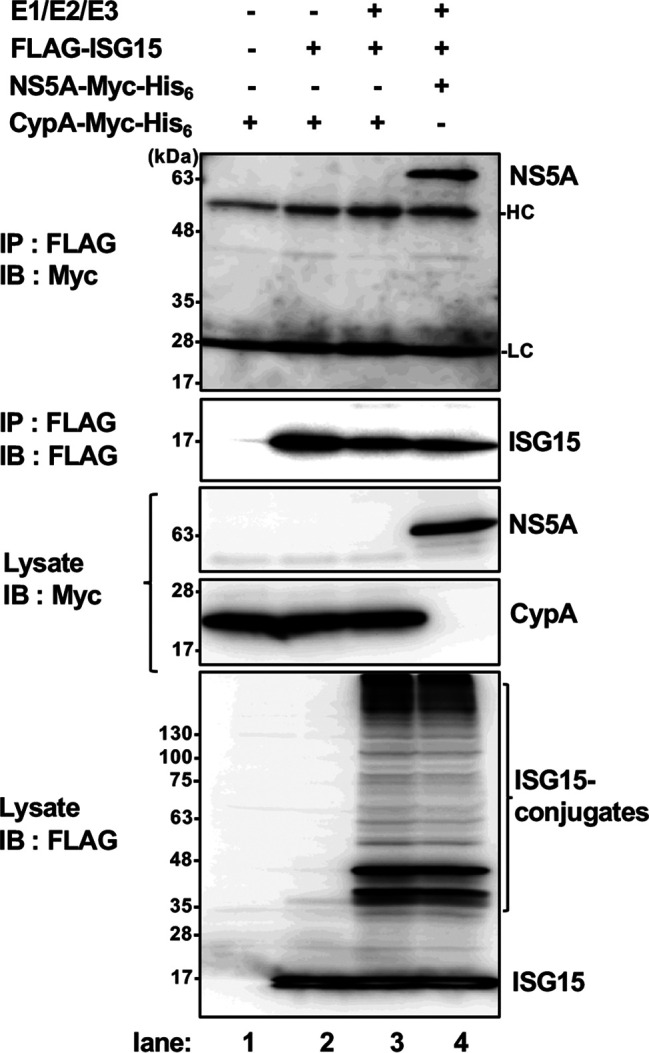
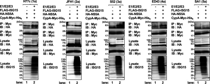
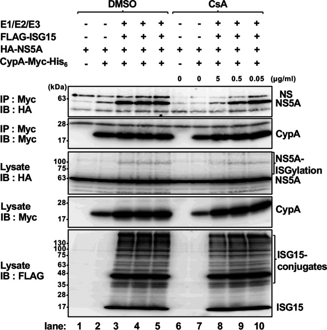
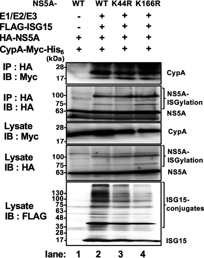
Interferon-stimulated gene 15 (ISG15) is a ubiquitin-like protein that is covalently conjugated to many substrate proteins in order to modulate their functions; this conjugation is called ISGylation. Several groups reported that the ISGylation of hepatitis C virus (HCV) NS5A protein affects HCV replication. However, the ISG15 conjugation sites on NS5A are not well determined, and it is unclear whether the role of NS5A ISGylation in HCV replication is proviral or antiviral. Here, we investigated the role of NS5A ISGylation in HCV replication by using HCV RNA replicons that encode a mutation at each lysine (Lys) residue of the NS5A protein. Immunoblot analyses revealed that 5 Lys residues (K44, K68, K166, K215, and K308) of the 14 Lys residues within NS5A (genotype 1b, Con1) have the potential to accept ISGylation. We tested the NS5A ISGylation among different HCV genotypes and observed that the NS5A proteins of all of the HCV genotypes accept ISGylation at multiple Lys residues. Using an HCV luciferase reporter replicon assay revealed that residue K308 of NS5A is important for HCV (1b, Con1) RNA replication. We observed that K308, one of the Lys residues for NS5A ISGylation, is located within the binding region of cyclophilin A (CypA), which is the critical host factor for HCV replication. We obtained evidence derived from all of the HCV genotypes suggesting that NS5A ISGylation enhances the interaction between NS5A and CypA. Taken together, these results suggest that NS5A ISGylation functions as a proviral factor and promotes HCV replication via the recruitment of CypA.IMPORTANCE Host cells have evolved host defense machinery (such as innate immunity) to eliminate viral infections. Viruses have evolved several counteracting strategies for achieving an immune escape from host defense machinery, including type I interferons (IFNs) and inflammatory cytokines. ISG15 is an IFN-inducible ubiquitin-like protein that is covalently conjugated to the viral protein via specific Lys residues and suppresses viral functions and viral propagation. Here, we demonstrate that HCV NS5A protein accepts ISG15 conjugation at specific Lys residues and that the HERC5 E3 ligase specifically promotes NS5A ISGylation. We obtained evidence suggesting that NS5A ISGylation facilitates the recruitment of CypA, which is the critical host factor for HCV replication, thereby promoting HCV replication. These findings indicate that E3 ligase HERC5 is a potential therapeutic target for HCV infection. We propose that HCV hijacks an intracellular ISG15 function to escape the host defense machinery in order to establish a persistent infection.

Keywords: ISGylation; cyclophilin A; hepatitis C virus; interferon; viral replication.


Figures

FIG 1
FIG 1
HCV NS5A is a target substrate of ISGylation. (A to D) Expression vector encoding HA-NS5A (1b, Con1) was coexpressed with FLAG-ISG15 together with E1 (UBE1L), E2 (UbcH8), and E3 ligase (HERC5) in 293T cells, followed by immunoprecipitation with anti-FLAG mouse MAb or anti-HA rabbit PAb and detection with anti-HA rabbit PAb (A), anti-FLAG mouse MAb (B), and specific antibodies against NS5A (C) and ISG15 (D), respectively. (E) Schematic diagrams of ISG15 and ISG15 mutant ISG15-AA. ISG15 was mutated by replacing the amino acid glycine (G) with alanine (A) within the C-terminal LRLRGG motif sequence. UBL1, ubiquitin-like domain 1; LRLRGG, Leu-Arg-Leu-Arg-Gly-Gly. (F) Expression vector encoding HA-NS5A was coexpressed with FLAG-ISG15 or FLAG-ISG15-AA together with E1, E2, and E3 ligase in 293T cells, followed by immunoprecipitation with anti-HA rabbit PAb and detection with anti-ISG15 mouse MAb or anti-HA rabbit PAb. Input samples (Lysate) were detected by anti-HA rabbit PAb, anti-FLAG mouse MAb, anti-NS5A mouse MAb, or anti-ISG15 mouse MAb. The asterisks indicate the various ISG15-conjugated NS5A proteins. IP, immunoprecipitation; IB, immunoblotting; HC, immunoglobulin heavy chain.
FIG 2
FIG 2
Detection of NS5A ISGylation in the HCV-replicating cells. Huh-7.5 cells were electroporated with in vitro-transcribed chimeric JFH1/5A-Con1 RNA together with E1 (UBE1L), E2 (UbcH8), and E3 ligase (FLAG-tagged HERC5), followed by immunoprecipitation analysis coupled with immunoblotting using NS5A-specific antibody. Input samples (Lysate) were detected with anti-NS5A mouse MAb, anti-FLAG mouse MAb, or anti-ISG15 mouse MAb. The asterisk indicates the 100-kDa conjugates of NS5A ISGylation. IP, immunoprecipitation; IB, immunoblotting.
FIG 3
FIG 3
HERC5 is an E3 ligase that is specific for NS5A ISGylation. (A) Expression vector encoding NS5A-Myc-His6 was coexpressed with FLAG-ISG15 and each of three HA-tagged E3 ligases, including (a) HERC5, (b) TRIM25, and (c) HHARI, together with E1 (UBE1L) and E2 (UbcH8) in 293T cells, followed by detection with anti-NS5A antibody. (B) Expression vector encoding HA-NS5A (1b, Con1) was coexpressed with FLAG-ISG15 together with E1, E2, and E3 ligase (HERC5) or without each of the enzymes, as indicated, in 293T cells, followed by immunoprecipitation with anti-HA rabbit PAb and detection with anti-ISG15 mouse MAb or anti-HA rabbit PAb. (C) Expression vector encoding HA-NS5A was coexpressed with FLAG-ISG15 together with E1, E2, and E3 ligase (HERC5) or an inactive C994A HERC5 mutant in 293T cells, followed by immunoprecipitation with anti-HA rabbit PAb and detection with anti-ISG15 mouse MAb and anti-HA rabbit PAb. Input samples (Lysate) were detected with anti-HA rabbit PAb, anti-FLAG mouse MAb, or anti-HERC5 rabbit PAb. The asterisks indicate the various ISG15-conjugated NS5A proteins.
FIG 4
FIG 4
Identification of ISGylation sites on NS5A protein. (A) Schematic diagram of HCV NS5A (1b, Con1). NS5A possesses 14 Lys residues within the coding sequence. (B, D) Expression vector encoding HA-NS5A or HA-NS5A mutants in which all Lys residues except one (K44, K68, K166, K215, and K308) are mutated to Ala and HA-NS5A mutants containing a point mutation of Lys to Arg at corresponding Lys residues (K44R, K68R, K166R, K215R, and K308R) was coexpressed with FLAG-ISG15 together with E1 (UBE1L), E2 (UbcH8), and E3 ligase (HERC5) in 293T cells, followed by immunoprecipitation with anti-FLAG mouse MAb and detection with anti-HA rabbit PAb. Null indicates a mutant with mutation of all of the Lys residues on NS5A. (C) Expression vector encoding either HA-NS5A (WT) or HA-NS5A mutants (K44, K68, K166, K215 and K308) was coexpressed with FLAG-ISG15 or FLAG-ISG15-AA together with E1, E2, and E3 ligase (HERC5) in 293T cells, followed by immunoprecipitation with anti-HA rabbit PAb and detection with anti-FLAG mouse MAb. (E) Expression vector encoding either HA-NS5A (WT) or NS5A Lys double mutants (K44/68R) was coexpressed with FLAG-ISG15 together with E1, E2, and E3 ligase (HERC5) in 293T cells, followed by immunoprecipitation with anti-FLAG mouse MAb and detection with anti-HA rabbit PAb. Input samples (Lysate) were detected with anti-HA rabbit PAb and anti-FLAG mouse MAb. The asterisks indicate the various ISG15-conjugated NS5A proteins.
FIG 5
FIG 5
NS5A ISGylation via K308 participates in the positive regulation of HCV RNA replication. (A) Experimental procedures for the HCV replication assay. Huh-7.5 cells were electroporated with either HCV subgenomic replicon (SGR) (genotype 1b, Con1) RNA possessing the firefly luciferase gene (HCV-SGR-Luc RNA) or the replicon possessing an NS5A Lys mutant in which a Lys is replaced with Arg. The luciferase activity was measured at the indicated time points after electroporation. The luciferase activity measured at 4 h after electroporation was used to normalize for the input RNA. IRES, internal ribosomal entry site; UTR, untranslated region; EMCV, encephalomyocarditis virus. (B to D) HCV-SGR-Luc RNAs derived from Con1 WT, a replication-defective mutant (GND), or a series of NS5A Lys mutants were electroporated into Huh-7.5 cells. At 48, 72, and 96 h postelectroporation of RNA, cells were harvested, lysed, and subjected to a luciferase assay. Results shown are the mean normalized relative light unit (RLU) values from triplicates ± standard errors (SE) at the indicated time points. *, P < 0.05, and **, P < 0.01, versus the results for cells transduced with SGR-Luc Con1 WT RNAs.
FIG 6
FIG 6
NS5A ISGylation in HCV genotypes. (A) Schematic diagrams of the locations of Lys (K) residues on HCV NS5A of different genotypes (1b, Con1; 1a, H77c; 2a, JFH1; 3a, S52; 4a, ED43; 5a, SA1). (B) Expression vector encoding HA-NS5A of different genotypes (1a [H77c], 2a [JFH1], 3a [S52], 4a [ED43], and 5a [SA1]) was coexpressed with FLAG-ISG15 together with E1 (UBE1L), E2 (UbcH8), and E3 ligase (HERC5) in 293T cells, followed by immunoprecipitation with anti-HA rabbit PAb and detection with anti-ISG15 mouse MAb and anti-HA antibody. Input samples (Lysate) were detected with anti-HA rabbit PAb and anti-FLAG mouse MAb. The slowly migrating bands indicate various ISG15-conjugated NS5A proteins.
FIG 7
FIG 7
The silencing of ISG15 suppresses HCV propagation in human hepatoma Huh-7/scr cells. (A) Stable knockdown cell lines based on the Huh-7/scr cells expressing shRNA targeted to ISG15 or its scrambled sequence (sc-ISG15) were infected with HCVcc (J6/JFH1) at a multiplicity of infection (MOI) of 1. At 2 or 3 days postinoculation, cell lysates were harvested and subjected to immunoblot analysis with the indicated antibodies. (B to D) The extracellular HCV titers (B), the amounts of extracellular core protein (C), and the viral RNA expression levels (D) were determined at 3 days postinoculation by a focus-forming assay (FFA), core ELISA, and real-time PCR, respectively. Results are the mean values from triplicates ± SE. *, P < 0.05 versus the results for Huh-7/scr cells (sc-ISG15). Data from the real-time PCR were normalized to the amount of GAPDH mRNA. (E) HCV full-genomic replicon (FGR) RNAs possessing the firefly luciferase gene (HCV-FGR-Luc RNA) and derived from J6/JFH1 WT or a replication-defective mutant (GND) were electroporated into Huh-7.5 cells. At 72 h postelectroporation of RNA, cells were harvested, lysed, and subjected to a luciferase assay (left) or IB analysis with the indicated antibodies (right). Results shown are the mean normalized RLU values ± SE from triplicates at 72 h after electroporation. (F) Huh7/scr cells expressing shRNA targeted to ISG15 and sc-ISG15 were infected with HCVcc (J6/JFH1) at an MOI of 1. At 2 days postinoculation, the cells were transfected with pCAG-FLAG-ISG15 (WT) or pCAG-FLAG-ISG15 (shR) and subjected to immunoblot analysis with the indicated antibodies at 72 h after transfection.
FIG 8
FIG 8
Reduced expression of NS5A ISGylation in the ISG15 knockdown cells. Huh7/scr cells expressing shRNA targeted to ISG15 and sc-ISG15 were electroporated with the in vitro-transcribed chimeric JFH1/5A-Con1 RNA together with E1 (UBE1L), E2 (UbcH8), and E3 ligase (HA-tagged HERC5), followed by immunoprecipitation analysis coupled with immunoblotting using NS5A-specific antibody. Input samples (Lysate) were detected with anti-NS5A mouse MAb, anti-HA rabbit PAb, anti-GAPDH mouse MAb, or anti-ISG15 mouse MAb. The asterisk indicates the 100-kDa conjugates of NS5A ISGylation. NS, nonspecific bands.
FIG 9
FIG 9
NS5A ISGylation promotes the association of NS5A with cyclophilin A (CypA). (A) The CypA-binding region on NS5A (1b, Con1) domain II. The amino acid regions from 305 to 311, 308 to 324, and 308 to 329 comprising the RNA-binding motif, the CypA-binding region (highlighted in pink), and the NS5B-binding region, respectively, are indicated. W316 is one of the key residues on NS5A for the protein-protein interactions with CypA and NS5B. (B) Expression vector encoding HA-NS5A (1b, Con1) was coexpressed with C-terminal Myc-His6-CypA in the presence or absence of FLAG-ISG15 together with E1 (UBE1L), E2 (UbcH8), and E3 ligase (HERC5) in 293T cells, followed by immunoprecipitation with anti-Myc mouse MAb or anti-HA rabbit PAb and detection with anti-HA rabbit PAb and anti-Myc mouse MAb, respectively. (C) HA-NS5A was coexpressed with CypA-Myc-His6 in the presence or absence of FLAG-ISG15 together with E1, E2, and E3 ligase in 293T cells. At 24 h posttransfection, the cells were treated with 5 μg/ml of CsA for 24 h, followed by immunoprecipitation with anti-Myc mouse MAb and detection with anti-HA rabbit PAb and anti-Myc mouse MAb. Input samples (Lysate) were detected with anti-Myc mouse MAb, anti-HA rabbit PAb, and anti-FLAG mouse MAb. NS, nonspecific bands. The binding ratio of HA-NS5A and CypA-Myc-His6 was determined to measure the band intensities (HA-NS5A [first panel]/CypA-Myc-His6 [second panel], lanes 3 and 6), using ImageQuant TL software (version 7). Results are the mean values from triplicates ± SE. *, P < 0.05 versus the results for the cells treated with dimethyl sulfoxide (DMSO). HC, immunoglobulin heavy chain. (D) The proximity ligation assay (PLA). Huh-7.5 cells were coexpressed with HA-NS5A and CypA-Myc-His6 together with pEGFP or pEGFP-ISG15 plus E1, E2, and E3 ligase, respectively. Cells were fixed, permeabilized, and incubated with anti-HA rabbit PAb and anti-Myc mouse MAb, followed by staining with PLA probes. Red fluorescent spots indicate an interaction with NS5A and CypA. Nuclei were stained with Hoechst 33342. Scale bars, 10 μm.
FIG 10
FIG 10
NS5A, but not CypA, associates with ISG15. Expression vector encoding either CypA-Myc-His6 or NS5A-Myc-His6 was coexpressed in the presence or absence of FLAG-ISG15 together with or without E1 (UBE1L), E2 (UbcH8), and E3 ligase (HERC5) in 293T cells, followed by immunoprecipitation with anti-FLAG mouse MAb and detection with anti-Myc mouse MAb or anti-FLAG mouse MAb. Input samples (Lysate) were detected by anti-Myc mouse MAb and anti-FLAG mouse MAb. HC, immunoglobulin heavy chain; LC, immunoglobulin light chain.
FIG 11
FIG 11
Enhancement of the interaction between NS5A and CypA by ISGylation in an HCV genotype-dependent manner. Expression vector encoding each of the HA-NS5A genotypes (1a [H77c], 2a [JFH1], 3a [S52], 4a [ED43], and 5a [SA1]) was coexpressed with CypA-Myc-His6 in the presence or absence of FLAG-ISG15 together with E1 (UBE1L), E2 (UbcH8), and E3 ligase (HERC5) in 293T cells, followed by immunoprecipitation with anti-Myc mouse MAb and detection with anti-HA rabbit PAb and anti-Myc mouse MAb. Input samples (Lysate) were detected with anti-Myc mouse MAb, anti-HA rabbit PAb, and anti-ISG15 mouse MAb.
FIG 12
FIG 12
CsA interferes with the ISGylation-promoted association of NS5A with CypA in a dose-dependent manner. Expression vector encoding HA-NS5A was coexpressed with CypA-Myc-His6 in the presence or absence of FLAG-ISG15 together with E1 (UBE1L), E2 (UbcH8), and E3 ligase (HERC5) in 293T cells. At 24 h posttransfection, the cells were treated with 5, 0.5, or 0.05 μg/ml of CsA for 24 h, followed by immunoprecipitation with anti-Myc mouse MAb and detection with anti-HA rabbit PAb and anti-Myc mouse MAb. Input samples (Lysate) were detected with anti-Myc mouse MAb, anti-HA rabbit PAb, and anti-FLAG mouse MAb. NS, nonspecific bands.
FIG 13
FIG 13
Enhancement of the interaction between NS5A and CypA in an ISGylation-dependent manner. (A) Expression vector encoding HA-NS5A (1b, Con1) was coexpressed with CypA-Myc-His6 in the presence or absence of FLAG-ISG15 or its conjugation-defective mutant (FLAG-ISG15-AA) together with E1 (UBE1L), E2 (UbcH8), and E3 ligase (HERC5) in 293T cells, followed by immunoprecipitation with anti-Myc mouse MAb and detection with anti-HA rabbit PAb and anti-Myc mouse MAb. (B, C) Expression vector encoding either HA-NS5A (1b, Con1) or NS5A Lys mutant K-Null (B) or K308R (C) was coexpressed with CypA-Myc-His6 in the presence or absence of FLAG-ISG15 together with E1, E2, and E3 ligase in 293T cells, followed by immunoprecipitation with anti-Myc mouse MAb or anti-HA rabbit PAb and detection with anti-HA rabbit PAb or anti-Myc mouse MAb, respectively. Input samples (Lysate) were detected by anti-Myc mouse MAb, anti-HA rabbit PAb, and anti-FLAG mouse MAb. NS, nonspecific bands. The binding ratios of HA-NS5A and CypA-Myc-His6 were determined to measure the band intensities of HA-NS5A (first panel)/CypA-Myc-His6 (second panel), lanes 3 and 4 (A), HA-NS5A (first panel)/CypA-Myc-His6 (second panel), lanes 3 and 6 (B), and CypA-Myc-His6 (first panel)/HA-NS5A (second panel), lanes 3 and 4 (C), using ImageQuant TL software (version 7). Results are the mean values from triplicates ± SE. *, P < 0.05, and **, P < 0.01, versus the results for the cells transduced with WT NS5A.
FIG 14
FIG 14
The K44 and K166 residues on NS5A are not involved in the enhanced association with CypA. Expression vector encoding HA-NS5A (1b, Con1) or an NS5A Lys mutant (K44R or K166R) was coexpressed with CypA-Myc-His6 in the presence or absence of FLAG-ISG15 together with E1 (UBE1L), E2 (UbcH8), and E3 ligase (HERC5) in the 293T cells, followed by immunoprecipitation with anti-HA rabbit PAb and detection with anti-Myc mouse MAb and anti-HA rabbit PAb. Input samples (Lysate) were detected by anti-Myc mouse MAb, anti-HA rabbit PAb, and anti-FLAG mouse MAb.
FIG 15
FIG 15
A model of the functional role of NS5A ISGylation in efficient HCV RNA replication. An intracellular ISG15 is covalently conjugated to HCV NS5A protein by the action of three enzymes, such as E1 (UBE1L), E2 (UbcH8) and E3 ligase (HERC5). The K308 residue within domain II (DII) of NS5A (genotype 1b, Con1) is the acceptor site for ISGylation. The ISGylated NS5A DII may allow its association with CypA, facilitating efficient HCV RNA replication.

References

    1. Moradpour D, Penin F. 2013. Hepatitis C virus proteins: from structure to function. Curr Top Microbiol Immunol 369:113–142. doi:10.1007/978-3-642-27340-7_5. - DOI - PubMed
    1. Moradpour D, Penin F, Rice CM. 2007. Replication of hepatitis C virus. Nat Rev Microbiol 5:453–463. doi:10.1038/nrmicro1645. - DOI - PubMed
    1. Lohmann V. 2013. Hepatitis C virus RNA replication. Curr Top Microbiol Immunol 369:167–198. doi:10.1007/978-3-642-27340-7_7. - DOI - PMC - PubMed
    1. Neufeldt CJ, Cortese M, Acosta EG, Bartenschlager R. 2018. Rewiring cellular networks by members of the Flaviviridae family. Nat Rev Microbiol 16:125–142. doi:10.1038/nrmicro.2017.170. - DOI - PMC - PubMed
    1. Harak C, Lohmann V. 2015. Ultrastructure of the replication sites of positive-strand RNA viruses. Virology 479-480:418–433. doi:10.1016/j.virol.2015.02.029. - DOI - PMC - PubMed

Publication types

LinkOut - more resources