. 2020 Jul 29;10(1):12661.
doi: 10.1038/s41598-020-69630-7.

Running towards amblyopia recovery

Affiliations

Running towards amblyopia recovery

Gabriele Sansevero et al. Sci Rep. .

Abstract

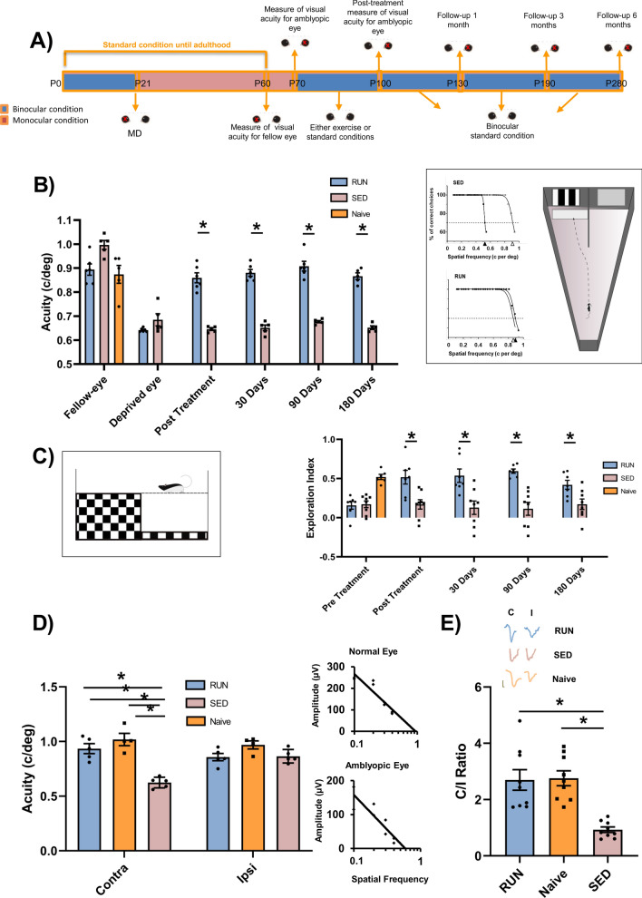
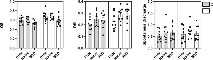
Amblyopia is a neurodevelopmental disorder of the visual cortex arising from abnormal visual experience early in life which is a major cause of impaired vision in infants and young children (prevalence around 3.5%). Current treatments such as eye patching are ineffective in a large number of patients, especially when applied after the juvenile critical period. Physical exercise has been recently shown to enhance adult visual cortical plasticity and to promote visual acuity recovery. With the aim to understand the potentialities for translational applications, we investigated the effects of voluntary physical activity on recovery of depth perception in adult amblyopic rats with unrestricted binocular vision; visual acuity recovery was also assessed. We report that three weeks of voluntary physical activity (free running) induced a marked and long-lasting recovery of both depth perception and visual acuity. In the primary visual cortex, ocular dominance recovered both for excitatory and inhibitory cells and was linked to activation of a specific intracortical GABAergic circuit.


Conflict of interest statement

The authors declare no competing interests.

Figures

Figure 1
Figure 1
Voluntary physical activity induces long-term recovery of visual functions. (A) Schematic diagram of the protocol. (B) Visual acuity through the long-term deprived and the fellow eye was measured using the visual water box task (right, inset). At the end of the physical activity period, visual acuity of the previously deprived eye was different from that of the fellow eye in SED rats (Two Way RM ANOVA, p < 0.001), but not in RUN animals (p = 0.403). Two-Way RM ANOVA revealed that, in RUN rats, the visual acuity of the previously deprived eye measured immediately after the end of the three weeks of physical activity was significant increased with respect to that measured before treatment (p < 0.001) and remained unaltered 30, 90 and 180 days after the end of the treatment (p = 0.760, p = 0.178 and p = 0.996, respectively). In contrast, the visual acuity of the long-term deprived eye did not change throughout the study in SED rats (Two-Way RM ANOVA with Holm-Sidak method, pre-treatment vs. post-treatment, p = 0.403; pre-treatment vs. 1 month after treatment, p = 0.575; pre-treatment vs 3 months, p = 0.992 and pre-treatment vs 6 months, p = 0.566). (C) Visual depth perception was assessed using the visual cliff task (left, inset), as the exploration preference for the shallow and depth side of the arena. One-way ANOVA showed a significant preference for the shallow side in RUN animals, which exhibited an exploration index statistically higher, respectively, than that of SED rats (Holm-Sidak method, p < 0.05). All animals were tested after restoration of binocular vision. (D) Electrophysiological recordings of visual evoked potentials form the primary visual cortex. In SED rats, visual acuity of the deprived eye remained significantly lower with respect to the other eye (One Way RM ANOVA, Holm-Sidak method, p < 0.001); in contrast, a full visual acuity recovery was achieved by RUN rats (p = 0.101) that showed values not different from those of naïve animals (p = 0.127). (E) Ocular dominance was assessed through the C/I VEP ratio in response to a low spatial frequency grating. The C/I VEP ratio was significantly higher in RUN than in SED rats (One-way ANOVA on ranks, Tukey Test, p < 0.05), but not different from that of naïve rats (n = 9; p > 0.05). Error bars indicate s.e.m.; * indicates statistical significance.
Figure 2
Figure 2
Spike analysis of single units for ocular dominance assessment in long-term deprived rats. (A) Assessment of ocular dominance in V1. Plots indicate average and individual ODI values of single animals. While SED rats had a reduced ocular dominance index (ODI) (One-Way ANOVA vs. control, F = 3.429, Holm-Sidak method, p < 0.05.), reflecting a shift toward the non-deprived eye, no significant difference was found between naïve and RUN animals (p = 0.1445). (B) Raster plots (based on spike counts) for three example neurons, one for each group of animals, in response to stimulation of either eye. (Inset) A schematic of the experimental setup for electrophysiological recordings of single units in V1. (C) Spike waveforms for all units analyzed, aligned to minimum and normalized by trough depth, demonstrating narrow-spiking (blue; n = 81) and broad-spiking (green; n = 437) units. (D) Assessment of ocular dominance in broad (excitatory) and narrow (inhibitory) spiking cells in V1. Long-term deprivation induced an ocular dominance shift in both classes of cells (broad-spiking, SED, n = 137, Naïve, n = 159, One-Way ANOVA, p < 0.001; narrow-spiking, SED, n = 23, Naïve, n = 32, One-Way ANOVA, p < 0.05). Physical activity induced a full recovery of OD in both classes of cells, and no differences were detectable between naïve and RUN animals (broad-spiking: RUN, n = 141; One-Way ANOVA, p = 0.55) (and narrow-spiking cells: RUN, n = 26; One-Way ANOVA, p < 0.69). * indicates statistical significance.
Figure 3
Figure 3
Assessment of direction and orientation selectivity and spontaneous discharge in V1. No difference was found in either the direction selectivity index (DSI), the orientation selectivity index (OSI), or in the spontaneous discharge among the three groups of animals (Two-Way RM ANOVA, Holm-Sidak method, F = 0.150, p = 0.861; F = 0.200, p = 0.820; F = 0.523, p = 0.599; respectively). Error bars indicate s.e.m.; * indicates statistical significance.
Figure 4
Figure 4
Phenotypical dissection of active GABAergic neurons in amblyopia recovery induced by voluntary physical activity. (A) Schematic diagram of the protocol employed for c-fos immunohistochemistry. (B) Schematic diagram of interneuron staining in the primary visual cortex . (CE) Activation of vasoactive intestinal peptide positive (VIP+, C) somatostatin positive (SOM+, D), and parvalbumin positive (PARVA+, E) interneurons in the primary visual cortex of adult amblyopic animals. The number of SOM+ cells was significantly increased with respect to naïve animals in SED rats, but it returned to normal levels in RUN animals (One-way ANOVA on ranks, Dunn’s Method, q = 2.687 and q = 0.109, respectively). The number of VIP+ cells was significantly increased in the visual cortex of RUN rats with respect to SED animals (One-way ANOVA on ranks, Dunns’ Method, q = 2.453, p < 0.05).The number of PARVA+ cells did not change among the three different groups (One-way ANOVA on ranks, q = 0.587). Error bars indicate s.e.m.; * indicates statistical significance. Right panels: representative images of double immunostaining for VIP+/cFos, SOM+/cFos+, and PARVA+/cFos+ cells in the primary visual cortex of RUN, SED and naïve rats. Scale-bar: 12 µm.

References

    1. Morishita H, Hensch TK. Critical period revisited: impact on vision. Curr. Opin. Neurobiol. 2008;18:101–107. doi: 10.1016/j.conb.2008.05.009. - DOI - PubMed
    1. Bavelier D, Levi DM, Li RW, Dan Y, Hensch TK. Removing brakes on adult brain plasticity: from molecular to behavioral interventions. J. Neurosci. 2010;30:14964–14971. doi: 10.1523/JNEUROSCI.4812-10.2010. - DOI - PMC - PubMed
    1. Sale A, Berardi N. Active training for amblyopia in adult rodents. Front. Behav. Neurosci. 2015;9:281. doi: 10.3389/fnbeh.2015.00281. - DOI - PMC - PubMed
    1. Holmes JM, Clarke MP. Amblyopia. Lancet. 2006;367:1343–1351. doi: 10.1016/S0140-6736(06)68581-4. - DOI - PubMed
    1. Sale A, et al. Environmental enrichment in adulthood promotes amblyopia recovery through a reduction of intracortical inhibition. Nat. Neurosci. 2007;10:679–681. doi: 10.1038/nn1899. - DOI - PubMed

Publication types