Review
. 2020 Oct;140(4):417-447.
doi: 10.1007/s00401-020-02196-w. Epub 2020 Jul 29.

The physiological roles of tau and Aβ: implications for Alzheimer's disease pathology and therapeutics

Affiliations
Review

The physiological roles of tau and Aβ: implications for Alzheimer's disease pathology and therapeutics

Sarah A Kent et al. Acta Neuropathol. 2020 Oct.

Abstract

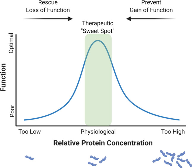
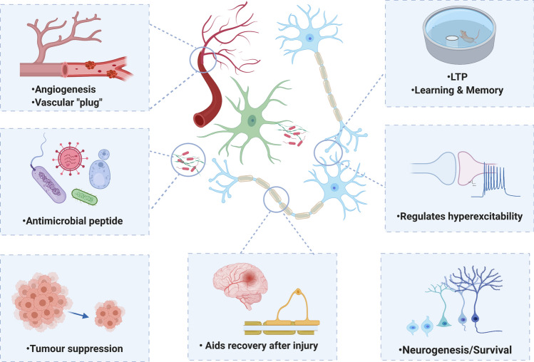
Tau and amyloid beta (Aβ) are the prime suspects for driving pathology in Alzheimer's disease (AD) and, as such, have become the focus of therapeutic development. Recent research, however, shows that these proteins have been highly conserved throughout evolution and may have crucial, physiological roles. Such functions may be lost during AD progression or be unintentionally disrupted by tau- or Aβ-targeting therapies. Tau has been revealed to be more than a simple stabiliser of microtubules, reported to play a role in a range of biological processes including myelination, glucose metabolism, axonal transport, microtubule dynamics, iron homeostasis, neurogenesis, motor function, learning and memory, neuronal excitability, and DNA protection. Aβ is similarly multifunctional, and is proposed to regulate learning and memory, angiogenesis, neurogenesis, repair leaks in the blood-brain barrier, promote recovery from injury, and act as an antimicrobial peptide and tumour suppressor. This review will discuss potential physiological roles of tau and Aβ, highlighting how changes to these functions may contribute to pathology, as well as the implications for therapeutic development. We propose that a balanced consideration of both the physiological and pathological roles of tau and Aβ will be essential for the design of safe and effective therapeutics.

Keywords: Memory; Microtubule dynamics; Myelination; Synapse; Therapeutics; Vasculature.


Figures

Fig. 1
Fig. 1
a The two pathways through which APP can be cleaved. The non-amyloidogenic (α) pathway (left-hand side of diagram) involves the cleavage of APP by α-secretase, within the Aβ sequence, to form C-terminal fragment α (CTFα) and soluble APP α (sAPPα). γ-secretase then cleaves the resulting CTFα, releasing the Aβ intracellular domain (AICD) and the extracellular p3 fragment. The amyloidogenic (β) pathway (right-hand side of the diagram) involves the cleavage of APP by BACE1 to form CTFβ and sAPPβ. γ-secretase then cleaves the resulting CTFβ, releasing the AICD and Aβ. b Aβ monomers can assemble to form higher order structures, from oligomers, to protofibrils and eventually mature fibrils containing β-sheets which form the core component of amyloid plaques. Created with https://biorender.com/
Fig. 2
Fig. 2
a Tau is encoded by the MAPT gene on chromosome 17. A total of 6 tau protein isoforms are generated via alternative splicing of exons 2, 3, and 10. Inclusion of exon 10 produces tau with 4 microtubule-binding (MTB) domains (4R), with omission of exon 10 producing tau with 3 MTB domains (3R). Tau can include (2 N or 1 N) or exclude (0 N) amino-terminal inserts through regulation of exons 2 and 3. Only 0N3R tau is expressed in the foetal human or mouse brain, with all 6 tau isoforms being expressed in adult humans. Adult mice and rats show almost exclusive expression of 4R tau. b Phosphorylated tau monomers can assemble to form oligomers, filaments (both straight and paired helical) and eventually tangles. N N-terminus, PRD proline-rich domain, MTB microtubule-binding domains, C C-terminus. Created with https://biorender.com/
Fig. 3
Fig. 3
Tissue-level protein expression of APP [297], BACE1 [298], and presenilin-1 [299] (indicating potential for Aβ production) and tau (MAPT) [296] according to the Human Protein Atlas [262]. Created with https://biorender.com/
Fig. 4
Fig. 4
Schematic representation of the hormetic responses to tau and Aß concentration. There is an optimal concentration of tau or Aβ for a number of physiological functions. Too little protein (or loss of function modifications) or too much protein (or gain of function modifications) can both disrupt normal function. Effectively rescuing loss of function or preventing gain of function to maintain optimal physiological conditions should be the ultimate goal of therapeutics. Created with https://biorender.com/
Fig. 5
Fig. 5
A schematic representation of the suggested physiological roles of tau in the brain and body. Created with https://biorender.com/
Fig. 6
Fig. 6
A schematic representation of the suggested physiological roles of Aβ in the brain and body. Created with https://biorender.com/

References

    1. Adalbert R, Milde S, Durrant C, Ando K, Stygelbout V, Yilmaz Z, et al. Interaction between a MAPT variant causing frontotemporal dementia and mutant APP affects axonal transport. Neurobiol Aging. 2018;68:68–75. doi: 10.1016/j.neurobiolaging.2018.03.033. - DOI - PMC - PubMed
    1. Adams JN, Lockhart SN, Li L, Jagust WJ. Relationships between tau and glucose metabolism reflect alzheimer’s disease pathology in cognitively normal older adults. Cereb Cortex N Y NY. 2019;29:1997–2009. doi: 10.1093/cercor/bhy078. - DOI - PMC - PubMed
    1. Ahmed T, Van der Jeugd A, Blum D, Galas M-C, D’Hooge R, Buee L, et al. Cognition and hippocampal synaptic plasticity in mice with a homozygous tau deletion. Neurobiol Aging. 2014;35:2474–2478. doi: 10.1016/j.neurobiolaging.2014.05.005. - DOI - PubMed
    1. Allen B, Ingram E, Takao M, Smith MJ, Jakes R, Virdee K, et al. Abundant tau filaments and nonapoptotic neurodegeneration in transgenic mice expressing human p301s tau protein. J Neurosci. 2002;22:9340–9351. doi: 10.1523/JNEUROSCI.22-21-09340.2002. - DOI - PMC - PubMed
    1. Alzheimer’s Association 2019 Alzheimer’s disease facts and figures. Alzheimers Dement. 2019;15:321–387. doi: 10.1016/j.jalz.2019.01.010. - DOI

Publication types