

Comparative study of lung ultrasound and chest computed tomography scan in the assessment of severity of confirmed COVID-19 pneumonia
- PMID: 32728966
- PMCID: PMC7388119
- DOI: 10.1007/s00134-020-06186-0
Comparative study of lung ultrasound and chest computed tomography scan in the assessment of severity of confirmed COVID-19 pneumonia
Abstract
Purpose: The relationship between lung ultrasound (LUS) and chest computed tomography (CT) scans in patients with severe acute respiratory syndrome coronavirus 2 (SARS-CoV-2) pneumonia is not clearly defined. The primary objective of our study was to assess the performance of LUS in determining severity of SARS-CoV-2 pneumonia compared with chest CT scan. Secondary objectives were to test the association between LUS score and location of the patient, use of mechanical ventilation, and the pulse oximetry (SpO2)/fractional inspired oxygen (FiO2) ratio.
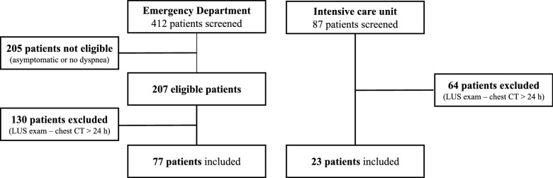
Methods: A multicentre observational study was performed between 15 March and 20 April 2020. Patients in the Emergency Department (ED) or Intensive Care Unit (ICU) with acute dyspnoea who were PCR positive for SARS-CoV-2, and who had LUS and chest CT performed within a 24-h period, were included.
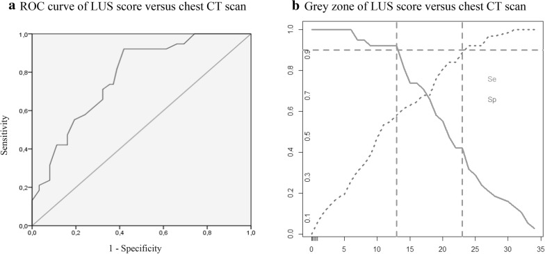
Results: One hundred patients were included. LUS score was significantly associated with pneumonia severity assessed by chest CT and clinical features. The AUC of the ROC curve of the relationship of LUS versus chest CT for the assessment of severe SARS-CoV-2 pneumonia was 0.78 (CI 95% 0.68-0.87; p < 0.0001). A high LUS score was associated with the use of mechanical ventilation, and with a SpO2/FiO2 ratio below 357.
Conclusion: In known SARS-CoV-2 pneumonia patients, the LUS score was predictive of pneumonia severity as assessed by a chest CT scan and clinical features. Within the limitations inherent to our study design, LUS can be used to assess SARS-CoV-2 pneumonia severity.
Keywords: Chest computed tomography; Diagnostic accuracy; Lung ultrasound; SARS-CoV-2.
Conflict of interest statement
LZ and TM received fees for teaching ultrasound to GE healthcare customers.
Figures
Similar articles
-
Correlation between Chest Computed Tomography and Lung Ultrasonography in Patients with Coronavirus Disease 2019 (COVID-19).Ultrasound Med Biol. 2020 Nov;46(11):2918-2926. doi: 10.1016/j.ultrasmedbio.2020.07.003. Epub 2020 Jul 13. Ultrasound Med Biol. 2020. PMID: 32771222 Free PMC article.
-
Lung Ultrasound in COVID-19 Pneumonia: Correlations with Chest CT on Hospital admission.Respiration. 2020;99(7):617-624. doi: 10.1159/000509223. Epub 2020 Jun 22. Respiration. 2020. PMID: 32570265 Free PMC article.
-
Lung ultrasound is a reliable diagnostic technique to predict abnormal CT chest scan and to detect oxygen requirements in COVID-19 pneumonia.Aging (Albany NY). 2020 Oct 30;12(20):19945-19953. doi: 10.18632/aging.104150. Epub 2020 Oct 30. Aging (Albany NY). 2020. PMID: 33136555 Free PMC article.
-
Thoracic imaging tests for the diagnosis of COVID-19.Cochrane Database Syst Rev. 2020 Sep 30;9:CD013639. doi: 10.1002/14651858.CD013639.pub2. Cochrane Database Syst Rev. 2020. Update in: Cochrane Database Syst Rev. 2020 Nov 26;11:CD013639. doi: 10.1002/14651858.CD013639.pub3. PMID: 32997361 Updated.
-
Lung Ultrasound May Support Diagnosis and Monitoring of COVID-19 Pneumonia.Ultrasound Med Biol. 2020 Nov;46(11):2908-2917. doi: 10.1016/j.ultrasmedbio.2020.07.018. Epub 2020 Jul 20. Ultrasound Med Biol. 2020. PMID: 32807570 Free PMC article. Review.
Cited by
-
Thoracic UltrasONOgraphy Reporting: The TUONO Study.J Clin Med. 2022 Nov 30;11(23):7126. doi: 10.3390/jcm11237126. J Clin Med. 2022. PMID: 36498700 Free PMC article.
-
Diagnostic Modalities in Critical Care: Point-of-Care Approach.Diagnostics (Basel). 2021 Nov 25;11(12):2202. doi: 10.3390/diagnostics11122202. Diagnostics (Basel). 2021. PMID: 34943438 Free PMC article. Review.
-
Combining lung ultrasound and Wells score for diagnosing pulmonary embolism in critically ill COVID-19 patients.J Thromb Thrombolysis. 2021 Jul;52(1):76-84. doi: 10.1007/s11239-020-02323-0. Epub 2020 Nov 3. J Thromb Thrombolysis. 2021. PMID: 33145663 Free PMC article.
-
Assessing COVID-19 pneumonia-Clinical extension and risk with point-of-care ultrasound: A multicenter, prospective, observational study.J Am Coll Emerg Physicians Open. 2021 May 1;2(3):e12429. doi: 10.1002/emp2.12429. eCollection 2021 Jun. J Am Coll Emerg Physicians Open. 2021. PMID: 33969350 Free PMC article.
-
Lung Ultrasound Score in COVID-19 Patients Correlates with PO2/FiO2, Intubation Rates, and Mortality.West J Emerg Med. 2024 Jan;25(1):28-39. doi: 10.5811/westjem.59975. West J Emerg Med. 2024. PMID: 38205982 Free PMC article.
References
Publication types
MeSH terms
LinkOut - more resources
Full Text Sources
Medical
Miscellaneous
